Embed Size (px)
Citation preview
8/2/2019 Anatomía (Esp)
http://slidepdf.com/reader/full/anatomia-esp 1/58
Anatomía para el mejor uso de
nosotros
U l t r a s o n o g r a f í a
8/2/2019 Anatomía (Esp)
http://slidepdf.com/reader/full/anatomia-esp 2/58
. 2
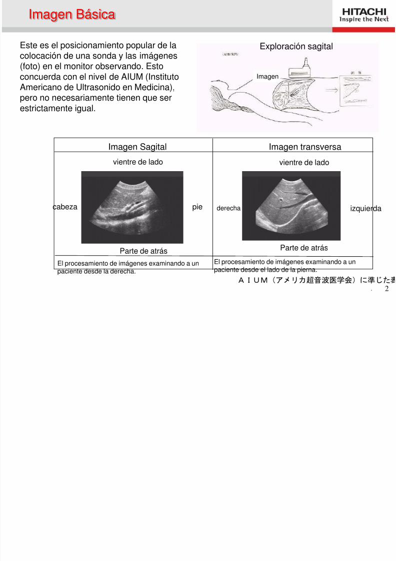
Este es el posicionamiento popular de lacolocación de una sonda y las imágenes
(foto) en el monitor observando. Estoconcuerda con el nivel de AIUM (InstitutoAmericano de Ultrasonido en Medicina),pero no necesariamente tienen que serestrictamente igual.
Imagen Sagital Imagen transversa
El procesamiento de imágenes examinando a unpaciente desde la derecha.
El procesamiento de imágenes examinando a unpaciente desde el lado de la pierna.
vientre de lado
Parte de atrás
pie
vientre de lado
derecha
Parte de atrás
izquierda
AIUM(アメリカ超音波医学会)に準じた
cabeza
Imagen
Imagen Básica
Exploración sagital
8/2/2019 Anatomía (Esp)
http://slidepdf.com/reader/full/anatomia-esp 3/58
. 3
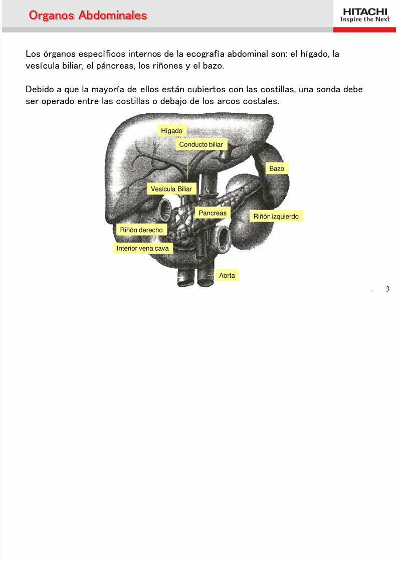
Conducto biliar
Hígado
Riñón derecho
Vesícula Biliar
Riñón izquierdo
Bazo
Pancreas
Aorta
Organos Abdominales
Los órganos específicos internos de la ecografía abdominal son: el hígado, la
vesícula biliar, el páncreas, los riñones y el bazo.
Debido a que la mayoría de ellos están cubiertos con las costillas, una sonda debeser operado entre las costillas o debajo de los arcos costales.
Interior vena cava
8/2/2019 Anatomía (Esp)
http://slidepdf.com/reader/full/anatomia-esp 4/58
. 4
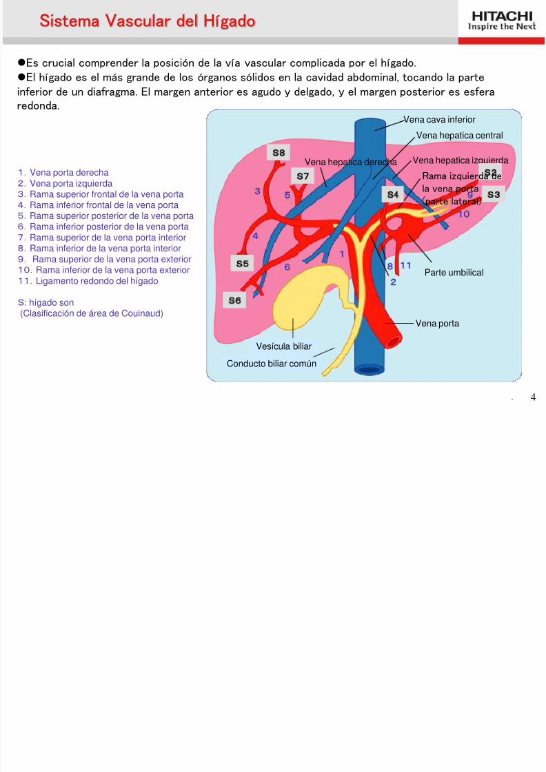
1.Vena porta derecha2.Vena porta izquierda
3.Rama superior frontal de la vena porta4.Rama inferior frontal de la vena porta5.Rama superior posterior de la vena porta6.Rama inferior posterior de la vena porta7.Rama superior de la vena porta interior8.Rama inferior de la vena porta interior9. Rama superior de la vena porta exterior10.Rama inferior de la vena porta exterior11.
Ligamento redondo del hígado
S: hígado son(Clasificación de área de Couinaud)
S
Vena cava inferior
Vena hepatica central
Vena hepatica izquierdaVena hepatica derecha
Parte umbilical
Vena porta
S6
S5
S8
S7 S2
S3
9
10
8 11
2
1
6
4
5 3 S4
Sistema Vascular del Hígado
Es crucial comprender la posición de la vía vascular complicada por el hígado.El hígado es el más grande de los órganos sólidos en la cavidad abdominal, tocando la parte
inferior de un diafragma. El margen anterior es agudo y delgado, y el margen posterior es esferaredonda.
Vesícula biliar
Conducto biliar común
Rama izquierda de
la vena porta(parte lateral)
8/2/2019 Anatomía (Esp)
http://slidepdf.com/reader/full/anatomia-esp 5/58
. 5
Couinaud Segment
Right hepatic vein
Umbilical portion
IVC
S1
S2
S3
S5
S6
S7
S8
Middle hepatic vein
S4
Higado
Segmento de hígado: En el diagnóstico de ultrasonido, el hígado se divide en ocho segmentos, teniendo lavena porta, la vena hepática, el ligamento, la fisura, etc como indicadores. Ya que la ubicación de la lesióndescrita se indica a menudo por los ocho segmentos, que vale la pena conocer. (Segmento de Couinaud)
Vesicula biliar
S1
S2
S2
S3
S3 S4
S4
S5
S5
S6
S6
S7
S7 S8
IVC
Portal vein
S1: Caudado del lóbulo + Proceso de caudadoS2:Left lateral superior sub segmentS3:Left lateral inferior sub segmentS4:Left medial segment
Left lateral segment
S5:Right anterior inferior sub segmentS6:Right posterior inferior sub segmentS7:Right posterior superior sub segment
S8:Right anterior superior sub segment
Rightposterior
segment
Rightanteriorsegment
Right lobe
Left lobe
Liver district classification:
8/2/2019 Anatomía (Esp)
http://slidepdf.com/reader/full/anatomia-esp 6/58
. 6
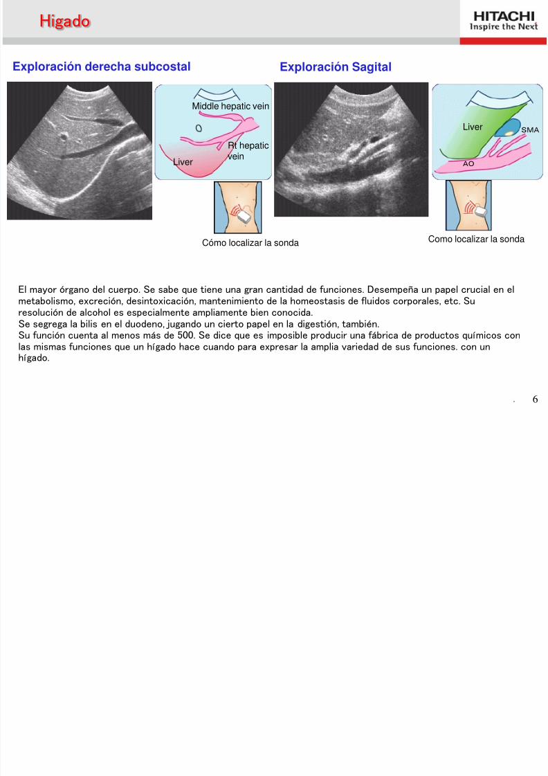
Exploración derecha subcostal Exploración Sagital
Cómo localizar la sonda
Rt hepaticvein
Liver AO
Liver SMA
Higado
Middle hepatic vein
El mayor órgano del cuerpo. Se sabe que tiene una gran cantidad de funciones. Desempeña un papel crucial en elmetabolismo, excreción, desintoxicación, mantenimiento de la homeostasis de fluidos corporales, etc. Suresolución de alcohol es especialmente ampliamente bien conocida.Se segrega la bilis en el duodeno, jugando un cierto papel en la digestión, también.Su función cuenta al menos más de 500. Se dice que es imposible producir una fábrica de productos químicos conlas mismas funciones que un hígado hace cuando para expresar la amplia variedad de sus funciones. con unhígado.
Como localizar la sonda
8/2/2019 Anatomía (Esp)
http://slidepdf.com/reader/full/anatomia-esp 7/58. 7
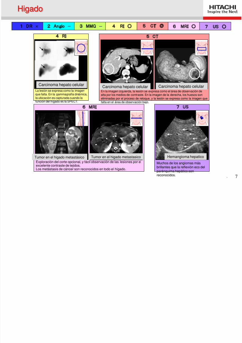
1 DR × 2 Angio - 3 MMG - 4 RI ○ 5 CT ◎ 6 MRI ○ 7 US ○
7 US
肝血管腫
6 MRI
転移性肝癌
Exploración del corte opcional, y fácil observación de las lesiones por elexcelente contraste de tejidos.
Los metástasis de cáncer son reconocidos en todo el hígado.
5 CT
肝細胞癌 肝細胞癌
En la imagen izquierda, la lesión se expresa como el área de observación dealta por los medios de contraste. En la imagen de la derecha, los huesos soneliminadas por el proceso de retoque, y la lesión se expresa como la imagen quefalta en el área de observación bajo.
4 RI
La lesión se expresa como la imagenque falta. En la gammagrafía dinámica,la ubicación es capturada cuando lafunción del hígado es la SPECT.
Higado
Carcinoma hepato celular Carcinoma hepato celular Carcinoma hepato celular
Tumor en el higado metastásico Tumor en el higado metastasico Hemangioma hepatico
Muchos de los angiomas másbrillantes que la reflexión eco delparénquima hepático sonreconocidos.
8/2/2019 Anatomía (Esp)
http://slidepdf.com/reader/full/anatomia-esp 8/58. 8
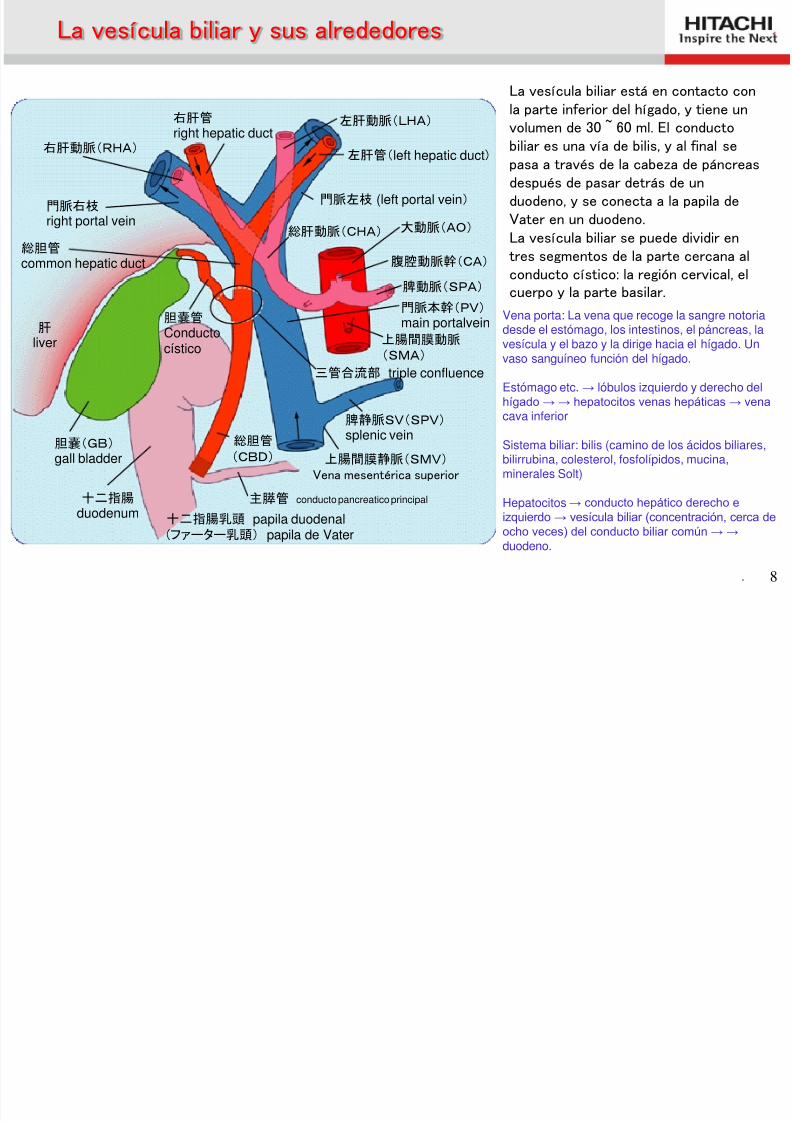
Vena porta: La vena que recoge la sangre notoriadesde el estómago, los intestinos, el páncreas, lavesícula y el bazo y la dirige hacia el hígado. Unvaso sanguíneo función del hígado.
Estómago etc. → lóbulos izquierdo y derecho delhígado → → hepatocitos venas hepáticas → vena
cava inferior
Sistema biliar: bilis (camino de los ácidos biliares,bilirrubina, colesterol, fosfolípidos, mucina,minerales Solt)
Hepatocitos → conducto hepático derecho e
izquierdo → vesícula biliar (concentración, cerca deocho veces) del conducto biliar común → →
duodeno.
左肝動脈(LHA)
左肝管(left hepatic duct)
門脈左枝 (left portal vein)
大動脈(AO) 総肝動脈(CHA)
腹腔動脈幹(CA)
脾動脈(SPA)
門脈本幹(PV) main portalvein
上腸間膜動脈 (SMA)
三管合流部 triple confluence
脾静脈SV(SPV) splenic vein
上腸間膜静脈(SMV) Vena mesentérica superior
総胆管
(CBD)
主膵管 conducto pancreatico principal
十二指腸乳頭 papila duodenal(ファーター乳頭) papila de Vater
十二指腸 duodenum
胆嚢(GB) gall bladder
胆嚢管 Conductocístico
右肝管
right hepatic duct右肝動脈(RHA)
門脈右枝 right portal vein
総胆管 common hepatic duct
肝 liver
La vesícula biliar y sus alrededores
La vesícula biliar está en contacto conla parte inferior del hígado, y tiene unvolumen de 30 ~ 60 ml. El conducto
biliar es una vía de bilis, y al final sepasa a través de la cabeza de páncreasdespués de pasar detrás de unduodeno, y se conecta a la papila deVater en un duodeno.La vesícula biliar se puede dividir entres segmentos de la parte cercana al
conducto cístico: la región cervical, elcuerpo y la parte basilar.
8/2/2019 Anatomía (Esp)
http://slidepdf.com/reader/full/anatomia-esp 9/58. 9
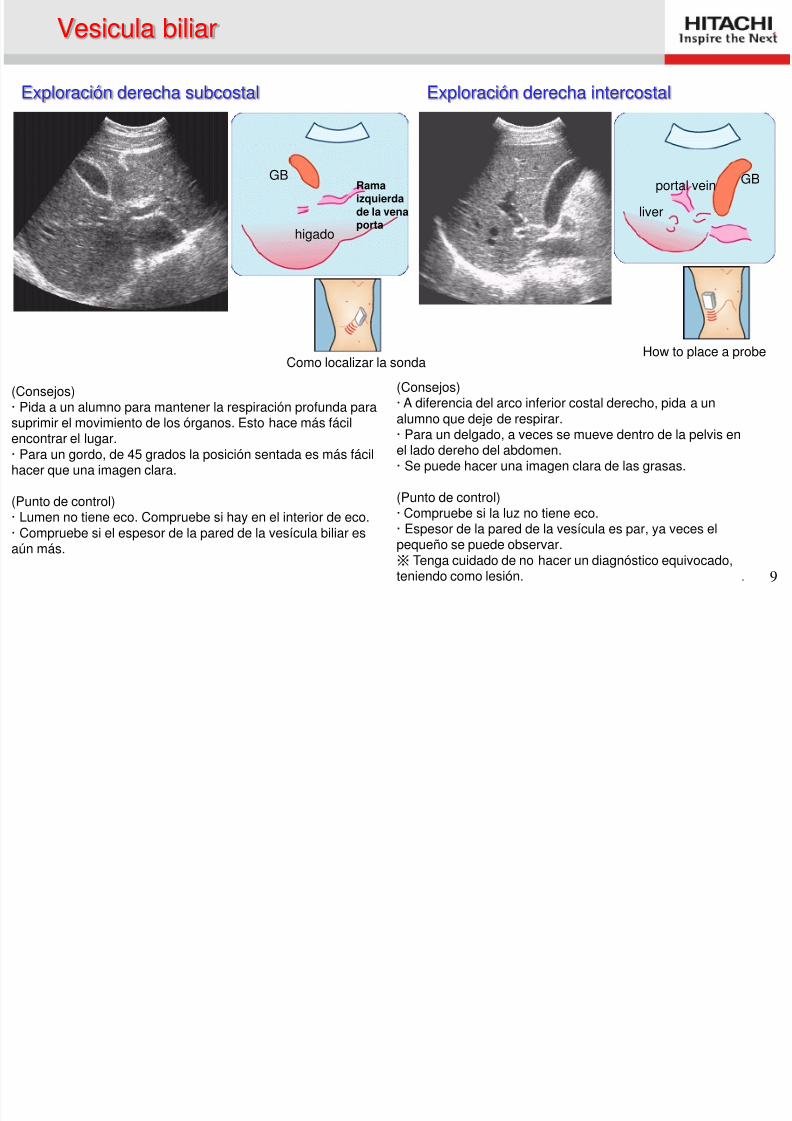
(Consejos)· Pida a un alumno para mantener la respiración profunda parasuprimir el movimiento de los órganos. Esto hace más fácilencontrar el lugar.· Para un gordo, de 45 grados la posición sentada es más fácilhacer que una imagen clara.
(Punto de control)· Lumen no tiene eco. Compruebe si hay en el interior de eco.· Compruebe si el espesor de la pared de la vesícula biliar es
aún más.
(Consejos)· A diferencia del arco inferior costal derecho, pida a un
alumno que deje de respirar.· Para un delgado, a veces se mueve dentro de la pelvis enel lado dereho del abdomen.· Se puede hacer una imagen clara de las grasas.
(Punto de control)· Compruebe si la luz no tiene eco.· Espesor de la pared de la vesícula es par, ya veces elpequeño se puede observar.
※ Tenga cuidado de no hacer un diagnóstico equivocado,teniendo como lesión.
Como localizar la sonda
GB
higado
Ramaizquierdade la venaporta
GB
liver
portal vein
Vesicula biliar
Exploración derecha subcostal Exploración derecha intercostal
How to place a probe
8/2/2019 Anatomía (Esp)
http://slidepdf.com/reader/full/anatomia-esp 10/58. 10
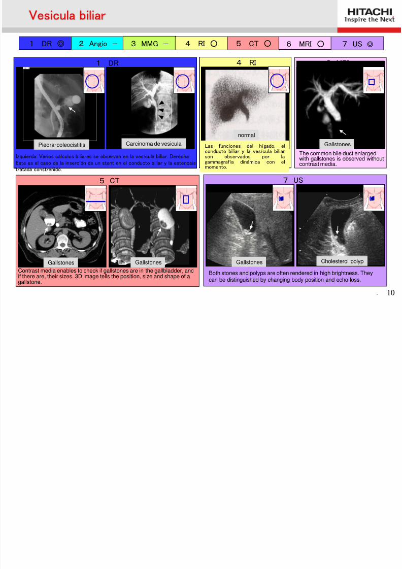
1 DR ◎ 2 Angio - 3 MMG - 4 RI ○ 5 CT ○ 6 MRI ○ 7 US ◎
7 US
Both stones and polyps are often rendered in high brightness. They
can be distinguished by changing body position and echo loss.
胆石 胆嚢ポリープ
5 CT
胆石症 胆石症 Contrast media enables to check if gallstones are in the gallbladder, andif there are, their sizes. 3D image tells the position, size and shape of a
gallstone.
6 MRI
胆石
The common bile duct enlargedwith gallstones is observed withoutcontrast media.
5 CT
1 DR
胆石・慢性胆嚢炎
Izquierda: Varios cálculos biliares se observan en la vesícula biliar. Derecha:Este es el caso de la inserción de un stent en el conducto biliar y la estenosistratada constreñido.
胆管癌
4 RI
健常例 Las funciones del hígado, elconducto biliar y la vesícula biliarson observados por lagammagrafía dinámica con elmomento.
Vesicula biliar
Piedra・coleocistitis Carcinoma de vesicula
Cholesterol polyp
normal
Gallstones Gallstones Gallstones
Gallstones
8/2/2019 Anatomía (Esp)
http://slidepdf.com/reader/full/anatomia-esp 11/58. 11
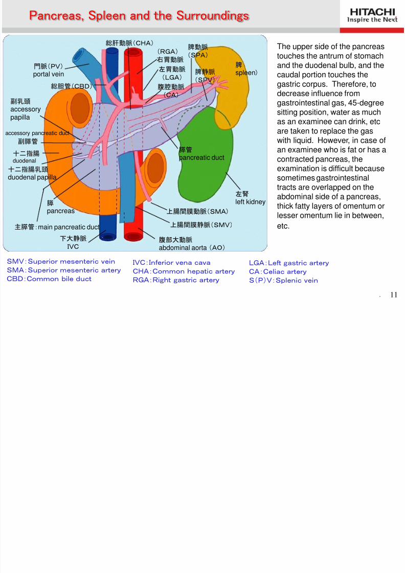
SMV:Superior mesenteric vein SMA:Superior mesenteric artery CBD:Common bile duct
脾
spleen)
総肝動脈(CHA)
腹腔動脈 (CA)
左胃動脈 (LGA)
(RGA) 右胃動脈
脾静脈 (SPV)
脾動脈 (SPA)
膵管 pancreatic duct
左腎 left kidney
上腸間膜動脈(SMA)
上腸間膜静脈(SMV)
腹部大動脈 abdominal aorta (AO)
下大静脈 IVC
門脈(PV) portal vein
総胆管(CBD)
副乳頭 accessorypapilla
十二指腸 duodenal
十二指腸乳頭 duodenal papilla
膵
pancreas
主膵管:main pancreatic duct
副膵管
accessory pancreatic duct
IVC:Inferior vena cava CHA:Common hepatic artery
RGA:Right gastric artery
LGA:Left gastric artery CA:Celiac artery
S(P)V:Splenic vein
Pancreas, Spleen and the Surroundings
The upper side of the pancreastouches the antrum of stomachand the duodenal bulb, and thecaudal portion touches thegastric corpus. Therefore, todecrease influence fromgastrointestinal gas, 45-degreesitting position, water as muchas an examinee can drink, etcare taken to replace the gas
with liquid. However, in case ofan examinee who is fat or has acontracted pancreas, theexamination is difficult becausesometimes gastrointestinaltracts are overlapped on theabdominal side of a pancreas,thick fatty layers of omentum or
lesser omentum lie in between,
etc.
8/2/2019 Anatomía (Esp)
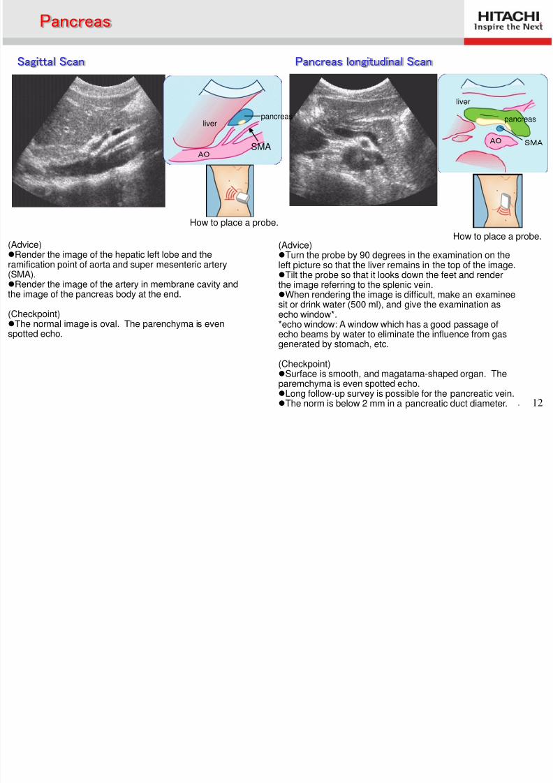
http://slidepdf.com/reader/full/anatomia-esp 12/58. 12
(Advice)Render the image of the hepatic left lobe and theramification point of aorta and super mesenteric artery(SMA).Render the image of the artery in membrane cavity andthe image of the pancreas body at the end.
(Checkpoint)The normal image is oval. The parenchyma is evenspotted echo.
(Advice)Turn the probe by 90 degrees in the examination on theleft picture so that the liver remains in the top of the image.Tilt the probe so that it looks down the feet and renderthe image referring to the splenic vein.When rendering the image is difficult, make an examineesit or drink water (500 ml), and give the examination asecho window*.*echo window: A window which has a good passage ofecho beams by water to eliminate the influence from gasgenerated by stomach, etc.
(Checkpoint)Surface is smooth, and magatama-shaped organ. The
paremchyma is even spotted echo.Long follow-up survey is possible for the pancreatic vein.The norm is below 2 mm in a pancreatic duct diameter.
How to place a probe.
liver
AO
pancreas pancreas
AO SMA
liver
Pancreas
Sagittal Scan Pancreas longitudinal Scan
SMA
How to place a probe.
8/2/2019 Anatomía (Esp)
http://slidepdf.com/reader/full/anatomia-esp 13/58. 13
How to place a probe.
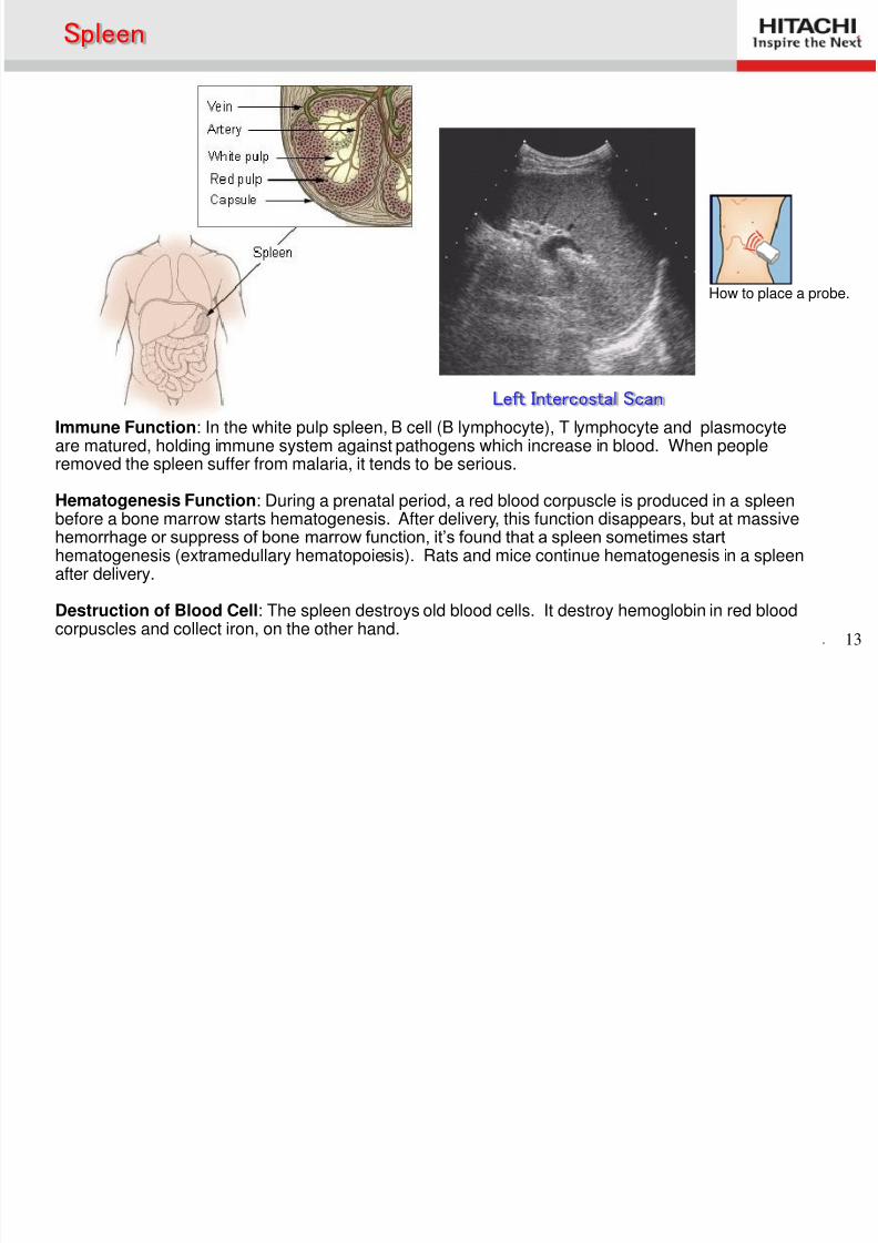
Spleen
Left Intercostal Scan
Immune Function: In the white pulp spleen, B cell (B lymphocyte), T lymphocyte and plasmocyteare matured, holding immune system against pathogens which increase in blood. When people
removed the spleen suffer from malaria, it tends to be serious.
Hematogenesis Function: During a prenatal period, a red blood corpuscle is produced in a spleenbefore a bone marrow starts hematogenesis. After delivery, this function disappears, but at massivehemorrhage or suppress of bone marrow function, it’s found that a spleen sometimes starthematogenesis (extramedullary hematopoiesis). Rats and mice continue hematogenesis in a spleenafter delivery.
Destruction of Blood Cell: The spleen destroys old blood cells. It destroy hemoglobin in red bloodcorpuscles and collect iron, on the other hand.
8/2/2019 Anatomía (Esp)
http://slidepdf.com/reader/full/anatomia-esp 14/58. 14
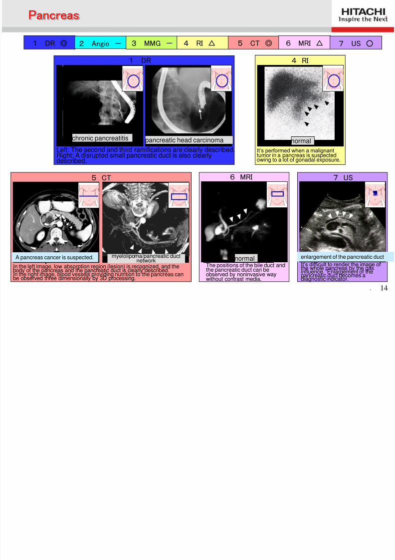
1 DR ◎ 2 Angio - 3 MMG - 4 RI △ 5 CT ◎ 6 MRI △ 7 US ○
7 US
It’s difficult to render the image of the whole pancreas by the gasinfluence. Enlargement of the
pancreatic duct becomes adiagnostic indicator.
5 CT
In the left image, low absorption region (lesion) is recognized, and thebody of the pancreas and the pancreatic duct is clearly described.
In the right image, blood vessels providing nutrition to the pancreas canbe observed three dimensionally by 3D processing.
myelolipoma/pancreatic ductnetwork
6 MRI
normalThe positions of the bile duct andthe pancreatic duct can be
observed by noninvasive waywithout contrast media.
1 DR
chronic pancreatitis
Left: The second and third ramifications are clearly described.Right: A disrupted small pancreatic duct is also clearlydescribed.
pancreatic head carcinoma
4 RI
normal
It’s performed when a malignanttumor in a pancreas is suspectedowing to a lot of gonadal exposure.
Pancreas
A pancreas cancer is suspected. enlargement of the pancreatic duct
8/2/2019 Anatomía (Esp)
http://slidepdf.com/reader/full/anatomia-esp 15/58. 15
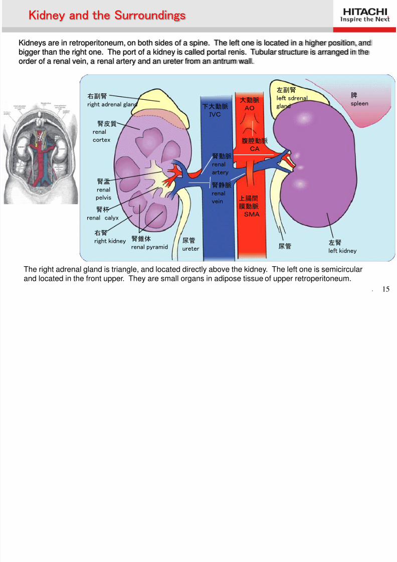
The right adrenal gland is triangle, and located directly above the kidney. The left one is semicircular
and located in the front upper. They are small organs in adipose tissue of upper retroperitoneum.
大動脈 AO 下大動脈
IVC
左副腎 left sdrenalgland
脾 spleen
左腎 left kidney
尿管
腹腔動脈 CA
腎動脈 renalartery
腎静脈 renalvein
尿管 ureter
腎錐体 renal pyramid
右腎 right kidney
腎杯 renal calyx
腎盂 renalpelvis
腎皮質 renal
cortex
右副腎 right adrenal gland
上腸間
膜動脈 SMA
Kidney and the Surroundings
Kidneys are in retroperitoneum, on both sides of a spine. The left one is located in a higher position, andbigger than the right one. The port of a kidney is called portal renis. Tubular structure is arranged in theorder of a renal vein, a renal artery and an ureter from an antrum wall.
8/2/2019 Anatomía (Esp)
http://slidepdf.com/reader/full/anatomia-esp 16/58. 16
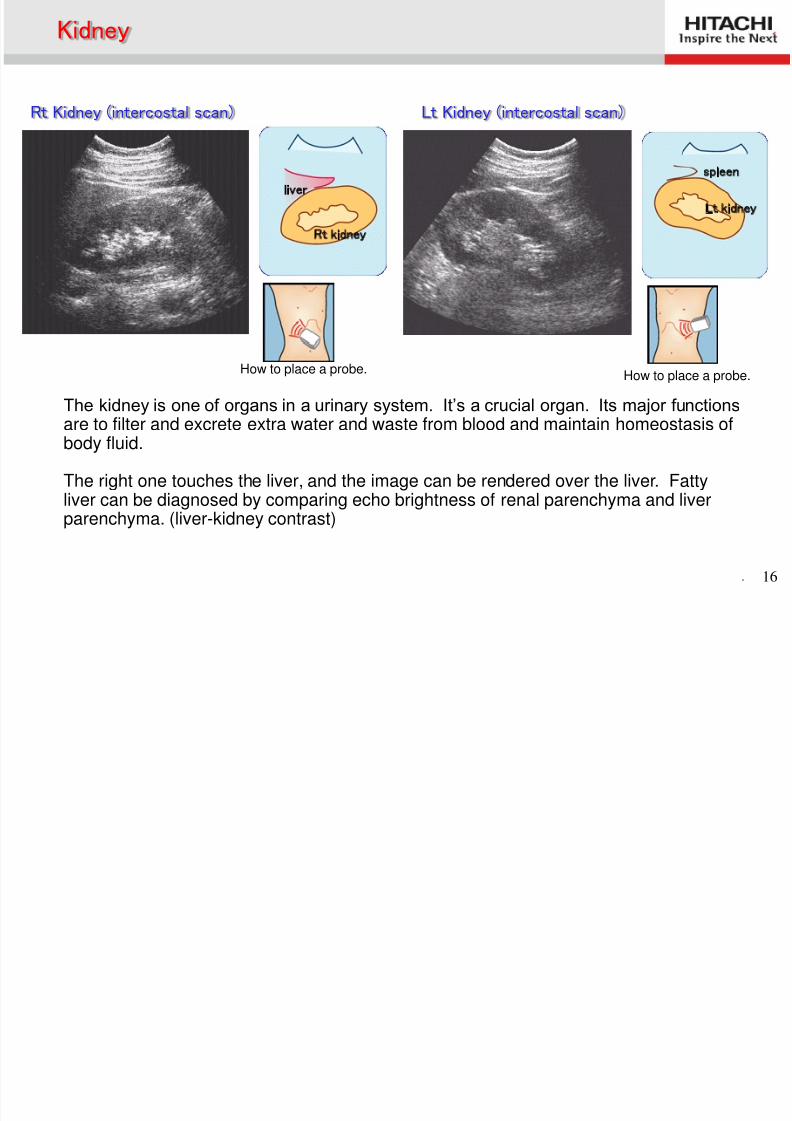
How to place a probe.
liver
Rt kidney
spleen
Lt kidney
Kidney
Rt Kidney (intercostal scan) Lt Kidney (intercostal scan)
The kidney is one of organs in a urinary system. It’s a crucial organ. Its major functionsare to filter and excrete extra water and waste from blood and maintain homeostasis ofbody fluid.
The right one touches the liver, and the image can be rendered over the liver. Fattyliver can be diagnosed by comparing echo brightness of renal parenchyma and liverparenchyma. (liver-kidney contrast)
How to place a probe.
8/2/2019 Anatomía (Esp)
http://slidepdf.com/reader/full/anatomia-esp 17/58. 17
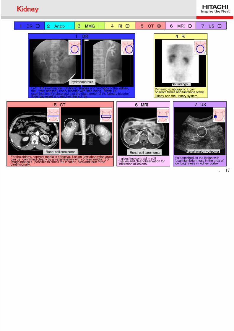
1 DR ○ 2 Angio - 3 MMG - 4 RI ○ 5 CT ◎ 6 MRI ○ 7 US ○
7 US
It’s described as the lesion withfocal high brightness in the area of
low brightness in kidney cortex.
Renal angiomyolipoma
6 MRI
腎細胞癌
It gives fine contrast in softtissues and clear observation for
infiltration of lesions.
5 CT
For the kidney, contrast media is effective. Lesion (low absorption area)can be confirmed clearly by an examination with contrast media. 3Dimage makes it possible to check the location, size and form threedimensionally.
1 DR
Left: DIP examination. Checking shapes and functions of the kidney,the ureter and the urinary bladder with time being. Right: RPexamination. It’s observed that the ritght ureter of the urinary bladder flows backward and reaches the kidney.
4 RI
No findings
Dynamic scintigraphy: it canobserve forms and functions of thekidney and the urinary system.
Kidney
Renal cell carcinoma
hydronephrosis
Renal cell carcinoma
8/2/2019 Anatomía (Esp)
http://slidepdf.com/reader/full/anatomia-esp 18/58. 18
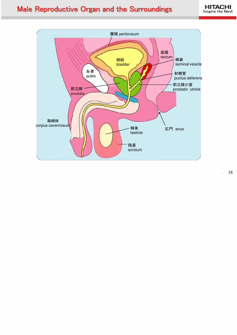
腹膜 peritoneum
直腸 rectum
精嚢 seminal vesicle
射精管 puctus deferens
肛門 anus精巣 testicle
陰嚢 scrotum
海綿体
corpus cavernosum
前立腺 prostate
恥骨 pubis
膀胱 bladder
前立腺小室 prostatic utricle
Male Reproductive Organ and the Surroundings
8/2/2019 Anatomía (Esp)
http://slidepdf.com/reader/full/anatomia-esp 19/58. 19
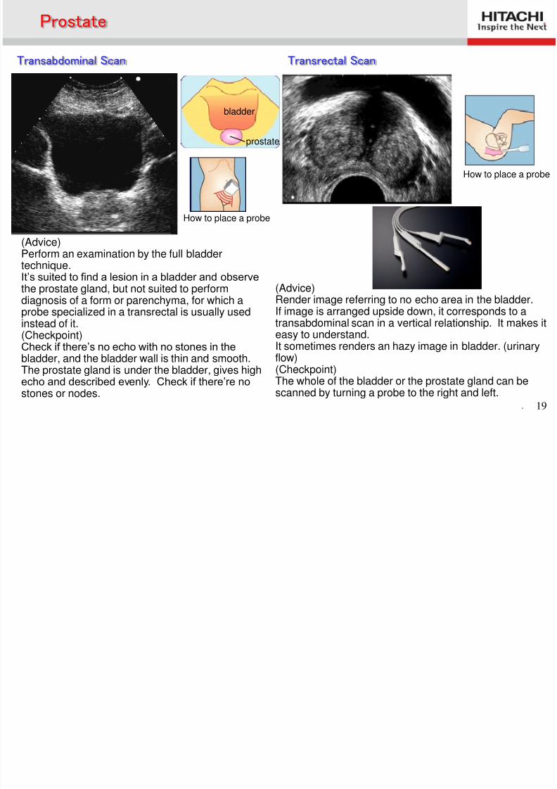
(Advice)Perform an examination by the full bladdertechnique.It’s suited to find a lesion in a bladder and observe
the prostate gland, but not suited to performdiagnosis of a form or parenchyma, for which aprobe specialized in a transrectal is usually usedinstead of it.(Checkpoint)Check if there’s no echo with no stones in thebladder, and the bladder wall is thin and smooth.The prostate gland is under the bladder, gives highecho and described evenly. Check if there’re no
stones or nodes.
(Advice)Render image referring to no echo area in the bladder.If image is arranged upside down, it corresponds to atransabdominal scan in a vertical relationship. It makes iteasy to understand.It sometimes renders an hazy image in bladder. (urinaryflow)(Checkpoint)The whole of the bladder or the prostate gland can be
scanned by turning a probe to the right and left.
How to place a probe
bladder
Prostate
Transabdominal Scan Transrectal Scan
prostate
How to place a probe
8/2/2019 Anatomía (Esp)
http://slidepdf.com/reader/full/anatomia-esp 20/58
. 20
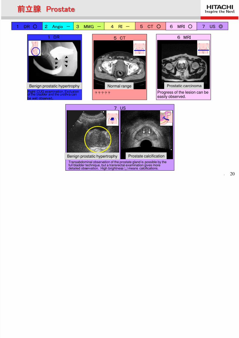
1 DR ○ 2 Angio - 3 MMG - 4 RI - 5 CT ○ 6 MRI ○ 7 US ◎
6 MRI
Progress of the lesion can beeasily observed.
前立腺癌
5 CT
健常例
?????
1 DR
前立腺肥大
7 US
Transabdominal observation of the prostate gland is possible by thefull bladder technique, but a transrectal examination gives moredetailed observation. High brightness(↓)means calcifications.
前立腺肥大 前立腺石灰化
前立腺 Prostate
Prostate calcification
Normal range Prostatic carcinomaBenign prostatic hypertrophy
Benign prostatic hypertrophy
Right: CUG examination. Exclusionof the bladder and the urethra canbe well observed.
8/2/2019 Anatomía (Esp)
http://slidepdf.com/reader/full/anatomia-esp 21/58
. 21
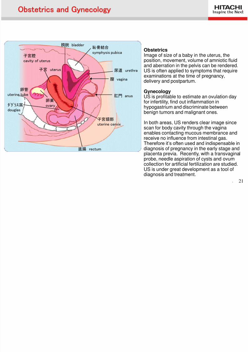
膀胱 bladder 恥骨結合 symphysis pubica
尿道 urethra
膣 vagina
肛門 anus
子宮頚部 uterine cervix
直腸 rectum
子宮 uterus
子宮腔 cavity of uterus
卵管 uterine tube
卵巣 ovaryダグラス窩
douglas
Obstetrics and Gynecology
ObstetricsImage of size of a baby in the uterus, theposition, movement, volume of amniotic fluidand aberration in the pelvis can be rendered.US is often applied to symptoms that requireexaminations at the time of pregnancy,delivery and postpartum.
GynecologyUS is profitable to estimate an ovulation dayfor infertility, find out inflammation inhypogastrium and discriminate betweenbenign tumors and malignant ones.
In both areas, US renders clear image sincescan for body cavity through the vaginaenables contacting mucous membrance andreceive no influence from intestinal gas.Therefore it’s often used and indispensable indiagnosis of pregnancy in the early stage andplacenta previa. Recently, with a transvaginalprobe, needle aspiration of cysts and ovumcollection for artificial fertilization are studied.US is under great development as a tool of
diagnosis and treatment.
8/2/2019 Anatomía (Esp)
http://slidepdf.com/reader/full/anatomia-esp 22/58
. 22
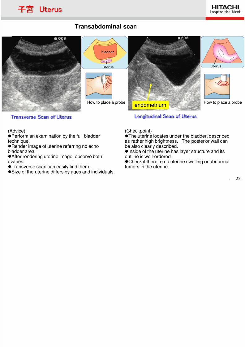
(Advice)Perform an examination by the full bladdertechnique.Render image of uterine referring no echobladder area.After rendering uterine image, observe bothovaries.Transverse scan can easily find them.
Size of the uterine differs by ages and individuals.
(Checkpoint)The uterine locates under the bladder, describedas rather high brightness. The posterior wall canbe also clearly described.Inside of the uterine has layer structure and itsoutline is well-ordered.Check if there’re no uterine swelling or abnormaltumors in the uterine.
How to place a probe
uterus
bladder
uterus
子宮 Uterus
Transverse Scan of Uterus Longitudinal Scan of Uterus
endometrium
Transabdominal scan
How to place a probe
8/2/2019 Anatomía (Esp)
http://slidepdf.com/reader/full/anatomia-esp 23/58
. 23
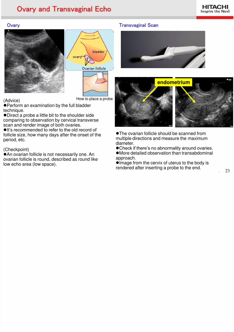
(Advice)Perform an examination by the full bladdertechnique.Direct a probe a little bit to the shoulder sidecomparing to observation by cervical transversescan and render image of both ovaries.It’s recommended to refer to the old record of follicle size, how many days after the onset of theperiod, etc.
(Checkpoint)An ovarian follicle is not necessarily one. Anovarian follicle is round, described as round like
low echo area (low space).
The ovarian follicle should be scanned frommultiple directions and measure the maximumdiameter.Check if there’s no abnormality around ovaries. More detailed observation than transabdominalapproach.
Image from the cervix of uterus to the body isrendered after inserting a probe to the end.
How to place a probe
Ovarian follicle
ovary
bladder
Ovary and Transvaginal Echo
Ovary Transvaginal Scan
endometrium
8/2/2019 Anatomía (Esp)
http://slidepdf.com/reader/full/anatomia-esp 24/58
. 24
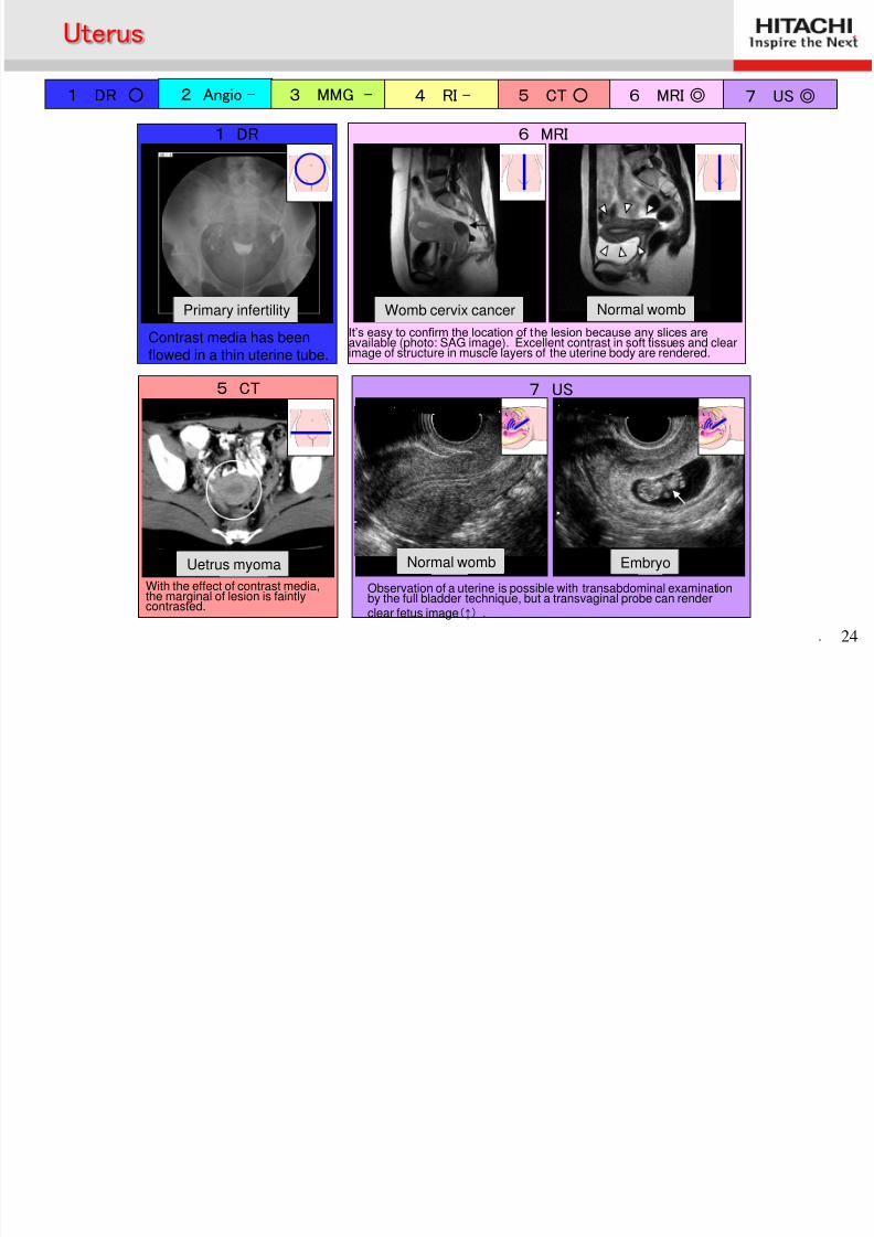
1 DR ○ 2 Angio - 3 MMG - 4 RI - 5 CT○ 6 MRI◎ 7 US◎
6 MRI
子宮頸癌 健常例
It’s easy to confirm the location of the lesion because any slices areavailable (photo: SAG image). Excellent contrast in soft tissues and clearimage of structure in muscle layers of the uterine body are rendered.
1 DR
原発性不妊症
Contrast media has beenflowed in a thin uterine tube.
5 CT
子宮筋腫
With the effect of contrast media,the marginal of lesion is faintlycontrasted.
7 US
Observation of a uterine is possible with transabdominal examinationby the full bladder technique, but a transvaginal probe can render
clear fetus image(↑) .
胎児 健常例
Uterus
Womb cervix cancerPrimary infertility
EmbryoNormal womb
Normal womb
Uetrus myoma
8/2/2019 Anatomía (Esp)
http://slidepdf.com/reader/full/anatomia-esp 25/58
Ultrasonography of Mammaly Gland
and Thyroid Gland
8/2/2019 Anatomía (Esp)
http://slidepdf.com/reader/full/anatomia-esp 26/58
. 26
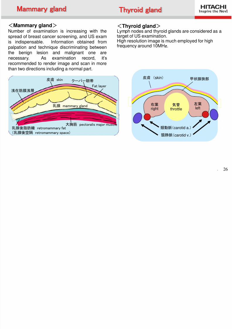
皮膚 skin
Fat layer浅在筋膜浅層
乳腺 mammary gland
大胸筋 pectoralis major muscle乳腺後脂肪織 retromammary fat(乳腺後空隔 retromammary space)
クーパー靭帯
<Mammary gland> Number of examination is increasing with thespread of breast cancer screening, and US exam
is indispensable. Information obtained frompalpation and technique discriminating betweenthe benign lesion and malignant one arenecessary. As examination record, it’s recommended to render image and scan in morethan two directions including a normal part.
<Thyroid gland> Lymph nodes and thyroid glands are considered as atarget of US examination.
High resolution image is much employed for highfrequency around 10MHz.
右葉 right
左葉 left
気管 throttle
皮膚 (skin)
頚動脈(carotid a.)
頸静脈(carotid v.)
甲状腺狭部
Mammary gland Thyroid gland
8/2/2019 Anatomía (Esp)
http://slidepdf.com/reader/full/anatomia-esp 27/58
. 27
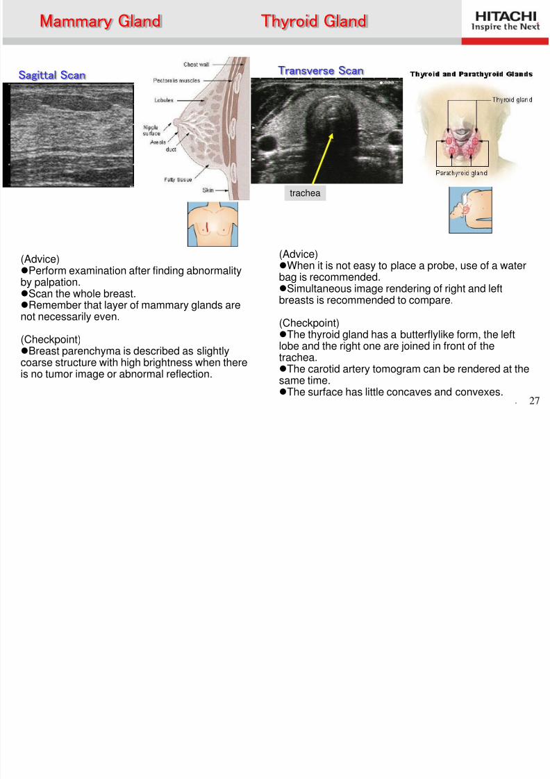
(Advice)Perform examination after finding abnormalityby palpation.Scan the whole breast.Remember that layer of mammary glands arenot necessarily even.
(Checkpoint)Breast parenchyma is described as slightlycoarse structure with high brightness when thereis no tumor image or abnormal reflection.
(Advice)When it is not easy to place a probe, use of a waterbag is recommended.
Simultaneous image rendering of right and leftbreasts is recommended to compare.
(Checkpoint)The thyroid gland has a butterflylike form, the leftlobe and the right one are joined in front of thetrachea.The carotid artery tomogram can be rendered at thesame time.The surface has little concaves and convexes.
Sagittal Scan Transverse Scan
Mammary Gland Thyroid Gland
trachea
8/2/2019 Anatomía (Esp)
http://slidepdf.com/reader/full/anatomia-esp 28/58
. 28
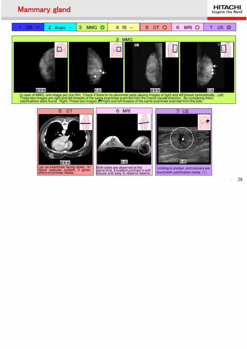
1 DR × 2 Angio - 3 MMG ◎ 4 RI - 5 CT ○ 6 MRI ○ 7 US ◎
7 US
乳癌
Limiting is unclear, and cancers are
found with calcification inside (↑).
6 MRI
乳癌
Both sides are observed at thesame time. Excellent contrast in soft
tissues and easy to observe lesions.
5 CT
健常例
Lay an examinee facing down. Inblood vascular system, it gives
effect of contrast media.
3 MMG
乳癌 乳癌 健常例 健常例
In case of MMG, one image per one film. Check if there’re no abnormal parts placing images of right and left breast symmetrically. Left:These two images are right and left breasts of the same examinee scanned from the cranio-caudal direction. By comparing them,calcifications were found. Right: These two images are right and left breasts of the same examinee scanned from the side.
Mammary gland
甲状腺
8/2/2019 Anatomía (Esp)
http://slidepdf.com/reader/full/anatomia-esp 29/58
. 29
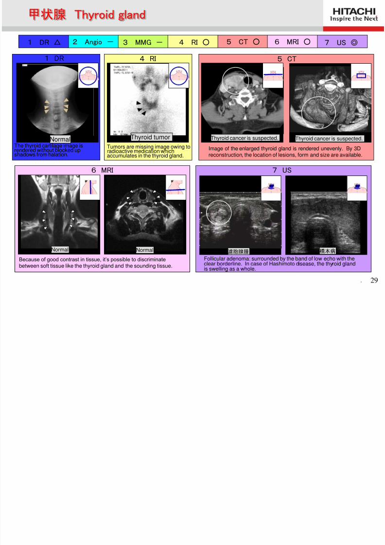
1 DR △ 2 Angio - 3 MMG - 4 RI ○ 5 CT ○ 6 MRI ○ 7 US ◎
7 US
橋本病
Follicular adenoma: surrounded by the band of low echo with theclear borderline. In case of Hashimoto disease, the thyroid gland
is swelling as a whole.
濾胞腺腫
6 MRI
Normal Normal
Because of good contrast in tissue, it’s possible to discriminate
between soft tissue like the thyroid gland and the sounding tissue.
5 CT
Image of the enlarged thyroid gland is rendered unevenly. By 3Dreconstruction, the location of lesions, form and size are available.
1 DR
NormalThe thyroid cartilage image isrendered without blocked upshadows from halation.
4 RI
Thyroid tumorTumors are missing image owing toradioactive medication whichaccumulates in the thyroid gland.
甲状腺 Thyroid gland
Thyroid cancer is suspected. Thyroid cancer is suspected.
O th di
8/2/2019 Anatomía (Esp)
http://slidepdf.com/reader/full/anatomia-esp 30/58
. 30
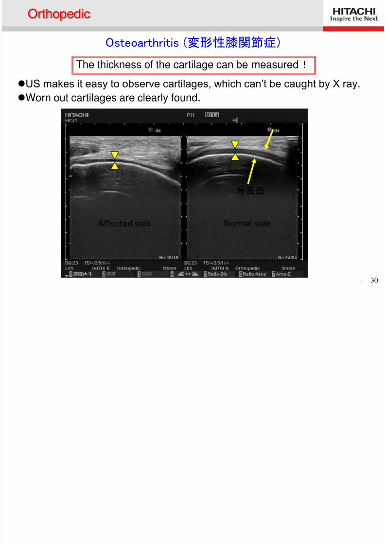
Osteoarthritis (変形性膝関節症)
US makes it easy to observe cartilages, which can’t be caught by X ray.
Worn out cartilages are clearly found.
Affected side Normal side
Knee cartilage
骨表面
The thickness of the cartilage can be measured!
Orthopedic
O h di
8/2/2019 Anatomía (Esp)
http://slidepdf.com/reader/full/anatomia-esp 31/58
. 31
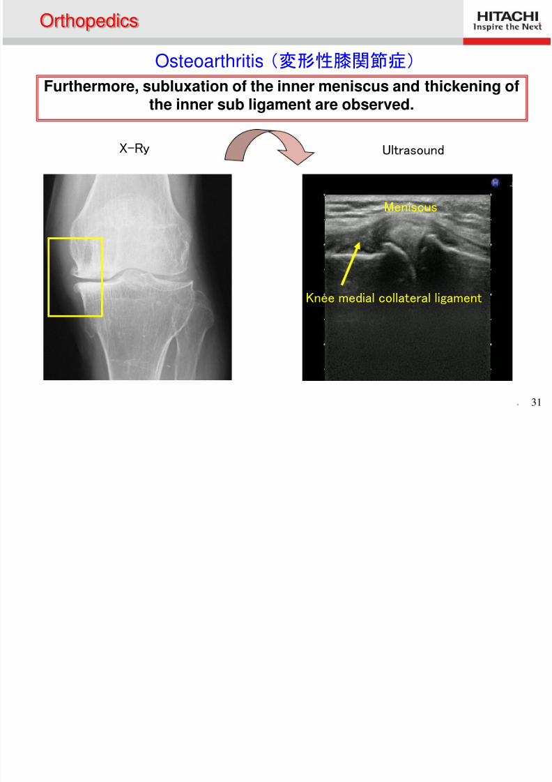
Furthermore, subluxation of the inner meniscus and thickening of
the inner sub ligament are observed.
Meniscus
Knee medial collateral ligament
X-Ry Ultrasound
Osteoarthritis (変形性膝関節症)
Orthopedics
O h di
8/2/2019 Anatomía (Esp)
http://slidepdf.com/reader/full/anatomia-esp 32/58
. 32
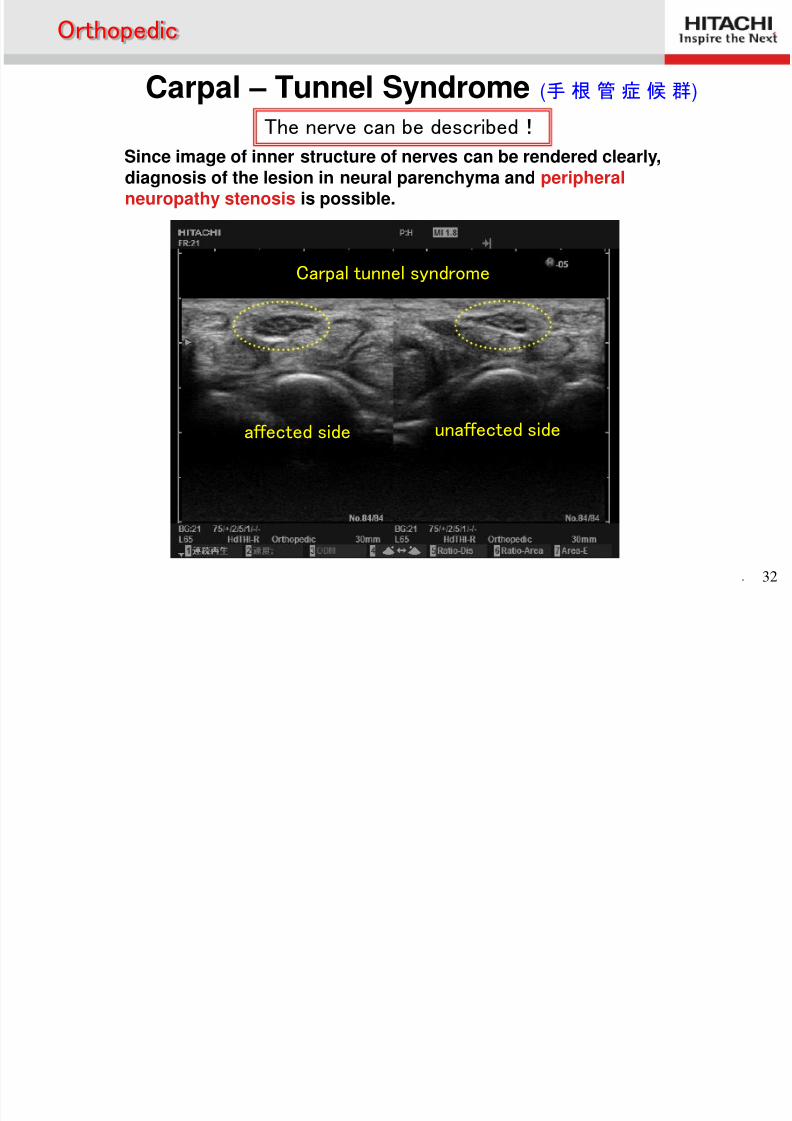
Since image of inner structure of nerves can be rendered clearly,diagnosis of the lesion in neural parenchyma and peripheralneuropathy stenosis is possible.
The nerve can be described!
Carpal tunnel syndrome
unaffected sideaffected side
Orthopedic
Carpal – Tunnel Syndrome (手 根 管 症 候 群)
O h di
8/2/2019 Anatomía (Esp)
http://slidepdf.com/reader/full/anatomia-esp 33/58
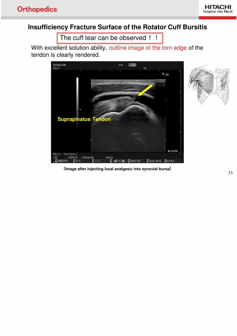
. 33(Image after injecting local analgesic into synovial bursa)
GreaterTubercle
棘上筋腱
The cuff tear can be observed!!
With excellent solution ability, outline image of the torn edge of thetendon is clearly rendered.
Insufficiency Fracture Surface of the Rotator Cuff Bursitis
Orthopedics
Suprapinatus Tendon
O th di
8/2/2019 Anatomía (Esp)
http://slidepdf.com/reader/full/anatomia-esp 34/58
. 34
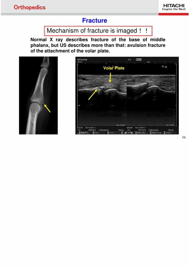
Mechanism of fracture is imaged!!
Normal X ray describes fracture of the base of middlephalanx, but US describes more than that: avulsion fractureof the attachment of the volar plate.
Normal SideLesion Side
Volar Plate
Fracture
Orthopedics
8/2/2019 Anatomía (Esp)
http://slidepdf.com/reader/full/anatomia-esp 35/58
Clinical Echocardiography
E i ti f H t
8/2/2019 Anatomía (Esp)
http://slidepdf.com/reader/full/anatomia-esp 36/58
. 36
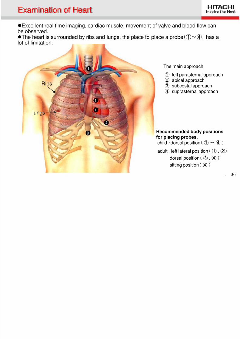
Excellent real time imaging, cardiac muscle, movement of valve and blood flow canbe observed.The heart is surrounded by ribs and lungs, the place to place a probe(①~④) has a
lot of limitation.
The main approach
① left parasternal approach② apical approach
③ subcostal approach④ suprasternal approach
Recommended body positionsfor placing probes.child :dorsal position( ① ~ ④ )
adult :left lateral position( ① ,②)
dorsal position( ③ ,④ )
sitting position( ④ )
Ribs
lungs
Examination of Heart
Blood Flow of Heart
8/2/2019 Anatomía (Esp)
http://slidepdf.com/reader/full/anatomia-esp 37/58
. 37
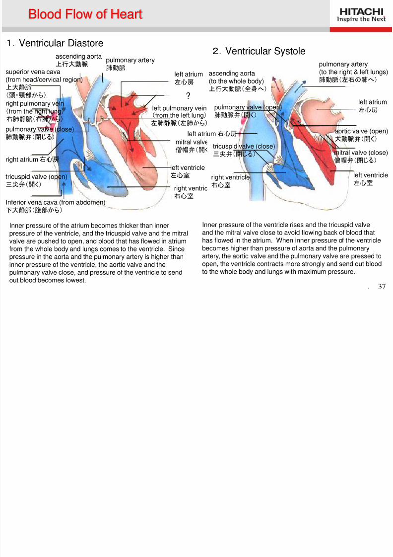
1.Ventricular Diastore2.Ventricular Systole
left atrium左心房
pulmonary artery肺動脈
ascending aorta上行大動脈
left pulmonary vein(from the left lung) 左肺静脈(左肺から)
mitral valve (open)僧帽弁(開く)
left ventricle左心室
right ventricle右心室
Inferior vena cava (from abdomen)
下大静脈(腹部から)
tricuspid valve (open)三尖弁(開く)
right atrium 右心房
pulmonary valve (close)肺動脈弁(閉じる)
right pulmonary vein(from the right lung) 右肺静脈(右肺から)
superior vena cava(from head/cervical region)上大静脈
(頭・頸部から)
ascending aorta(to the whole body)上行大動脈(全身へ)
pulmonary valve (open)肺動脈弁(開く)
left atrium 右心房
tricuspid valve (close)三尖弁(閉じる)
right ventricle右心室
pulmonary artery
(to the right & left lungs)肺動脈(左右の肺へ)
left atrium左心房
aortic valve (open)
大動脈弁(開く)
mitral valve (close)僧帽弁(閉じる)
left ventricle左心室
Inner pressure of the atrium becomes thicker than innerpressure of the ventricle, and the tricuspid valve and the mitralvalve are pushed to open, and blood that has flowed in atriumfrom the whole body and lungs comes to the ventricle. Sincepressure in the aorta and the pulmonary artery is higher thaninner pressure of the ventricle, the aortic valve and thepulmonary valve close, and pressure of the ventricle to sendout blood becomes lowest.
Inner pressure of the ventricle rises and the tricuspid valveand the mitral valve close to avoid flowing back of blood thathas flowed in the atrium. When inner pressure of the ventriclebecomes higher than pressure of aorta and the pulmonaryartery, the aortic valve and the pulmonary valve are pressed toopen, the ventricle contracts more strongly and send out bloodto the whole body and lungs with maximum pressure.
Blood Flow of Heart
?
P t l L ft V t i l L A i Vi
8/2/2019 Anatomía (Esp)
http://slidepdf.com/reader/full/anatomia-esp 38/58
. 38
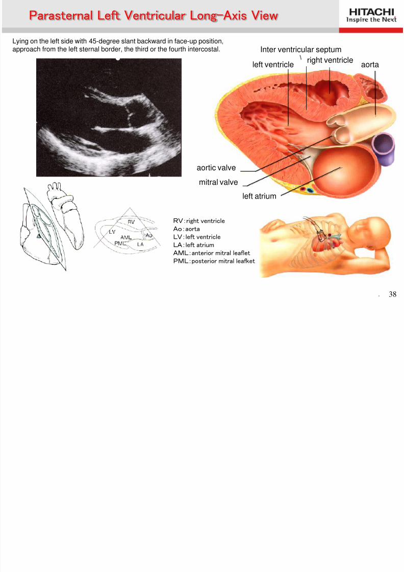
Lying on the left side with 45-degree slant backward in face-up position,approach from the left sternal border, the third or the fourth intercostal.
RV:right ventricleAo:aortaLV:left ventricleLA:left atriumAML:anterior mitral leafletPML:posterior mitral leafket
右室
Parasternal Left Ventricular Long-Axis View
left atrium
mitral valve
aortic valve
left ventricle
Inter ventricular septum
aortaright ventricle
Parasternal Left Ventric lar Short A is Vie
8/2/2019 Anatomía (Esp)
http://slidepdf.com/reader/full/anatomia-esp 39/58
. 39
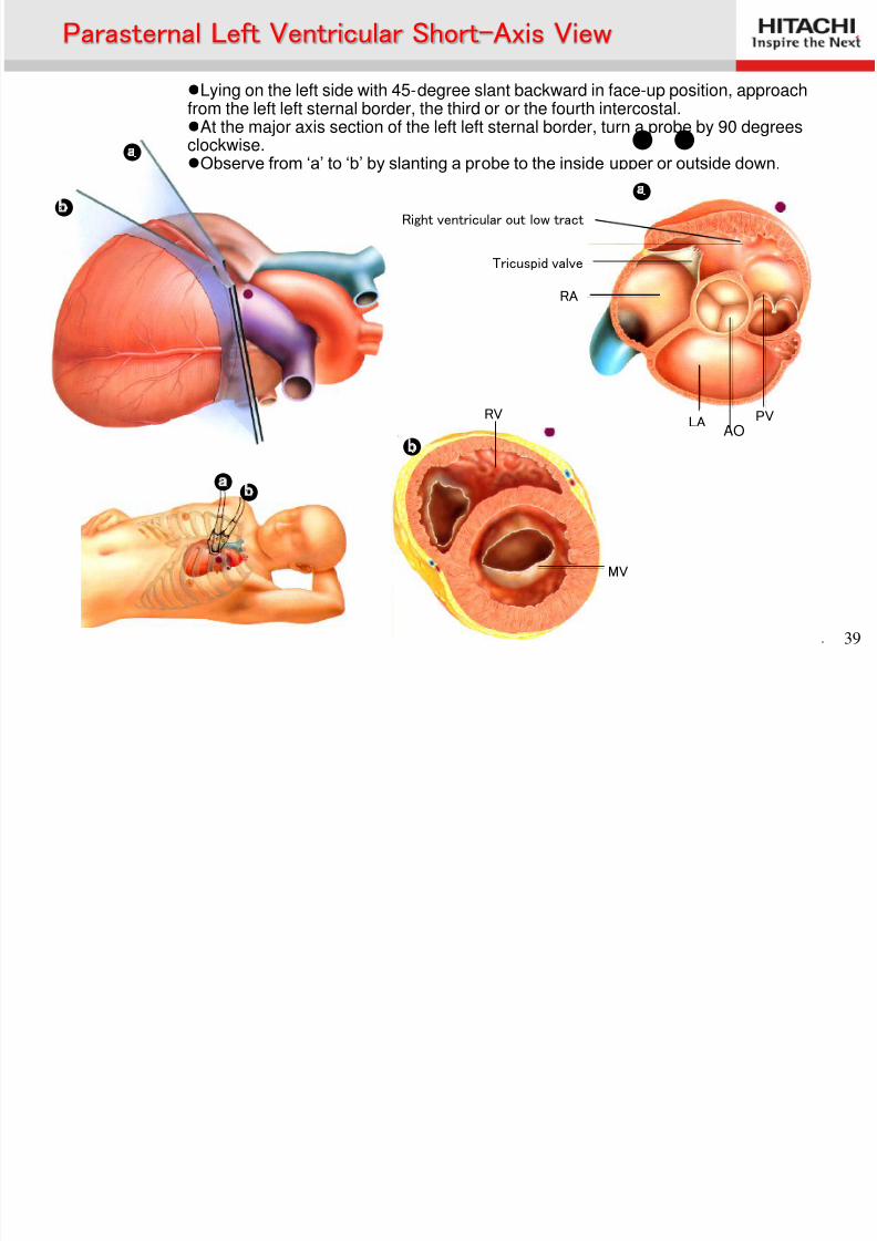
●
●
● ●
● ●
Lying on the left side with 45-degree slant backward in face-up position, approachfrom the left left sternal border, the third or or the fourth intercostal.At the major axis section of the left left sternal border, turn a probe by 90 degreesclockwise.Observe from ‘a’ to ‘b’ by slanting a probe to the inside upper or outside down.
Right ventricular outf low tract
Tricuspid valve
RA
LA PV
●
RV
MV
●
Parasternal Left Ventricular Short-Axis View
AO
P t l L ft V t i l Sh t A i Vi
8/2/2019 Anatomía (Esp)
http://slidepdf.com/reader/full/anatomia-esp 40/58
. 40
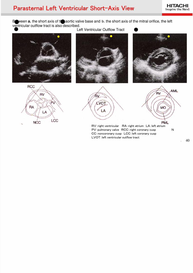
● ● Between a. the short axis of the aortic valve base and b. the short axis of the mitral orifice, the leftventricular outflow tract is also described.● ● Left Ventricular Outflow Tract
RV:right ventricular RA:right atrium LA:left atriumPV:pulmonary valve RCC:right coronary cusp NCC:noncoronary cusp LCC:left coronary cusp
LVOT:left ventricular outflow tract
Parasternal Left Ventricular Short-Axis View
Parasternal Left Ventricular Short Axis View
8/2/2019 Anatomía (Esp)
http://slidepdf.com/reader/full/anatomia-esp 41/58
. 41
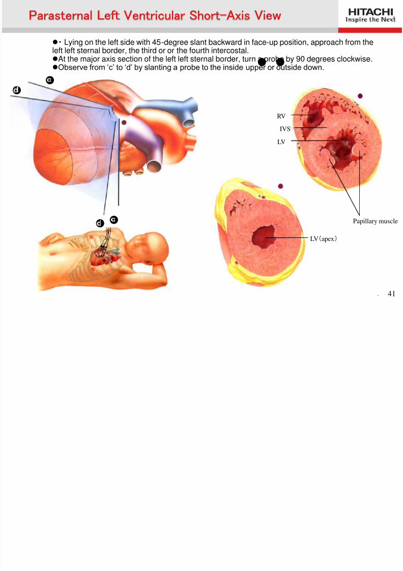
● ●
・ Lying on the left side with 45-degree slant backward in face-up position, approach from theleft left sternal border, the third or or the fourth intercostal.At the major axis section of the left left sternal border, turn a probe by 90 degrees clockwise.Observe from ‘c’ to ‘d’ by slanting a probe to the inside upper or outside down.
● ●
RV
LV
IVS
Papillary muscle
LV(apex)
● ●
Parasternal Left Ventricular Short-Axis View
Parasternal Left Ventricular Short-Axis View
8/2/2019 Anatomía (Esp)
http://slidepdf.com/reader/full/anatomia-esp 42/58
. 42
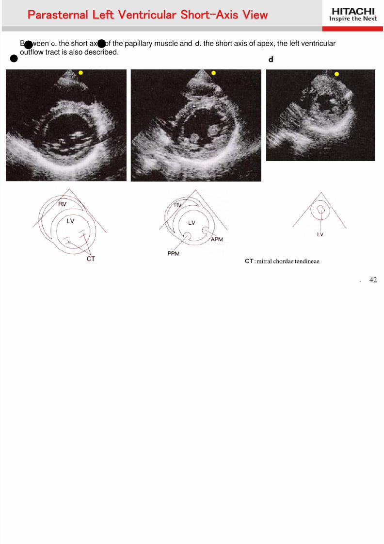
● ● Between c. the short axis of the papillary muscle and d. the short axis of apex, the left ventricularoutflow tract is also described.
CT:mitral chordae tendineae
● ●
Parasternal Left Ventricular Short-Axis View
Apical Four-Chamber View
8/2/2019 Anatomía (Esp)
http://slidepdf.com/reader/full/anatomia-esp 43/58
. 43
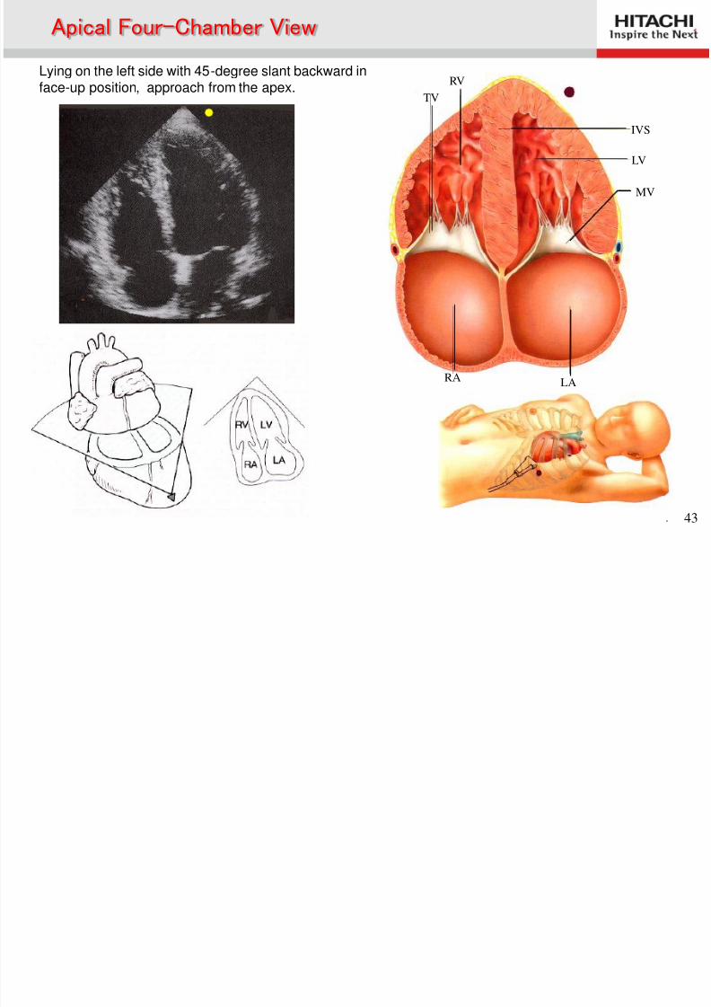
Lying on the left side with 45-degree slant backward inface-up position, approach from the apex.
RV
TV
IVS
LV
MV
LARA
Apical Four-Chamber View
Apical Two-chamber View
8/2/2019 Anatomía (Esp)
http://slidepdf.com/reader/full/anatomia-esp 44/58
. 44
LV
MV
LA
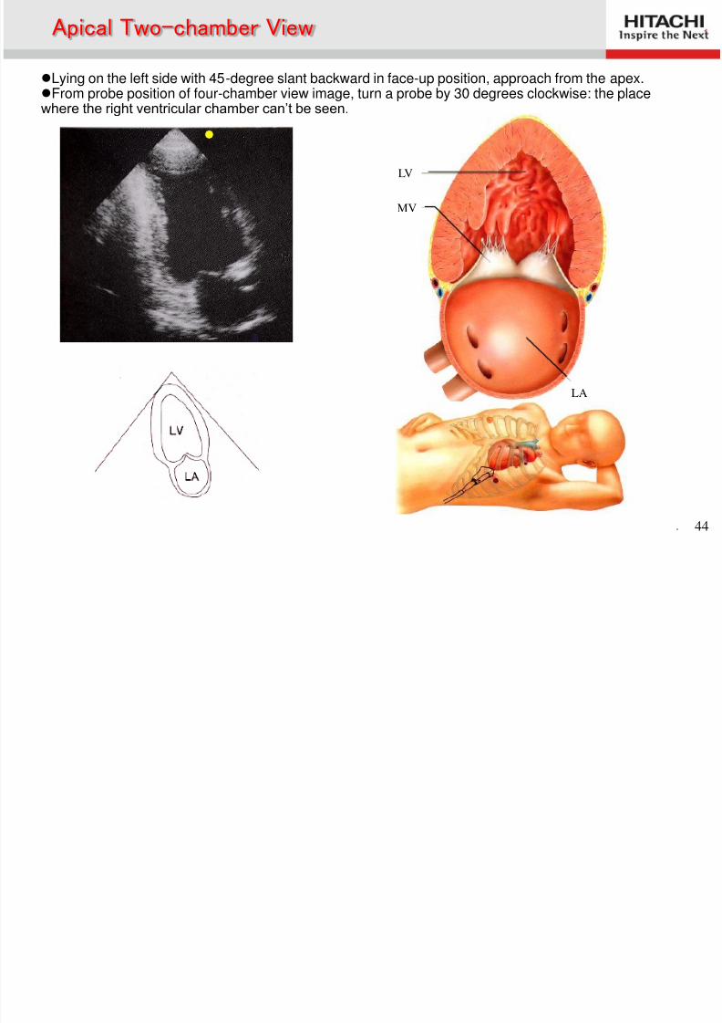
Lying on the left side with 45-degree slant backward in face-up position, approach from the apex.From probe position of four-chamber view image, turn a probe by 30 degrees clockwise: the placewhere the right ventricular chamber can’t be seen.
Apical Two chamber View
Apical Left Ventricular Long-Axis View
8/2/2019 Anatomía (Esp)
http://slidepdf.com/reader/full/anatomia-esp 45/58
. 45
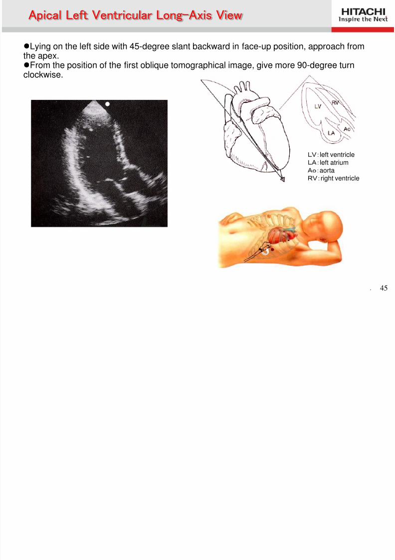
LV:left ventricle LA:left atriumAo:aortaRV:right ventricle
Lying on the left side with 45-degree slant backward in face-up position, approach fromthe apex.From the position of the first oblique tomographical image, give more 90-degree turn
clockwise.
Apical Left Ventricular Long Axis View
Cardiac
8/2/2019 Anatomía (Esp)
http://slidepdf.com/reader/full/anatomia-esp 46/58
. 46
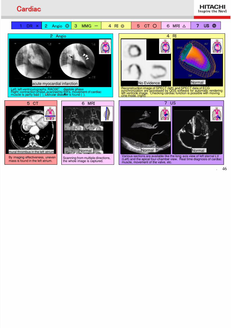
1 DR × 2 Angio ◎ 3 MMG - 4 RI ◎ 5 CT ○ 6 MRI △ 7 US ◎
2 Angio
Left: left ventriculography, RAO30°, diastole phaseRight: contraction phase, scanned by 60f/s, movement of cardiacmuscle is partly bad ( ), valvular disease is found ( ).
acute myocardial infarction
4 RI
No Evidence NormalReconstruction image of SPECT (left) and SPECT data of ECGsynchronization are processed by QGS software for automatic renderingleft ventricle image. Checking cardiac function is possible with movingcine mode. (right)
7 US
Normal Normal
Various sections are available like the long axis view of left sternal LV(Left) and the apical four-chamber view. Real time diagnosis of cardiac
muscle, movement of the valve, etc.
5 CT
By imaging effectiveness, unevenmass is found in the left atrium.
mural thrombus in the left atrium
6 MRI
Scanning from multiple directions,the whole image is captured.
Normal
Cardiac
8/2/2019 Anatomía (Esp)
http://slidepdf.com/reader/full/anatomia-esp 47/58
Ultrasonography of Carotid Artery
Carotid Artery
8/2/2019 Anatomía (Esp)
http://slidepdf.com/reader/full/anatomia-esp 48/58
. 48
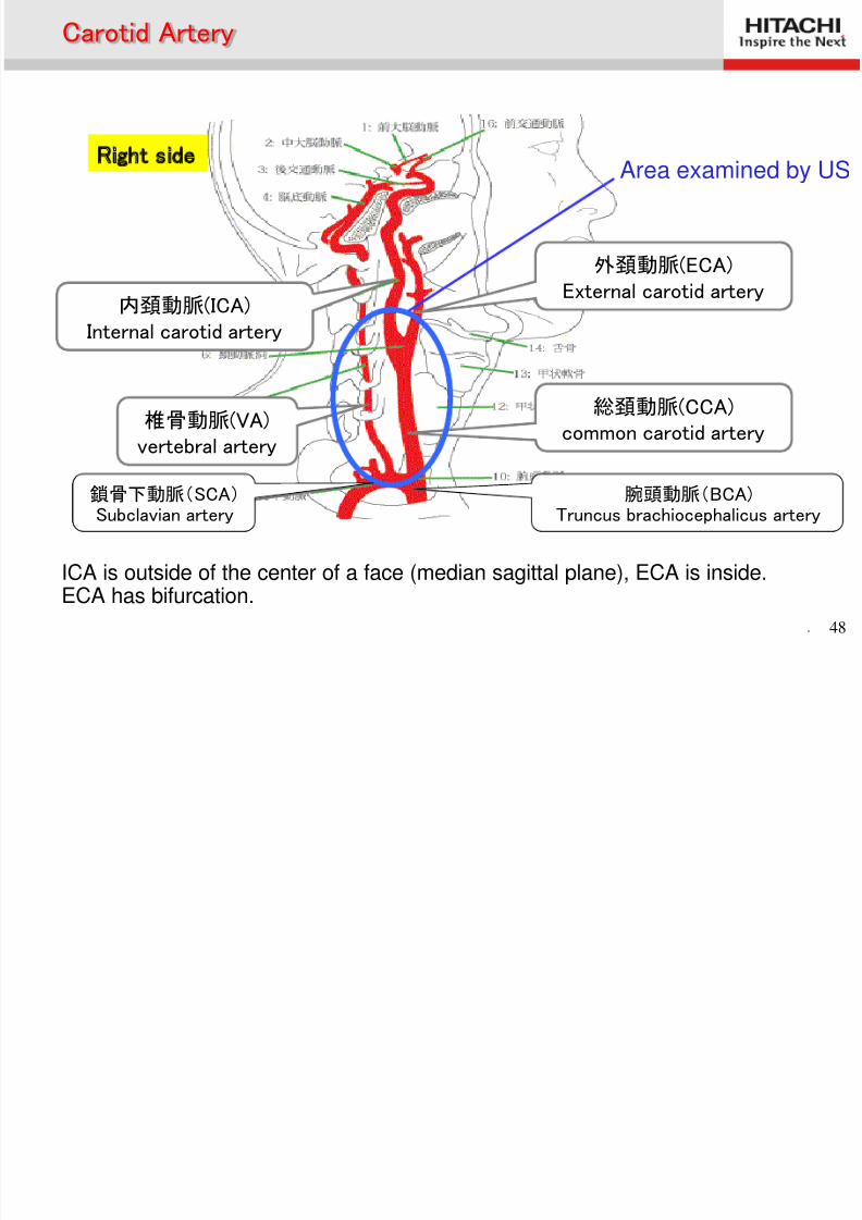
外頚動脈(ECA)External carotid artery
内頚動脈(ICA)Internal carotid artery
椎骨動脈(VA)vertebral artery
総頚動脈(CCA)common carotid artery
鎖骨下動脈 腕頭動脈
Right side Area examined by US
ICA is outside of the center of a face (median sagittal plane), ECA is inside.ECA has bifurcation.
Carotid Artery
腕頭動脈(BCA)Truncus brachiocephalicus artery
鎖骨下動脈(SCA) Subclavian artery
CCA (Common Carotid Artery)
8/2/2019 Anatomía (Esp)
http://slidepdf.com/reader/full/anatomia-esp 49/58
. 49
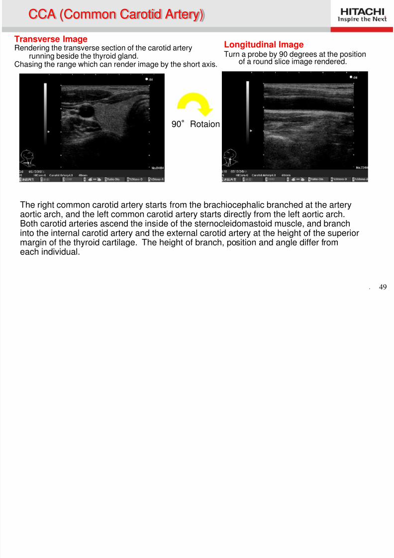
Transverse Image Rendering the transverse section of the carotid artery
running beside the thyroid gland.Chasing the range which can render image by the short axis.
Longitudinal Image Turn a probe by 90 degrees at the position
of a round slice image rendered.
CCA (Common Carotid Artery)
90°Rotaion
The right common carotid artery starts from the brachiocephalic branched at the arteryaortic arch, and the left common carotid artery starts directly from the left aortic arch.Both carotid arteries ascend the inside of the sternocleidomastoid muscle, and branchinto the internal carotid artery and the external carotid artery at the height of the superiormargin of the thyroid cartilage. The height of branch, position and angle differ fromeach individual.
ICA (Internal Carotid Artery) ECA (External Carotid Artery)
8/2/2019 Anatomía (Esp)
http://slidepdf.com/reader/full/anatomia-esp 50/58
. 50
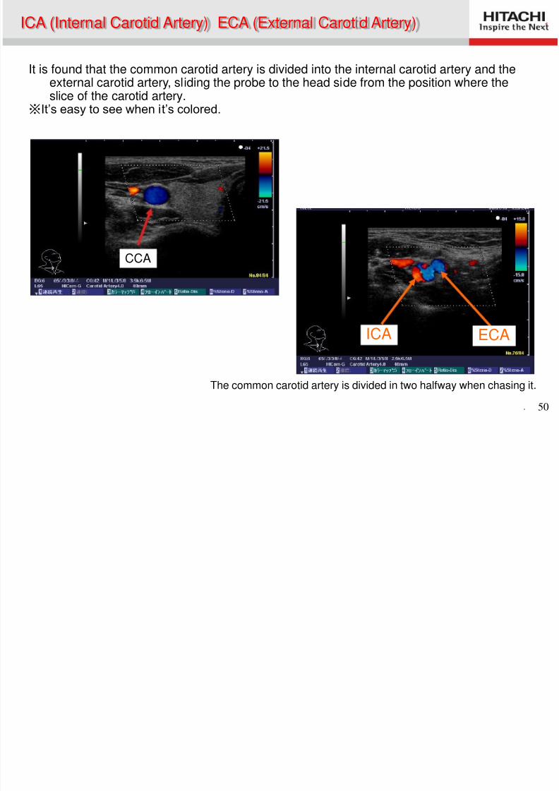
It is found that the common carotid artery is divided into the internal carotid artery and theexternal carotid artery, sliding the probe to the head side from the position where theslice of the carotid artery.
※It’s easy to see when it’s colored.
The common carotid artery is divided in two halfway when chasing it.
ICA (Internal Carotid Artery) ECA (External Carotid Artery)
CCA
ECAICA
ICA (Internal Carotid Artery)
8/2/2019 Anatomía (Esp)
http://slidepdf.com/reader/full/anatomia-esp 51/58
. 51
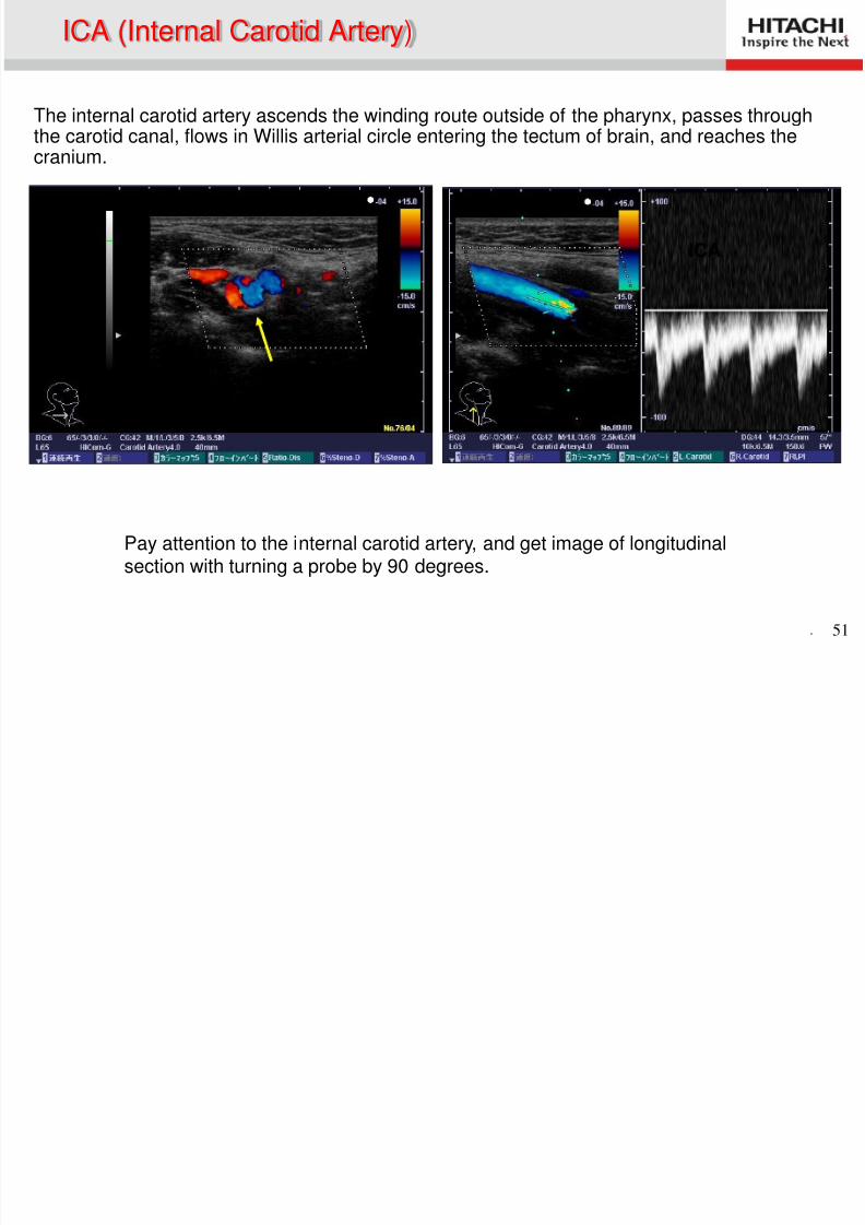
Pay attention to the internal carotid artery, and get image of longitudinalsection with turning a probe by 90 degrees.
ICA (Internal Carotid Artery)
The internal carotid artery ascends the winding route outside of the pharynx, passes throughthe carotid canal, flows in Willis arterial circle entering the tectum of brain, and reaches the
cranium.
ICA
ECA (External Carotid Artery)
8/2/2019 Anatomía (Esp)
http://slidepdf.com/reader/full/anatomia-esp 52/58
. 52
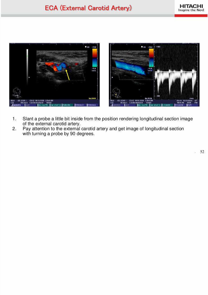
1. Slant a probe a little bit inside from the position rendering longitudinal section imageof the external carotid artery.
2. Pay attention to the external carotid artery and get image of longitudinal sectionwith turning a probe by 90 degrees.
ECA (External Carotid Artery)
ECA
Discrimination between Internal Carotid Arteryd E t l C tid A t
8/2/2019 Anatomía (Esp)
http://slidepdf.com/reader/full/anatomia-esp 53/58
. 53
ECAICA
and External Carotid Artery
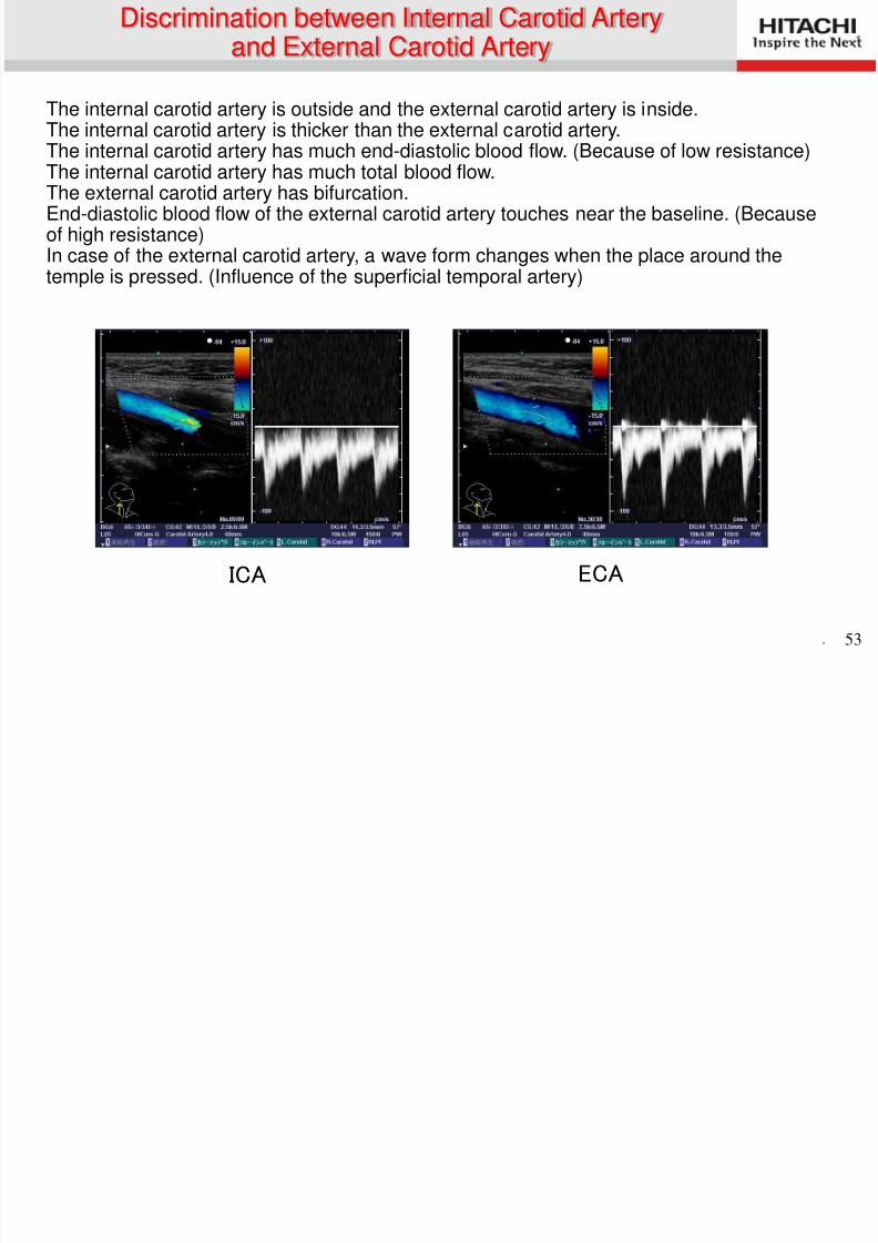
The internal carotid artery is outside and the external carotid artery is inside.The internal carotid artery is thicker than the external carotid artery.The internal carotid artery has much end-diastolic blood flow. (Because of low resistance)The internal carotid artery has much total blood flow.The external carotid artery has bifurcation.End-diastolic blood flow of the external carotid artery touches near the baseline. (Becauseof high resistance)In case of the external carotid artery, a wave form changes when the place around thetemple is pressed. (Influence of the superficial temporal artery)
Vertebral Artery
8/2/2019 Anatomía (Esp)
http://slidepdf.com/reader/full/anatomia-esp 54/58
. 54
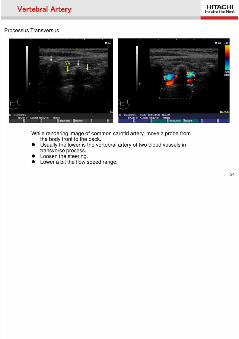
While rendering image of common carotid artery, move a probe fromthe body front to the back.
Usually the lower is the vertebral artery of two blood vessels intransverse process.
Loosen the steering. Lower a bit the flow speed range.
Vertebral Artery
Transverse process of vertebra
VA
Processus Transversus
Intima-Media Thickness (IMT)
8/2/2019 Anatomía (Esp)
http://slidepdf.com/reader/full/anatomia-esp 55/58
. 55
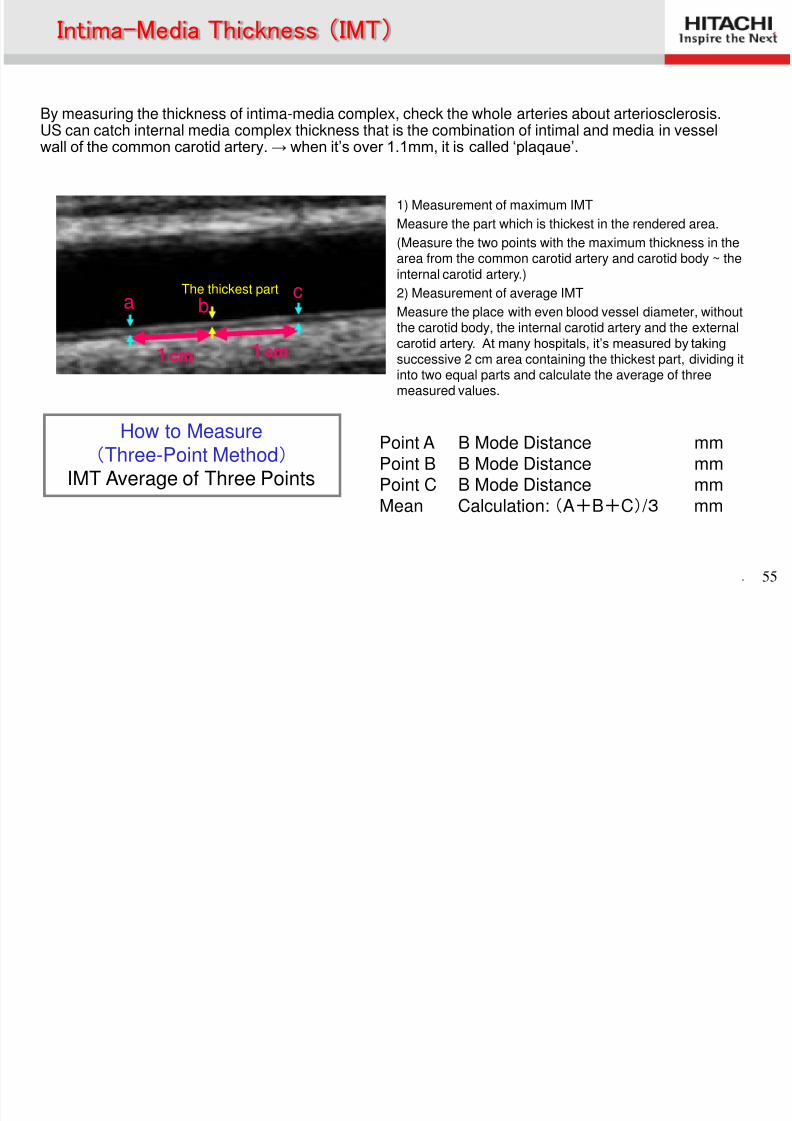
By measuring the thickness of intima-media complex, check the whole arteries about arteriosclerosis.US can catch internal media complex thickness that is the combination of intimal and media in vessel
wall of the common carotid artery. → when it’s over 1.1mm, it is called ‘plaqaue’.
1cm 1cm
a bc
How to Measure(Three-Point Method)
IMT Average of Three Points
Point A B Mode Distance mmPoint B B Mode Distance mmPoint C B Mode Distance mmMean Calculation: (A+B+C) / 3 mm
( )
1) Measurement of maximum IMT
Measure the part which is thickest in the rendered area.
(Measure the two points with the maximum thickness in thearea from the common carotid artery and carotid body ~ theinternal carotid artery.)
2) Measurement of average IMT
Measure the place with even blood vessel diameter, withoutthe carotid body, the internal carotid artery and the externalcarotid artery. At many hospitals, it’s measured by taking
successive 2 cm area containing the thickest part, dividing itinto two equal parts and calculate the average of threemeasured values.
The thickest part
IMT Measurement Point

8/2/2019 Anatomía (Esp)
http://slidepdf.com/reader/full/anatomia-esp 56/58
. 56
Normality (0.6mm)
Doubt of arteriosclerosis(1.3mm)Inside of low
brightness
Outside ofhigh brightness
Outer
Media
IntimalIntima-Media Complex
(IMC)
Carotid Artery
8/2/2019 Anatomía (Esp)
http://slidepdf.com/reader/full/anatomia-esp 57/58
. 57
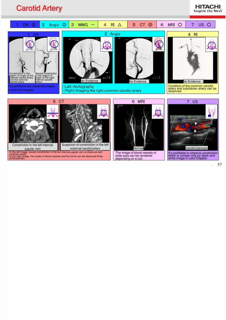
1 DR ◎ 2 Angio ◎ 3 MMG - 4 RI △ 5 CT ◎ 6 MRI ○ 7 US ○
7 US
It’s profitable to observe constrictionwhich is unclear only by black andwhite image in color Doppler.
Carotid stenosis
6 MRI
Normal
The image of blood vessels ofwide area can be rendered
depending on a coil.
5 CT
In the left image, severe constriction in the left internal jugular vein is observed withcontrast media.In the right image, the routes of blood vessels and the forms can be observed three-dimensionally.
Constriction in the left internal jugular vein
Suspicion of constriction in the leftexternal carotid artery
2 Angio
(Left)Aortography(Right)Imaging the right common carotid artery
No Evidence
1 DR
Constriction in theregion of origin of theinternal carotid arteryand the externalcarotid artery
Severe constrictionin the region oforigin of the rightinternal carotidartery
Constrictions are observed clearlyin both the images.
4 RI
Condition of the common carotidartery and subclavian artery can beobserved.
y
No Evidence No Evidence

8/2/2019 Anatomía (Esp)
http://slidepdf.com/reader/full/anatomia-esp 58/58