Embed Size (px)
Citation preview

Tesis doctoral
Estudio molecular y de apoptosis en ovocitos de cabras prepúberes y su relación con el desarrollo embrionario
Begoña Anguita Bustamante

La Dra. Maria Teresa Paramio Nieto, profesora titular de universidad del Departament
de Ciència Animal i dels Aliments de la Universitat Autònoma de Barcelona
Y
La Dra. Maria Dolors Izquierdo Tugas, profesora lector del Departament de Ciència
Animal i dels Aliments de la Universitat Autònoma de Barcelona
CERTIFICAN
Que el trabajo de investigación titulado “Estudio molecular y de apoptosis en ovocitos
de cabras prepúberes y su relación con el desarrollo embrionario”, realizado por Begoña
Anguita Bustamante, se ha llevado a cabo bajo su dirección en la Unidad de Producción
Animal del Departament de Ciència Animal i dels Aliments de la Universitat Autònoma
de Barcelona para optar al grado de doctor, y gracias a la financiación del Ministerio de
Ciencia y Tecnología (proyectos AGL-2000-0353 y AGL-2004304737-C03-01-GAN) y
a la beca predoctoral otorgada por la Generalitat de Catalunya (2003FI 00282).
Maria Teresa Paramio Nieto Maria Dolors Izquierdo Tugas
ÍNDICE
Capítulo1. Introducción 1
Capítulo 2. Revisión bibliográfica 5
2.1. Maduración del ovocito 7
2.1.1. Maduración nuclear o meiótica 7
2.1.2. Maduración citoplasmática 9
2.1.3. Papel del cumulus oophorus en la maduración del
ovocito
11
2.1.4. Regulación de la maduración ovocitaria 12
2.2. Apoptosis 17
2.2.1. Conceptos generales 17
2.2.2. Métodos de detección de la apoptosis 18
2.2.2.1. Evaluación morfológica 18
2.2.2.2. Expresión de proteínas 20
2.2.2.3. Tinción con Annexin-V 21
2.2.2.4. Visualización de la escalera de DNA 22
2.2.2.5. TUNEL (Tdt-mediated dUTP nick-end labelling) 23
2.2.3. Apoptosis en el ovario 24
2.2.3.1. En el período pre-natal 24
2.2.3.2. En el período post-natal 24
2.2.4. Apoptosis en el embrión pre-implantacional 27
2.3. Producción in vitro de embriones 29
2.3.1. Animales adultos vs. Prepúberes 29
2.3.2. Obtención de los ovocitos 30
2.3.2.1. Obtención de los ovarios 30
2.3.2.2. Métodos de obtención de los ovocitos 31
ÍNDICE
2.3.3. Selección de los ovocitos 32
2.3.3.1. Métodos no invasivos 32
a. Tamaño folicular 32
b. Diámetro ovocitario 33
c. Morfología del COC 34
d. Otros métodos no invasivos 35
2.3.3.2. Métodos invasivos 36
a. Contenido mitocondrial del ovocito 36
b. Abundancia de transcritos específicos 36
c. Grado de apoptosis 37
Capítulo 3. Objetivos 39
Capítulo 4. Efecto del diámetro del ovocito en la competencia
meiótica, el desarrollo embrionario, la expresión de p34cdc2 y
la actividad del MPF en ovocitos de cabras prepúberes
Effect of oocyte diameter on meiotic competence, embryo
development, p34 (cdc2) expression and MPF activity in prepubertal
goat oocytes.
Theriogenology (2007), Feb; 67(3): 526-536.
43
Capítulo 5. Contenido de RNA y proteína total, expresión de Ciclina
B1 y desarrollo embrionario en ovocitos de cabras prepúberes.
Total RNA and protein content, Cyclin B1 expression and developmental
competence in prepubertal goat oocytes.
Animal Reproduction Science (2007), doi: 10.1016/ j.animreprosci.
2006. 12.018.
57

ÍNDICE
Capítulo 6. La competencia para el desarrollo de ovocitos
bovinos no está relacionada con la incidencia de apoptosis en
ovocitos, células del cumulus y blastocistos.
Developmental competence of bovine oocytes is not related to
apoptosis incidence in oocytes, cumulus cells and blastocysts.
Theriogenology (2007), Feb; 67(3): 537-549.
85
Capítulo 7. Efecto de la incidencia de apoptosis en ovocitos
y células del cumulus, evaluado mediante TUNEL, en el desarrollo
embrionario a partir de ovocitos de cabras prepúberes.
Effect of prevalence of apoptosis in oocytes and cumulus cells, assessed
by TUNEL assay, on embryo development in prepubertal goats.
101
Capítulo 8. Efecto de la incidencia de apoptosis en ovocitos y células
del cumulus, evaluado mediante la tinción de Annexin V, en el
desarrollo embrionario a partir de ovocitos de cabras prepúberes.
Effect of prevalence of apoptosis in oocytes and cumulus cells, assessed
by Annexin V staining, on embryo development in prepubertal goats.
131
Capítulo 9. Discusión general 159
Capítulo 10. Conclusiones 169
Capítulo 11. Bibliografía 173

RESUMEN
En nuestro laboratorio siempre se ha trabajado con la finalidad de
conseguir el máximo número de blastocistos posibles a partir de ovocitos
recuperados de cabras prepúberes sacrificadas en matadero. La población
ovocitaria recuperada de estas hembras suele ser muy variable, debido
básicamente a factores fisiológicos. Por este motivo, los ovocitos deben
seleccionarse cuidadosamente con el fin de utilizar en el procedimiento de
producción in vitro de embriones sólo aquéllos capaces de madurar in vitro,
se fecundados y mantener el desarrollo embrionario. Hasta el momento, la
selección realizada en nuestro laboratorio se basaba en criterios morfológicos,
seleccionando sólo los ovocitos de gran tamaño, con citoplasma homogéneo y
rodeados de varias capas compactas de células del cumulus. Sin embargo, el
porcentaje de blastocistos obtenidos a partir de los ovocitos seleccionados
siguiendo estos criterios ha sido bajo, y no se ha conseguido superar el 10%
(Izquierdo et al., 1999). Esta tesis, por lo tanto, nació de la necesidad de
encontrar marcadores que pudieran indicarnos la competencia del ovocito, de
modo que podamos distinguir los ovocitos competentes para el desarrollo del
resto de ovocitos que no son capaces de desarrollarse hasta el estadío de
blastocisto.
Los dos primeros trabajos de esta tesis, que podemos englobar dentro
de un gran bloque, tiene como objetivo estudiar el papel que juegan la
expresión de las subunidades del MPF (Maturation Promoting Factor), la
Ciclina B1 y la p34cdc2, así como su actividad kinasa, y la acumulación de RNA
y proteínas en los ovocitos con la adquisición de competencia para el
desarrollo. Las técnicas utilizadas para evaluar estos parámetros son técnicas
invasivas, de modo que los ovocitos analizados no pueden ser utilizados
posteriormente para continuar el desarrollo embrionario. Por lo tanto, era
necesario relacionar las características moleculares estudiadas con un
parámetro que nos permitiera seleccionar los ovocitos de forma visual. En
estos trabajos el parámetro no invasivo escogido fue el diámetro ovocitario,
ya que en numerosas especies se ha observado que existe una relación

positiva entre la capacidad del ovocito para dar lugar a un embrión viable y el
diámetro folicular (bovino: Furher et al., 1989; caprino: Crozet et al., 1995;
porcino: Marchal et al., 2002), y éste, a su vez, se correlaciona con el
diámetro ovocitario (bovino: Arlotto et al., 1996; caprino: Crozet et al., 2000;
de Smedt et al., 1994). De este modo, los ovocitos de cabras prepúberes
recuperados de matadero fueron divididos en 4 grupos según su tamaño: <110
µm, 110-125 µm, 125-135 µm y >135 µm (de Smedt et al., 1994). Estos
ovocitos fueron madurados y fecundados in vitro, y los embriones resultantes
fueron cultivados in vitro para comprobar la competencia ovocitaria de cada
grupo de tamaño. A su vez, se evaluó el estadío nuclear de una muestra de
ovocitos de cada grupo antes y después de la MIV. El estudio de expresión de
las subunidades del MPF se realizó mediante RT-PCR y Western blot para la
p34cdc2. Además, se cuantificó la cantidad de proteínas y de RNA acumulados
en ovocitos de cada grupo mediante el método de Lowry modificado y por
espectrofotometría, respectivamente. Los resultados mostraron que en el
momento de recoger los ovocitos de los folículos, la mayoría ya había
reanudado la meiosis, y esta proporción era mayor a medida que aumentaba
el diámetro ovocitario. Además, los ovocitos de mayor tamaño fueron los que
alcanzaron mayor porcentaje de maduración nuclear tras la MIV, tuvieron
mayor tasa de fecundación normal, de división embrionaria a las 48 h post-
fecundación, y dieron lugar al mayor porcentaje de blastocistos. Nuestros
resultados mostraron que los ovocitos de mayor diámetro, que eran los más
competentes, no diferían del resto de grupos estudiados en cuanto a la
expresión de ARN y ARNm de p34cdc2, de ARNm de Ciclina B1 y de proteína
total, antes y después de la MIV; por el contrario, sí observamos mayor niveles
de ARN total acumulado y de ARN de Ciclina B1 después de la MIV, mayor nivel
de proteína p34cdc2 y de actividad del complejo MPF. Por lo tanto, nuestros
resultados indicaron que la competencia para el desarrollo estaba relacionada
con el MPF, sugiriendo un posible papel de este complejo en la maduración
citoplasmática.
El tercer trabajo surgió a raíz de los resultados obtenidos en los
primeros estudios. Nos sorprendió observar que la mayoría de ovocitos
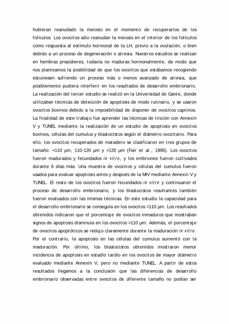
hubieran reanudado la meiosis en el momento de recuperarlos de los
folículos. Los ovocitos sólo reanudan la meiosis en el interior de los folículos
como respuesta al estímulo hormonal de la LH, previo a la ovulación, o bien
debido a un proceso de degeneración o atresia. Nuestros estudios se realizan
en hembras prepúberes, todavía no maduras hormonalmente, de modo que
nos planteamos la posibilidad de que los ovocitos que estábamos recogiendo
estuviesen sufriendo un proceso más o menos avanzado de atresia, que
posiblemente pudiera interferir en los resultados de desarrollo embrionario.
La realización del tercer estudio se realizó en la Universidad de Gante, donde
utilizaban técnicas de detección de apoptosis de modo rutinario, y se usaron
ovocitos bovinos debido a la imposibilidad de disponer de ovocitos caprinos.
La finalidad de este trabajo fue aprender las técnicas de tinción con Annexin
V y TUNEL mediante la realización de un estudio de apoptosis en ovocitos
bovinos, células del cumulus y blastocistos según el diámetro ovocitario. Para
ello, los ovocitos recuperados de matadero se clasificaron en tres grupos de
tamaño: <110 µm, 110-120 µm y >120 µm (Fair et al., 1995). Los ovocitos
fueron madurados y fecundados in vitro, y los embriones fueron cultivados
durante 6 días más. Una muestra de ovocitos y células del cumulus fueron
usados para evaluar apoptosis antes y después de la MIV mediante Annexin V y
TUNEL. El resto de los ovocitos fueron fecundados in vitro y continuaron el
proceso de desarrollo embrionario, y los blastocistos resultantes también
fueron evaluados con las mismas técnicas. En este estudio la capacidad para
el desarrollo embrionario se conseguía en los ovocitos >110 µm. Los resultados
obtenidos indicaron que el porcentaje de ovocitos inmaduros que mostraban
signos de apoptosis disminuía en los ovocitos >110 µm. Además, el porcentaje
de ovocitos apoptóticos se redujo claramente durante la maduración in vitro.
Por el contrario, la apoptosis en las células del cumulus aumentó con la
maduración. Por último, los blastocistos obtenidos mostraron menor
incidencia de apoptosis en estadío tardío en los ovocitos de mayor diámetro
evaluado mediante Annexin V, pero no mediante TUNEL. A partir de estos
resultados llegamos a la conclusión que las diferencias de desarrollo
embrionario observadas entre ovocitos de diferente tamaño no podían ser

explicados únicamente por una diferencia en la incidencia de apoptosis entre
diámetros.
Por último, el cuarto y quinto trabajo pretendían evaluar la apoptosis
en ovocitos caprinos de diferente diámetro mediante las dos técnicas
utilizadas en el estudio anterior. En estos trabajos quisimos introducir un
parámetro más a tener en cuenta, además del diámetro ovocitario, y por ello
clasificamos nuestros ovocitos también según criterios morfológicos en dos
grupos: los que no mostraban signos de atresia, es decir, que presentaban el
cumulus compacto y el citoplasma homogéneo; y aquellos que presentaban
signos de atresia temprana, es decir, con el cumulus ligeramente expandido
y/o el citoplasma del ovocito heterogéneo. Pensamos que era interesante
introducir el parámetro de la morfología porque podría ser utilizado como
criterio predictivo de la calidad de los embriones resultantes. Para ello, los
ovocitos fueron clasificados por diámetro siguiendo los criterios explicados en
el primer trabajo, y por morfología según los parámetros anteriormente
mencionados. Al igual que en el anterior estudio, la apoptosis fue evaluada en
ovocitos y células del cumulus antes y después de la maduración, y en los
blastocistos obtenidos. En estos estudios, al igual que el realizado en bovino,
también observamos una disminución de la apoptosis en los ovocitos y un
incremento en las células del cumulus durante la maduración in vitro. En
general, se observó que los ovocitos de mayor diámetro y de morfología sana
presentaban menos incidencia de apoptosis, aunque los resultados obtenidos
difirieron según la técnica utilizada. No se detectaron diferencias en cuanto a
incidencia de apoptosis en los blastocistos obtenidos a partir de ovocitos de
diferente diámetro. Por último, pudimos observar que la capacidad para el
desarrollo embrionario no dependía sólo del diámetro del ovocito, sino
también de su morfología. Por lo tanto, pudimos concluir que la apoptosis sí
puede influenciar la capacidad para el desarrollo del ovocito, pero no la
calidad del blastocisto obtenido.

SUMMARY
In our laboratory, we have always worked in order to achieve as many
blastocysts as possible from oocytes obtained from slaughtered prepubertal
goats. Oocyte population recovered from these females use to be very
variable, basically due to physiological factors. For this reason, we must
select oocytes very carefully in order to use only the oocytes capable to
mature in vitro, be fertilized and maintain embryonic development. So far,
selection performed in our laboratory was based on morphological criteria,
and we only selected large oocytes that were surrounded by several layers of
compact cumulus cells and had homogeneous cytoplasm. However, the
blastocyst rate obtained using the oocytes selected with that criteria has been
low, and we have not achieved more than 10% (Izquierdo et al., 1999). This
thesis, as a consequence, was born because we needed to find some markers
of oocyte competence, so we could distinguish developmental competent
oocytes from the rest of oocytes incapable to develop until blastocyst stage.
The aim of the first two works of this thesis, which can be grouped into
one chapter, was to study the role that plays the expression of the subunits of
MPF (Maturation Promoting Factor), Cyclin B1 and p34cdc2, MPF kinase activity
and the storage of RNA and proteins in oocytes in the acquisition of
developmental competence. The methods used to evaluate these parameters
were invasive ones, and therefore the oocytes evaluated could not longer be
used to continue the subsequent embryonic development. As a consequence,
it was necessary establish a relationship between the molecular
characteristics studied with a parameter that allowed us to select the oocytes
visually. Oocyte diameter was chosen as non-invasive parameter, because a
relationship between oocyte competence to develop into a viable embryo and
follicular diameter has been observed in many species (bovine: Furher et al.,
1989; caprine: Crozet et al., 1995; porcine: Marchal et al., 2002) and the
follicular diameter has been also related to oocyte diameter (bovine: Arlotto
et al., 1996; caprine: Crozet et al., 2000; de Smedt et al., 1994). That way,
prepubertal goat oocytes recovered from the slaughterhouse were divided in
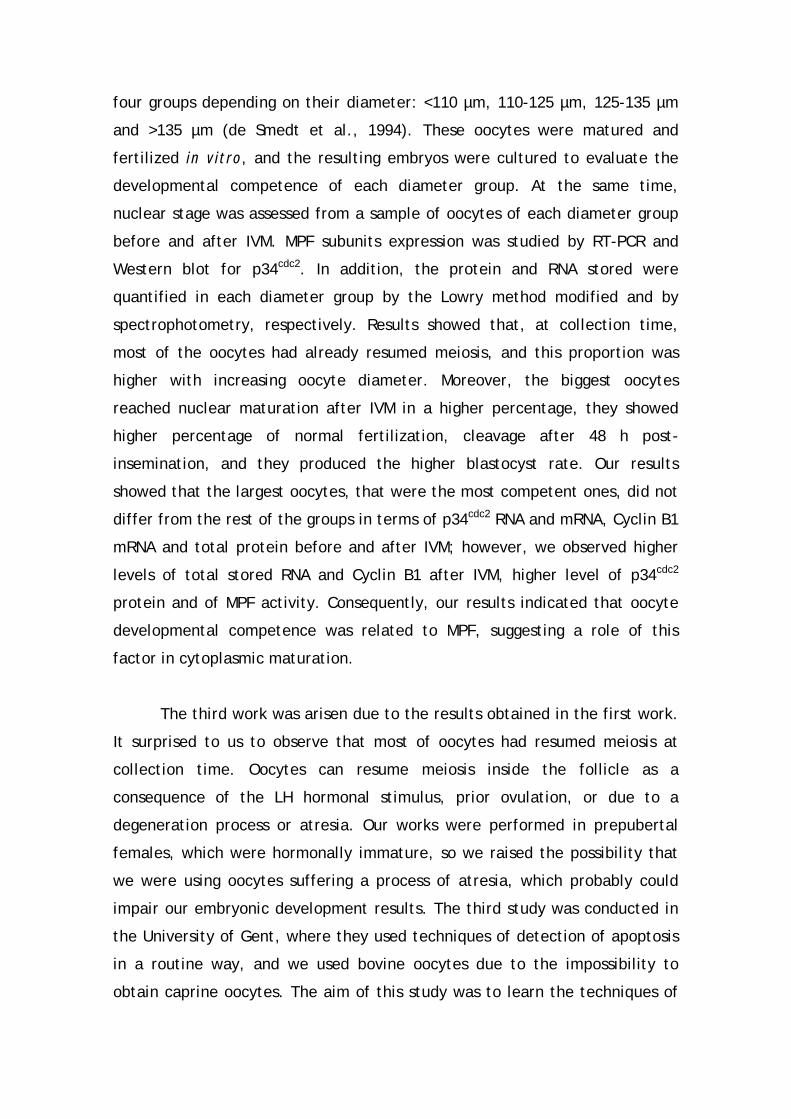
four groups depending on their diameter: <110 µm, 110-125 µm, 125-135 µm
and >135 µm (de Smedt et al., 1994). These oocytes were matured and
fertilized in vitro, and the resulting embryos were cultured to evaluate the
developmental competence of each diameter group. At the same time,
nuclear stage was assessed from a sample of oocytes of each diameter group
before and after IVM. MPF subunits expression was studied by RT-PCR and
Western blot for p34cdc2. In addition, the protein and RNA stored were
quantified in each diameter group by the Lowry method modified and by
spectrophotometry, respectively. Results showed that, at collection time,
most of the oocytes had already resumed meiosis, and this proportion was
higher with increasing oocyte diameter. Moreover, the biggest oocytes
reached nuclear maturation after IVM in a higher percentage, they showed
higher percentage of normal fertilization, cleavage after 48 h post-
insemination, and they produced the higher blastocyst rate. Our results
showed that the largest oocytes, that were the most competent ones, did not
differ from the rest of the groups in terms of p34cdc2 RNA and mRNA, Cyclin B1
mRNA and total protein before and after IVM; however, we observed higher
levels of total stored RNA and Cyclin B1 after IVM, higher level of p34cdc2
protein and of MPF activity. Consequently, our results indicated that oocyte
developmental competence was related to MPF, suggesting a role of this
factor in cytoplasmic maturation.
The third work was arisen due to the results obtained in the first work.
It surprised to us to observe that most of oocytes had resumed meiosis at
collection time. Oocytes can resume meiosis inside the follicle as a
consequence of the LH hormonal stimulus, prior ovulation, or due to a
degeneration process or atresia. Our works were performed in prepubertal
females, which were hormonally immature, so we raised the possibility that
we were using oocytes suffering a process of atresia, which probably could
impair our embryonic development results. The third study was conducted in
the University of Gent, where they used techniques of detection of apoptosis
in a routine way, and we used bovine oocytes due to the impossibility to
obtain caprine oocytes. The aim of this study was to learn the techniques of

Annexin V staining and TUNEL by the accomplishment of a study of apoptosis
in bovine oocytes, cumulus cells and blastocysts depending on oocyte
diameter. For this purpose, oocytes obtained from the slaughterhouse were
classified in three groups of size: <110 µm, 110-120 µm y >120 µm (Fair et al.,
1995). Oocytes were matured and fertilized in vitro, and the resulting
embryos were culture for additional 6 days. A sample of oocytes and cumulus
cells were used to assessed apoptosis before and after IVM by means of
Annexin V staining and TUNEL. The rest of the oocytes kept on developing,
and the resulting blastocysts were also evaluated with the same techniques.
In this study, oocyte developmental competence was achieved in oocytes
higher than 110 µm. Our results showed that the percentage of immature
oocytes showing signs of apoptosis decreased in oocytes > 110 µm. In addition,
the proportion of apoptotic oocytes was reduced clearly during IVM. On the
contrary, apoptosis in cumulus cells increased during maturation. Finally, the
blastocysts obtained showed less incidence of late apoptosis in the biggest
oocytes when evaluated by means of Annexin V, but not by means of TUNEL.
From these results we could conclude that differences observed in different
diameter groups in terms of embryonic development could not be explained
only because a differential incidence of apoptosis among diameter groups.
Finally, the purpose of the fourth and fifth studies was to evaluate
apoptosis in goat oocytes of different diameter by means of the two
techniques used in the previous work. We wanted to introduce a new
parameter in these studies, in addition to oocyte diameter, and so we
classified the oocytes by morphological criteria as well: the ones that did not
show any sign of atresia, that is, oocytes that had compact cumulus cells
layers and homogeneous cytoplasm, and the ones that showed early signs of
atresia, that is, oocytes with heterogeneous cytoplasm and/or cumulus cells
with initial expansion. We believed that it would be more interesting to
introduce the morphological classification because it could be use as a
predictive parameter of the quality of the resulting embryos. For that
purpose, oocytes were classified by diameter following the criteria explained
in the first work, and by morphology following the criteria explained before.

Like in the previous study, apoptosis was evaluated in oocytes and cumulus
cells before and after in vitro maturation, and in the blastocysts obtained.
Like in bovine, we also observed in this study a decrease of apoptosis in
oocytes and an increase in cumulus cells during IVM. In general, it was
observed that the biggest oocytes with healthy morphology showed fewer
incidences of apoptosis, although the results obtained differed depending on
the technique used. Finally, we could observe that oocyte developmental
competence did not only depend on oocyte diameter, but also in COC
morphology. Consequently, we could conclude that apoptosis can affect
oocyte developmental competence, but not blastocyst quality.

INTRODUCCIÓN
1. INTRODUCCIÓN
Durante los últimos años ha existido un interés creciente por las
técnicas de reproducción asistida, no sólo en su aplicación clínica en
humanos, sino también en el campo de la producción animal. El avance
logrado en las técnicas de maduración in vitro (MIV), fecundación in vitro
(FIV) y cultivo in vitro de embriones pre-implantacionales (CIV) ha permitido
el desarrollo de otras tecnologías, como la obtención de células madre, el
sexaje de embriones, la transgénesis o la clonación. La obtención de un mayor
número de embriones a menor coste mediante la producción in vitro de
embriones (PIV), en comparación con la técnica MOET (Multiovulation and
Embryo Transfer), ha hecho posible el avance de las nuevas tecnologías
anteriormente mencionadas. Por otro lado también ha permitido la
recuperación de especies en peligro de extinción, la reproducción de animales
muertos, acortar el intervalo generacional (de interés en programas de
mejora genética) con el uso de animales prepúberes como donantes de
ovocitos, etc. En general, la mayoría de los estudios sobre MIV, FIV y CIV
realizados hasta el momento en animales de producción se han centrado en el
bovino. Sin embargo, nuestro laboratorio ha optado por el caprino como
modelo experimental, ya que presenta diversas ventajas frente al bovino:
mayor manejabilidad, manutención menos costosa, período de gestación más
corto y mayor prolificidad. Además, la posibilidad de obtener productos de
interés farmacológico e industrial sintetizados en glándula mamaria posibilita
que la cabra presente un alto interés en el campo de la transgénesis.
Los ovarios de cabras sacrificadas en matadero se ha convertido en la
fuente más importante de ovocitos para la producción in vitro de embriones,
básicamente debido a que se obtienen un gran número de ovocitos a un coste
mucho menor que si se utilizaran animales vivos como donantes de ovocitos.
Sin embargo, la gran mayoría de cabras sacrificadas para el consumo en
nuestro país son prepúberes, de alrededor 1-2 meses de edad. Obtener los
ovocitos de animales prepúberes supone una desventaja respecto a las adultas
1
INTRODUCCIÓN
en cuanto a la eficiencia en la producción de blastocistos (Armstrong, 2001);
no obstante, la cantidad de ovocitos obtenidos por ovario en animales
prepúberes es mayor (Baldassarre et al., 1997). Además producir embriones y
descendencia de animales prepúberes intensifica la respuesta a la selección
genética al acortar el intervalo generacional.
El trabajo de esta tesis es la continuación del trabajo que viene
desarrollándose en nuestro laboratorio desde los años 90. Los primeros
estudios realizados comprobaron que los ovocitos obtenidos de cabras
prepúberes eran capaces de madurar in vitro (Martino et al., 1994), ser
fecundados (Martino et al., 1995) y desarrollarse hasta blastocisto (Mogas et
al., 1997). Sin embargo, también se observaron numerosas anomalías en la
fecundación, como la no descondensación de la cabeza del espermatozoide o
la polispermia (Martino et al., 1995; Mogas et al., 1997), y en el desarrollo
embrionario, como la parada en el desarrollo en el momento de activación del
genoma embrionario (Izquierdo et al., 1999) o una alta incidencia de haploidía
(Villamediana et al., 2001). Todos estos estudios indicaban que las anomalías
detectadas eran consecuencia de una deficiente maduración citoplasmática
de los ovocitos utilizados, de modo que los estudios posteriores trataron de
mejorarla mediante la selección de los ovocitos con Brilliant Cresyl Blue
(Rodríguez-González et al., 2003a; Rodríguez-González et al., 2002; Urdaneta
et al., 2003b), la adición de compuestos tioles en los medios de cultivo (Mayor
et al., 2001; Rodríguez-González et al., 2003a; Rodríguez-González et al.,
2003b; Urdaneta et al., 2003a; Urdaneta et al., 2004), o la premaduración
(Jiménez-Macedo et al., 2006). Estos estudios permitieron incrementar el
porcentaje de blastocistos obtenidos, aunque este porcentaje es más bajo que
el obtenido con ovocitos de cabras adultas. El siguiente paso, englobado en el
trabajo presentado en esta tesis, ha sido el estudio de la calidad ovocitaria a
través de la búsqueda de marcadores moleculares que nos permitieran la
selección únicamente de los ovocitos capaces de desarrollarse hasta el estadío
de blastocisto, ya que la calidad ovocitaria es el principal factor que
determina el éxito del desarrollo embrionario (Rizos et al., 2002).
2

INTRODUCCIÓN
Numerosos estudios han centrado sus esfuerzos en la búsqueda de
marcadores de calidad ovocitaria, tales como el contenido mitocondrial del
ovocito (Santos et al., 2006), la cantidad relativa de transcritos específicos en
el ovocito (De Sousa et al., 1998; Watson et al., 2000) o en el embrión, la
actividad transcripcional (Bilodeau-Goeseels y Panich, 2002) o la incidencia de
apoptosis en ovocitos (Liu et al., 2000; Yang y Rajamahendran, 2002; Yuan et
al., 2005), embriones (Liu et al., 2000; Pomar et al., 2005; Yang y
Rajamahendran, 2002) o células del cumulus (Corn et al., 2005; Lee et al.,
2001; Yuan et al., 2005; Zeuner et al., 2003). Sin embargo, la mayoría de
marcadores se han estudiado mediante técnicas invasivas, de modo que el
ovocito no puede utilizarse para su desarrollo posterior. Como consecuencia,
es importante relacionar estos marcadores con características no invasivas
(ej: morfología del cumulus, diámetro folicular, diámetro ovocitario). Existen
varios estudios que relacionan el diámetro del folículo con la competencia de
su ovocito para desarrollarse hasta blastocisto (bovino: Furher et al., 1989;
caprino: Crozet et al., 1995; porcino: Marchal et al., 2002); de esta forma
utilizan este criterio folicular, junto con la técnica de la aspiración, para
seleccionar los ovocitos que se utilizarán en el proceso de producción in vitro
de embriones. En ovarios de cabras prepúberes la textura del tejido y el
tamaño de los folículos hacen impracticable este método de selección
ovocitaria. El diámetro folicular ha sido relacionado de forma positiva con el
diámetro ovocitario en bovino (Arlotto et al., 1996) y caprino (Crozet et al.,
2000; de Smedt et al., 1994). Consecuentemente, en el presente trabajo de
investigación se escogió el uso del diámetro ovocitario como indicador no
invasivo.
Teniendo en cuenta las consideraciones anteriores, el objetivo de esta
tesis fue el estudio de características moleculares en ovocitos de diferente
tamaño que nos permitieran seleccionar aquéllos con mayor capacidad para
desarrollarse hasta el estadío de blastocisto. Los posibles marcadores
seleccionados para el estudio fueron:
3
INTRODUCCIÓN
a) La expresión a nivel transcripcional y traduccional del Maturation
Promoting Factor (MPF), así como su actividad. El MPF es un heterodímero
compuesto por la p34cdc2 y la Ciclina B1, y se considera el principal regulador
de la maduración nuclear del ovocito y un posible candidato a regular la
maduración citoplasmática (Naito et al., 1992). La acumulación de una
cantidad umbral de alguno de sus componentes ha sido descrita como el
factor limitante para la reanudación meiótica en el ovocito (p34cdc2: Chesnel y
Eppig, 1995; De Vantéry et al., 1996; de Vantery et al., 1997; Dedieu et al.,
1998; Mitra y Schultz, 1996; Ciclina B1: Levesque yd Sirard, 1996; Sun et al.,
2001). Por lo tanto, también puede jugar un papel importante en la
adquisición de competencia ovocitaria para el desarrollo (Vigneron et al.,
2004).
b) la incidencia de apoptosis en ovocitos y células del cumulus dependiendo,
no sólo del diámetro ovocitario, sino también en la morfología del complejo
cumulus-ovocito (COC). El uso de ovarios procedentes de matadero supone
obtener una población ovocitaria muy heterogénea, donde podemos encontrar
diferentes grados de desarrollo y de atresia. De hecho, el 85% de los folículos
presentes en un ovario están sufriendo atresia (Kruip y Dieleman, 1982). Por
lo tanto, era necesario establecer la incidencia de la apoptosis en COC de
cabras prepúberes y de qué modo podía influenciar el desarrollo embrionario.
Para llevar a cabo este estudio se realizó un trabajo previo con ovocitos de
vaca, con la finalidad de poner a punto los métodos que se iban a utilizar
posteriormente con ovocitos de cabras prepúberes. A su vez, este estudio
también nos sirvió como comparativa entre los ovocitos caprinos y bovinos.
4

REVISIÓN BIBLIOGRÁFICA
7
2. REVISIÓN BIBLIOGRÁFICA 2.1. MADURACIÓN DEL OVOCITO
Cuando los ovocitos se forman, durante el desarrollo fetal de la
hembra, quedan parados en la profase de la primera división meiótica después
de sufrir una serie de divisiones mitóticas. En este estado los ovocitos no son
capaces de ser fecundados y dar lugar a un embrión viable; es necesario que
el ovocito crezca, junto con el folículo, y que se produzcan una serie de
cambios morfológicos, ultraestructurales y moleculares que dotarán al ovocito
de capacidad para finalizar la meiosis, originando una célula haploide, ser
fecundado y dar lugar a un embrión viable. Este conjunto de cambios se
conoce como maduración ovocitaria, que engloba dos procesos: la maduración
nuclear o meiótica y la maduración citoplasmática.
2.1.1. Maduración nuclear o meiótica
La maduración nuclear en el ovocito es el proceso que tiene lugar
desde la reanudación de la primera división meiótica hasta la parada en
metafase de la segunda división meiótica. El ovocito solamente podrá
completar la meiosis cuando se dé su reactivación mediante la fecundación.
La maduración nuclear in vivo se inicia con el pico de la hormona luteinizante
(LH), cuando el ovocito ya ha completado su fase de crecimiento. In vitro, los
ovocitos también son capaces de madurar cuando se les libera de su folículo
(Pincus y Enzmann, 1935; revisado por Ponderato et al., 2001); en este caso
es necesario que el ovocito haya alcanzado el 80-90% de su tamaño final para
que pueda reanudar la meiosis.
Al recibir el estímulo pre-ovulatorio de la LH se produce la activación
de una cascada de reacciones en las que intervienen numerosas proteínas con

REVISIÓN BIBLIOGRÁFICA
8
actividad kinasa que conducirán a la ruptura de la vesícula germinal (GVBD =
Germinal vesicle breakdown). Los nucleolos desaparecen, se produce la
polimerización de los microtúbulos, y los cromosomas se condensan y se
alinean en la placa metafásica (Metafase I). Seguidamente se separan los
pares de cromosomas homólogos en una división asimétrica que origina dos
células de diferente tamaño: el primer corpúsculo polar (PBI), y el ovocito
secundario. Al iniciarse la meiosis II el ovocito no entra en profase y los
cromosomas permanecen condensados, en un período conocido como
interkinesis. El ovocito secundario, que ahora se encuentra en estadío de
metafase II, sufre una nueva parada meiótica, y es en este estadío cuando, en
la mayoría de mamíferos, el ovocito es ovulado. Sólo se completará la meiosis
en caso de que el ovocito se active por la penetración de un espermatozoide,
extruyendo al final el segundo corpúsculo polar (PBII).
A B
D C

REVISIÓN BIBLIOGRÁFICA
9
Figura 1. Diferentes estadios meióticos en el ovocito (tinción con lacmoide):
A) Vesícula germinal (GV); B) Ruptura de la vesícula germinal (GVBD); C)
Metafase I (MI); D) Metafase II (MII)
2.1.2. Maduración citoplasmática
La maduración citoplasmática comprende todos los procesos que tienen
lugar en el citoplasma y que van a posibilitar que el ovocito sea capaz de
descondensar la cabeza del espermatozoide tras la fecundación y mantener el
posterior desarrollo embrionario (Mermillod et al., 1999; Prather y Day, 1998).
A diferencia de la maduración nuclear, que puede ser fácilmente observada
con el microscopio óptico, la evaluación de la maduración citoplasmática es
mucho más complicada, y debería valorarse según la capacidad del ovocito
para dar lugar a un embrión viable, capaz de desarrollarse y dar lugar al
nacimiento de un animal vivo y sano. Sin embargo, debido a la dificultad de
llevar a cabo estos procesos a nivel experimental, generalmente se evalúa la
maduración citoplasmática de un ovocito mediante su capacidad para llegar al
estadío embrionario de blastocisto.
La acumulación de ARNm y proteínas en el citoplasma durante el
crecimiento del ovocito son esenciales para que éste puede mantener el
desarrollo embrionario hasta que se produzca la activación del genoma del
embrión (Bachvarova, 1992), que tiene lugar en embriones de 2 células en
ratón, de 4 células en cerdos, de 4-8 células en humanos, de 8 células en
conejo, y embriones de 8-16 células en vacas y ovejas (Telford et al., 1990).
En la especie caprina, igual que en los rumiantes citados, la activación del
genoma embrionario se produce a las 8-16 células. El almacenamiento del
ARNm en el ovocito se produce mayoritariamente por poliadenilación
citoplasmática selectiva (Gandolfi y Gandolfi, 2001), ya que se ha observado
que las colas poli-A son cortas cuando el ARNm materno se almacena, y se
alargan para que el ARNm sea reclutado para la traducción (Brevini-Gandolfi
et al., 1999; Huarte et al., 1992; Temeles y Schultz, 1997). La regulación de

REVISIÓN BIBLIOGRÁFICA
10
la transcripción del ARNm almacenado también se produce por
enmascaramiento de elementos específicos de determinados ARNm (Gandolfi
y Gandolfi, 2001), impidiendo así su traducción a proteínas. El ARN que se
transcribe durante la maduración citoplasmática es muy estable, con una vida
media aproximada de 28 días (Wassarman et al., 1996).
Al mismo tiempo, a medida que el ovocito va creciendo también se
produce la redistribución y maduración de orgánulos citoplasmáticos (Caralco,
1995; Ducibella et al., 1994; Hyttel et al., 1986). Las mitocondrias, que se
encuentran en el centro del ovocito al inicio de su crecimiento, migran hacia
la periferia a medida que aumenta el tamaño ovocitario (Cran, 1985; Fair et
al., 1995). Los gránulos corticales, importantes para prevenir fenómenos de
polispermia durante la fecundación (Guraya, 1982), migran del centro hasta
situarse por debajo de la membrana citoplasmática del ovocito (Assey et al.,
1994; Fair et al., 1995). El número de aparatos de Golgi, precursores de los
gránulos corticales, también aumenta con el diámetro del ovocito (Fair et al.,
1995). El Retículo Endoplasmático se distribuye de forma dispersa por el
citoplasma, formando sacos densos que avanzan hacia la periferia con el
crecimiento ovocitario.
Pocos días antes del pico de LH se dan en el ovocito los últimos cambios
a nivel citoplasmático que van a conducir a su “capacitación”, es decir, van a
dotarle de competencia para madurar y ser fecundado correctamente, y
mantener el desarrollo embrionario posterior (Hyttel et al., 1997). Se produce
una disminución del aparato de Golgi, y un reagrupamiento de los gránulos
corticales (Cran, 1985). Se inhibe la síntesis de ARN y proteínas, que era muy
activa durante el crecimiento del ovocito, mediante la condensación del
nucleolo y la eliminación de ribosomas (Fair et al., 1995; Hyttel et al., 1986,
1989). Justo antes de la ovulación aparece entre el ovocito y la zona pelúcida
el espacio perivitelino, que aumentará de volumen con el tiempo.

REVISIÓN BIBLIOGRÁFICA
11
2.1.3. Papel del cumulus oophorus en la maduración del ovocito
La unión de las células del cumulus al ovocito durante la maduración es
importante para completar con éxito no sólo la maduración, sino también la
fecundación y el posterior desarrollo embrionario (Atef et al., 2005;
Wongsrikeao et al., 2005). Las células del cumulus se comunican entre ellas y
con el ovocito mediante uniones tipo gap (Eppig, 1982; Furger et al., 1996;
Moor et al., 1980), lo que permite el intercambio de moléculas entre los dos
tipos celulares, produciéndose una regulación cumulus-ovocito recíproca
(Heikinheimo y Gibbons, 1998).
Las células del cumulus juegan un papel importante en la maduración
del ovocito mediante la regulación de varios procesos:
a) el mantenimiento del ovocito en parada meiótica: la transferencia
directa de sustancias desde las células del cumulus al ovocito
mantienen a éste en parada meiótica. Hay varias sustancias que se
consideran inhibidoras de la meiosis, como purinas (Eppig et al.,
1985) y el AMPc (cyclic Adenosine monophosphate) (Kumar y Gilula,
1996).
b) La reanudación de la meiosis: Una vez el ovocito ha completado su
crecimiento, la reanudación de la meiosis in vivo se produce por la
pérdida de uniones gap entre el cumulus y el ovocito a consecuencia
de la expansión de las células del cumulus (Wert y Larsen, 1989)
inducida por el pico de LH que tiene lugar antes de la ovulación. La
pérdida de las uniones impide el paso de sustancias inhibidoras de la
meiosis al ovocito.
c) la maduración citoplasmática: la presencia de células del cumulus
durante la maduración favorece la posterior descondensación de la
cabeza del espermatozoide y su transformación a pronúcleo
masculino, mediante el aumento de la concentración intraovocitaria
de glutatión (Calvin et al., 1986; Perreault et al., 1988; Yoshida et
al., 1993), además de favorecer la fecundación monospérmica y el
desarrollo embrionario (ovejas: Staigmiller y Moor, 1984; ratas:
REVISIÓN BIBLIOGRÁFICA
12
Vanderhyden y Armstrong, 1989; vacas: Chian et al., 1994; cerdos:
Yamauchi y Nagai, 1999). Esta acción de las células del cumulus se
produce mediante glicosaminoglicanos, hormonas esteroideas y
otros factores que promueven la maduración citoplasmática en el
ovocito (Brower y Schultz, 1982; Danforth, 1995; Dode y Graves,
2002; Yamauchi y Nagai, 1999). Además las células del cumulus
estabilizan los gránulos corticales, impidiendo que se produzca una
migración prematura o una exocitosis parcial (Galeati et al., 1991).
d) Protección del ovocito: las células del cumulus también intervienen
en la protección del ovocito promoviendo la reducción de la cistina
presente en el medio a cisteína, y la captura de cisteína por parte
del ovocito (Takahashi et al., 1993) de modo que aumenta la
concentración intraovocitaria de glutatión. El glutatión mantiene
del estado redox de las células, protegiendo al ovocito del estrés
oxidativo (Tatemoto et al., 2000).
2.1.4. Regulación de la maduración ovocitaria
El AMPc es continuamente transferido desde las células del cumulus a
los ovocitos en crecimiento para mantener su parada meiótica (Dekel, 1988).
Está ampliamente aceptado que el AMPc es la señal inhibidora de la meiosis
ovocitaria. Concentraciones altas de AMPc en el ovocito promueve la actividad
catalítica de PKA (AMPc dependent protein kinase), que impide la activación
del MPF (Maturation Promoting Factor, también conocido como M-phase
promoting factor) a través de la inactivación de la fosfatasa Cdc25 y de la
inhibición de la síntesis de Ciclina B1 (revisado por Dekel, 2005). A su vez, la
localización subcelular de PKA está regulada por APAK (A kinase anchoring
proteins), que actúa como un mecanismo complementario para la regulación
de la actividad de PKA y un mejor control de la parada meiótica (revisado por
Dekel, 2005). Cuando se produce el pico de LH se genera una disminución de
la cantidad de AMPc que llega al ovocito desde las células del cumulus, debido
a una pérdida de conexiones célula-célula. La disminución de la concentración

REVISIÓN BIBLIOGRÁFICA
13
ovocitaria de AMPc, junto con la translocación de la PKA de su lugar de
acción, inhibe la actividad catalítica de PKA. A consecuencia, se produce la
activación de la fosfatasa Cdc25 mediante fosforilación. Esta fosfatasa
provocará la activación del MPF que dará lugar a la reanudación de la meiosis
y la progresión hasta MII.
El MPF fue descrito por primera vez por Masui y Markert (1971) en
ovocitos de anfibios, y posteriormente ha sido descrito en numerosas
especies. El MPF está compuesto por dos subunidades: la subunidad catalítica
(p34cdc2) que presenta actividad seronina/treonina kinasa, y la subunidad
reguladora (Ciclina B1) (Gautier et al., 1990). Este heterodímero se forma
inicialmente como un pre-MPF inactivo, en el cual la subunidad catalítica
presenta los residuos Thr 161, Thr 14 y Tyr 15 fosforilados. La activación del
complejo MPF se produce por la defosforilación de la Thr 14 y Tyr 15 mediada
por la fosfatasa Cdc25 B (Gould y Nurse, 1989; revisado por Dekel, 2005),
quien a su vez es fosforilada por el MPF, dando lugar a una retro-alimentación
positiva requerida para una activación rápida del complejo (Hoffmann et al.,
1993).
La actividad del MPF sigue un patrón oscilatorio, que ha sido
demostrado en muchas especies: se produce un aumento de la actividad de
MPF justo antes de la GVBD, alcanza un pico máximo en MI, disminuye su
actividad durante la anafase-telofase para que puedan separarse los
cromosomas, y aumenta de nuevo para alcanzar un nuevo pico máximo en
estadío de MII (Ratón: Choi et al., 1991; Fulka et al., 1992; Cerdo: Naito y
Toyoda, 1991; Bovino: Collas et al., 1993; Conejo: Jelinkova et al., 1994;
Cabra: Dedieu et al., 1996). Entre la meiosis I y II, aunque la actividad del
MPF disminuye, se mantiene a un nivel básico para impedir la replicación del
ADN.

REVISIÓN BIBLIOGRÁFICA
14
Figura 2. Representación esquemática de la actividad de MPF a lo largo de la
maduración del ovocito, fecundación y primeros estadíos de desarrollo
embrionario (www.med.yale.edu).
La activación del MPF da lugar a la ruptura de la envoltura nuclear
(GVBD), la condensación de los cromosomas, su disposición en la placa
metafásica y la poliadenilación del ARNm mos para promover su traducción.
Niveles elevados de MPF activo inhiben la interfase y la extrusión del primer
corpúsculo polar. La actividad de p34cdc2 controla el complejo APC/C, que es
el responsable de la degradación de la Ciclina B1, de manera que el MPF
activo induce su propia inactivación a través de la degradación de la Ciclina
B1 para salir de la primera división meiótica (Frank-Vaillant et al., 2001) y
para que se libere el primer corpúsculo polar. Debe darse acumulación de
Ciclina B1 para que aumente de nuevo la actividad de MPF. La actividad
máxima de MPF se alcanza en MII, y sólo disminuirá en caso de que se
produzca la fecundación del ovocito. La disminución de la actividad del MPF
se produce por la disociación del heterodímero y la degradación proteosomal
de la Ciclina B1 (Josefsberg et al., 2000).
Aunque el MPF ha sido ampliamente estudiado, se conocen pocos
sustratos sobre los que actúa. La histona H1 es el clásico sustrato del MPF, y
su fosforilación por parte del complejo da lugar a la condensación de los
cromosomas. Las lamininas nucleares también son fosforiladas por el MPF, así

REVISIÓN BIBLIOGRÁFICA
15
como la proteína kinasa MELK, que interviene en el control del ciclo celular,
la proliferación celular y el splicing de ARNm (Badouel et al., 2006). Además,
se ha sugerido una regulación por parte del MPF de la actividad de la pp60c-
src y ARN polimerasa II, que conduciría a reorganizaciones del citoesqueleto y
a la inhibición de la transcripción que tiene lugar durante la división celular,
respectivamente (revisado por Heikinheimo y Gibbons, 1998).
El MPF se considera el principal regulador de la maduración nuclear,
pero no es la única molécula que interviene en este proceso. Otros factores
que juegan un papel importante son las proteínas ERK-1 y ERK-2 (Extracellular
signal-regulated kinases), que forman parte de la familia de las MAPK
(Mitogen activated Protein kinase) (Lazar et al., 2002). El MPF regula la
poliadenilación del protooncogen mos, de modo que cuando el MPF está
activo se produce síntesis de proteína mos. Mos da lugar a la activación de
MEK Kinasa, quien a su vez regula la actividad de MEK, la proteína kinasa que
activa la proteína MAPK. La expresión del protooncogen mos, y por lo tanto,
la actividad de MAPK, están reguladas por las concentraciones intraovocitarias
de AMPc (Lazar et al., 2002), a través de la actividad de MPF (Lazar et al.,
2004). A su vez, la activación de MAPK también conduce a la activación del
MPF, a través de la inhibición de la degradación de la Ciclina B1, lo que da
lugar a la acumulación de la subunidad reguladora durante el paso de meiosis
I a meiosis II, y mantiene los niveles de MPF elevados durante la parada
meiótica en metafase II (revisado por Heikinheimo y Gibbons, 1998). Aunque
se ha visto que en ovocitos de Xenopus la expresión de mos es necesaria para
la reanudación de la meiosis (revisado por Roy et al., 1996), en algunos
mamíferos se ha observado que la reanudación de la meiosis es un proceso
independiente de MAPK (Lazar et al., 2002). La principal función de MAPK en
el proceso meiótico es mantener la parada en estadío de metafase II (Colledge
et al., 1994; Hashimoto et al., 1994) hasta que se produzca la fecundación.
Debido al importante papel que juega el MPF en la maduración nuclear
del ovocito, numerosos autores han hipotetizado que la deficiencia en la
síntesis de alguna de las dos subunidades del complejo, o de su actividad,

REVISIÓN BIBLIOGRÁFICA
16
podían ser las causas de la incompetencia de los ovocitos para reanudar la
meiosis. Se ha observado que las posibles causas de la incompetencia meiótica
ovocitaria varía entre especies:
? En ratón, la adquisición de competencia meiótica se asocia en parte
con la síntesis de p34cdc2 (Chesnel y Eppig, 1995; de Vantéry et al.,
1996, 1997; Mitra and Schultz, 1996), y no con la de Ciclina B1 (Chesnel
y Eppig, 1995; de Vantéry et al., 1996).
? En cabras, mientras que la Ciclina B1 en forma de ARNm y proteína se
detecta en ovocitos competentes e incompetentes (Hue et al., 1997),
la p34cdc2 sólo se encuentra en ovocitos meióticamente competentes
(Dedieu et al., 1998). Esto indica que, en esta especie, la incapacidad
de los ovocitos incompetentes para reanudar la meiosis podría deberse
a la ausencia de p34cdc2 en estos ovocitos, necesitándose de esta
manera una concentración mínima de esta proteína para dar lugar a la
ruptura de la vesícula germinal (Crozet et al., 2000; Dedieu et al.,
1998). Además Dedieu et al. (1998) hipotetizan que la cantidad de
p34cdc2 en ovocitos parcialmente competentes no es suficiente para
reanudar la meiosis espontáneamente en el momento adecuado,
produciéndose un retraso en la maduración.
? En cerdos, al contrario, los 2 componentes del MPF se encuentran en
los ovocitos inmaduros, sugiriendo que en esta especie la parada
meiótica se controla por otros mecanismos (Christmann et al., 1994).
Sin embargo, estudios posteriores han determinado que la síntesis de
Ciclina B1 es necesaria para reanudar la meiosis (Sun et al., 2001).
? En bovino, la proteína Ciclina B1 no se detecta en el estadío de vesícula
germinal (Levesque y Sirard, 1996) pero sí se detecta su ARNm, cuyos
niveles disminuyen durante el crecimiento folicular (Robert et al.,
2002). La reanudación de la meiosis en bovino se inicia con la
acumulación de proteína ciclina B1 (Levesque y Sirard, 1996).
La regulación de la maduración citoplasmática, al contrario que la
regulación de la maduración nuclear, es bastante desconocida; sin embargo,
se cree que el MPF y la maduración citoplasmática podrían estar relacionados

REVISIÓN BIBLIOGRÁFICA
17
de algún modo, ya que se ha observado que ovocitos con baja actividad MPF
en metafase II son incapaces de formar el pronúcleo masculino después de la
fecundación (Naito et al., 1992), debido a una incapacidad para eliminar la
membrana nuclear del espermatozoide (Peter et al., 1990). Niveles bajos de
MPF en la segunda parada meiótica no serían suficientes para activar la
degradación de la Ciclina B1, y por lo tanto, la disociación del MPF que se
produce tras la fecundación.
2.2. APOPTOSIS
2.2.1. Conceptos generales
La apoptosis es el mecanismo que permite regular procesos biológicos,
como la morfogénesis y la homeostasis tisular, mediante la eliminación de
células defectuosas o que ya no son necesarias (Steller, 1995). Es un tipo de
muerte celular programada totalmente controlada por la expresión de
determinados genes, y caracterizada por una serie de cambios morfológicos y
bioquímicos (Wyllie et al., 1980).
Existen 2 rutas principales de señalización de la apoptosis: la
dependiente de receptor y la dependiente de mitocondria (Ashkenazi y Dixit,
1998; Green y Reed, 1998; Nagata, 1997; revisado por Hussein, 2005). En la
primera, la unión de diferentes ligandos, como TNF (tumor necrosis factor),
TRAIL (TNF- related apoptosis-inducing ligand), Fas o APO3L, a sus receptores
desencadena una cascada de reacciones que tendrá como consecuencia final
la activación de caspasas efectoras y la muerte de la célula. Esta ruta está
regulada por Flip (FLICE inhibitory protein), que previene la activación de
caspasas iniciadoras, y por IAP (inhibitor of apoptosis) (Hussein et al., 2003).
En la ruta dependiente de mitocondria, factores inductores de la apoptosis,
como la irradiación, la presencia o la ausencia de citokinas o determinados
factores de crecimiento, inducen la desestabilización de la membrana
mitocondrial y, por consiguiente, la liberación de citocromo-c al citosol, que
se unirá al Apaf-1 (apoptotic protease-activating factor 1) (Robles et al.,

REVISIÓN BIBLIOGRÁFICA
18
1999; Zou et al., 1997). Este complejo se une a la pro-caspasa-9, la activa y
se inicia la activación de caspasas efectoras, como la caspasa-3 (Grutter,
2000; Wang, 2001), dando lugar a la muerte celular.
Figura 3. Representación de las dos rutas que intervienen en la activación y
ejecución de la apoptosis (Hussein, 2005).
2.2.2. Métodos de detección de la apoptosis
2.2.2.1. Evaluación morfológica
La apoptosis fue descrita por primera vez por Kerr et al. (1972),
quienes describieron las distintas fases morfológicas que permiten distinguir
una célula que está sufriendo un proceso apoptótico: inicialmente se produce
la condensación nuclear debido a la redistribución de la cromatina junto a la
membrana nuclear, el citoplasma se condensa dando lugar a la pérdida de
volumen celular, y las membranas nuclear y citoplasmática pierden
integridad. Seguidamente el ADN se fragmenta, y se forman unas vacuolas
rodeadas de membrana que contienen parte del citoplasma, orgánulos
celulares y fragmentos nucleares, conocidos como cuerpos apoptóticos o

REVISIÓN BIBLIOGRÁFICA
19
picnóticos, que serán fagocitados por las células vecinas. La apoptosis,
además, se caracteriza porque no lleva asociada una respuesta inflamatoria,
al contrario que sucede con la necrosis (Kerr et al., 1994).
Figura 4. Representación esquemática de las principales características
morfológicas y bioquímicas de la apoptosis. (Hardy, 1999)
Sin embargo, estudios posteriores observaron que algunas de las
características morfológicas presentes en estadíos iniciales de la apoptosis
también se observaban en el inicio de la necrosis (Columbano, 1995;
Lemasters et al., 1998; Rosales-Torres et al., 2000; Zamai et al., 1996;
Zamzami et al., 1997). Se ha postulado que la apoptosis y la necrosis se
inician con una alteración de la integridad de la membrana mitocondrial
(Columbano, 1995; Lemasters et al., 1998; Zamzami et al., 1997), pero el tipo
de muerte celular que se llevará a cabo viene determinado según el contenido
energético de la célula (Vayssiere et al., 1994; Zamzami et al., 1995): si los
niveles de ATP son altos se desencadenará la apoptosis (Ankarcrona et al.,
1995; Leist et al., 1997), mientras que si son bajos se producirá la necrosis
(Ankarcrona et al., 1995). Además, la apoptosis se inicia antes de que las
señales morfológicas de degeneración sean visibles (Asselin et al., 2000). Todo

REVISIÓN BIBLIOGRÁFICA
20
ello, unido a la dificultad de detectar células apoptóticas mediante un
microscopio óptico, hace necesario combinar la evaluación morfológica de las
células con otras técnicas para poder identificar la apoptosis.
2.2.2.2. Expresión de proteínas
En la regulación y ejecución de la apoptosis intervienen numerosas
proteínas y factores, cuya expresión puede servir para evaluar la incidencia
de apoptosis en una célula determinada. Entre estos factores podemos
destacar los siguientes:
? Familia Bcl-2 (B-Cell lymphoma-leukemia-2): contiene proteínas
inductoras (Bax, Bak, Bok) e inhibidoras de la apoptosis (Bcl-2, Bcl-xL)
(revisado por Guthrie et al., 2000). La proteína Bcl-2 (anti-apoptótica)
previene la apoptosis manteniendo la integridad de la membrana
mitocondrial (Yang et al., 1997). En cambio, cuando Bax (pro-
apoptótica) se sobre-expresa, forma heterodímeros con Bcl-2,
contrarrestando de esa manera los efectos de esta molécula en la
supervivencia de la célula (Oltvai et al., 1993). Por lo tanto, la ratio
Bcl-2/Bax es determinante en la supervivencia o muerte de la célula
(Oltvai et al., 1993).
? Citocromo c: la activación de la apoptosis compromete la integridad de
la membrana mitocondrial, dando lugar a la liberación al citosol del
citocromo c, que se encontraba en el interior de la mitocondria
(Kelekar y Thompson, 1998).
? Caspasas: son cisteína-proteasas que intervienen en el proceso de
apoptosis. Encontramos dos tipos de caspasas, las iniciadoras (caspasa-
2, caspasa-8, caspasa-9, caspasa-10) que responden a un estímulo
proapoptótico dando lugar a la activación del otro tipo de caspasas, las
efectoras (caspasa-3, caspasa-6, caspasa-7), que se encargan de
ejecutar la muerte celular.

REVISIÓN BIBLIOGRÁFICA
21
? Proteína p53: es un factor de transcripción que controla la
proliferación celular. Esta proteína regula la apoptosis a nivel genético
cuando hay daño irreparable en el ADN de la célula, induciendo la
transcripción de Bax e inhibiendo la transcripción de Bcl-2 y
promoviendo así su muerte (Ding y Fisher, 1998; Evan y Littlewood,
1998; Ko y Prives, 1996).
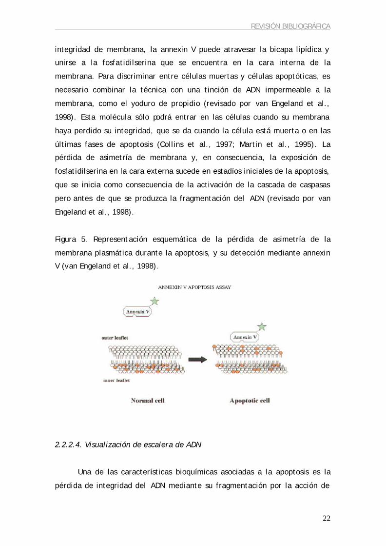
2.2.2.3. Tinción con Annexin V
Las células viables mantienen una asimetría entre la cara externa e
interna de la membrana plasmática (Bretscher, 1972), de modo que la
fosfatidilcolina y la esfingomielina se encuentran en la cara externa, mientras
que la fosfatidilserina y la fosfatidiletanolamina se observan sólo en la cara
interna. Esta asimetría de membrana se mantiene por la acción de unas
proteínas denominada flipasas (Higgins, 1994; revisado por Diaz y Schroit,
1996). Las células tienen la habilidad de translocar la fosfatidilserina a la cara
externa de la membrana citoplasmática en determinadas condiciones (Fadok
et al., 1992, 1993), sirviendo como diana de reconocimiento específica para
los macrófagos que deben fagocitar a las células en degeneración. Durante la
apoptosis se produce la activación de unas proteasas que degradan la fodrina,
responsable del ancoraje de la fosfatidilserina en la cara interna de la
membrana celular (revisado por van Engeland et al., 1998). De este modo, la
detección de la fosfatidilserina en la cara externa de la membrana puede
servir como indicador de apoptosis.
La annexin V es una molécula que se une específicamente a la
fosfatidilserina en presencia de calcio (Andree et al., 1990; Tait et al., 1989).
La combinación de la annexin V con biotina o fluorocromos permite su
detección mediante reacciones colorimétricas, citometría de flujo o
microscopía de fluorescencia, lo que facilita la detección de células
apoptóticas. La annexin V no es capaz de atravesar la membrana plasmática
en las células viables; sin embargo, en células muertas, que han perdido la
REVISIÓN BIBLIOGRÁFICA
22
integridad de membrana, la annexin V puede atravesar la bicapa lipídica y
unirse a la fosfatidilserina que se encuentra en la cara interna de la
membrana. Para discriminar entre células muertas y células apoptóticas, es
necesario combinar la técnica con una tinción de ADN impermeable a la
membrana, como el yoduro de propidio (revisado por van Engeland et al.,
1998). Esta molécula sólo podrá entrar en las células cuando su membrana
haya perdido su integridad, que se da cuando la célula está muerta o en las
últimas fases de apoptosis (Collins et al., 1997; Martin et al., 1995). La
pérdida de asimetría de membrana y, en consecuencia, la exposición de
fosfatidilserina en la cara externa sucede en estadíos iniciales de la apoptosis,
que se inicia como consecuencia de la activación de la cascada de caspasas
pero antes de que se produzca la fragmentación del ADN (revisado por van
Engeland et al., 1998).
Figura 5. Representación esquemática de la pérdida de asimetría de la
membrana plasmática durante la apoptosis, y su detección mediante annexin
V (van Engeland et al., 1998).
2.2.2.4. Visualización de escalera de ADN
Una de las características bioquímicas asociadas a la apoptosis es la
pérdida de integridad del ADN mediante su fragmentación por la acción de
REVISIÓN BIBLIOGRÁFICA
23
una endonucleasa (Williams et al., 1974). La fragmentación se produce a nivel
internucleosomal, de modo que los fragmentos de ADN que se originan tienen
una longitud múltiple de 180-200 pb. Este patrón en la fragmentación de ADN
puede ser fácilmente evaluado mediante electroforesis en un gel de agarosa
(Wyllie et al., 1980) a través de la aparición de “escaleras de ADN”
correspondientes a los oligonucleosomas. Sin embargo, es posible que se
produzca apoptosis sin la formación de estas “escaleras de ADN” (Cohen et
al., 1992; Collins et al., 1992), lo que limitaría la eficiencia de esta técnica
para la identificación de la apoptosis. Por otro lado, la necesidad de disponer
de gran cantidad de ADN para poder visualizarlo en un gel de agarosa hace
imposible el análisis cuando el número de células es limitado, como en los
embriones.
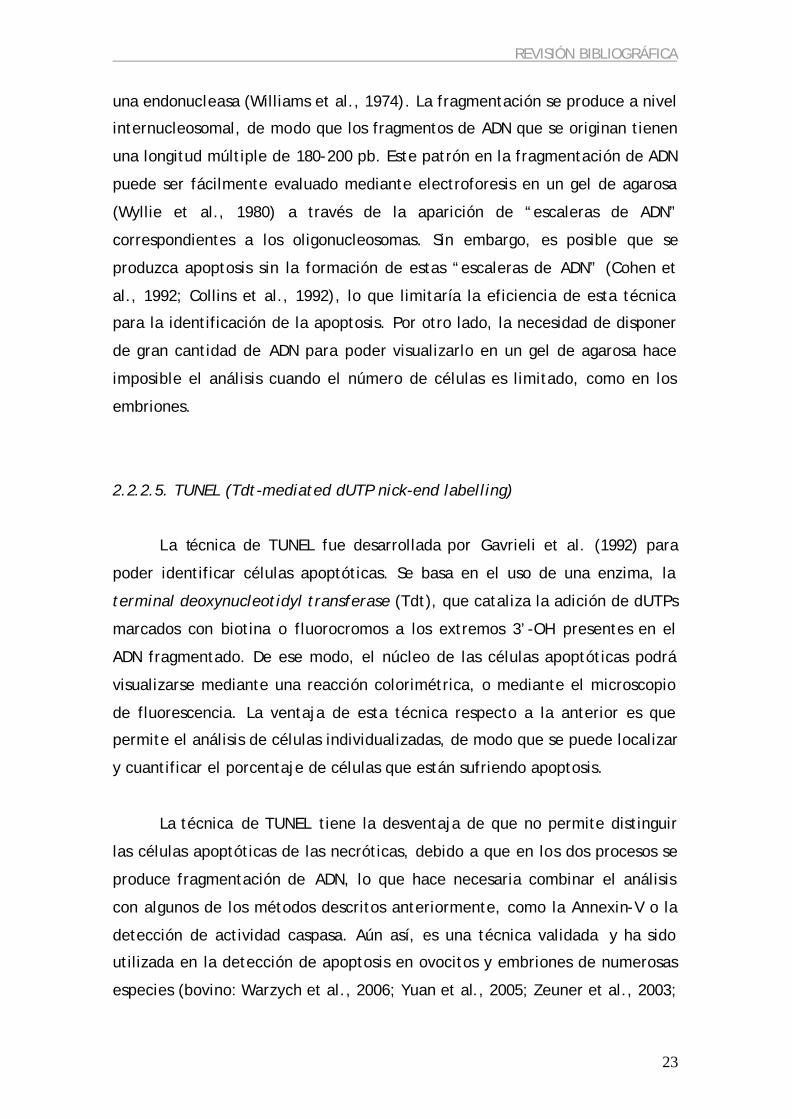
2.2.2.5. TUNEL (Tdt-mediated dUTP nick-end labelling)
La técnica de TUNEL fue desarrollada por Gavrieli et al. (1992) para
poder identificar células apoptóticas. Se basa en el uso de una enzima, la
terminal deoxynucleotidyl transferase (Tdt), que cataliza la adición de dUTPs
marcados con biotina o fluorocromos a los extremos 3’-OH presentes en el
ADN fragmentado. De ese modo, el núcleo de las células apoptóticas podrá
visualizarse mediante una reacción colorimétrica, o mediante el microscopio
de fluorescencia. La ventaja de esta técnica respecto a la anterior es que
permite el análisis de células individualizadas, de modo que se puede localizar
y cuantificar el porcentaje de células que están sufriendo apoptosis.
La técnica de TUNEL tiene la desventaja de que no permite distinguir
las células apoptóticas de las necróticas, debido a que en los dos procesos se
produce fragmentación de ADN, lo que hace necesaria combinar el análisis
con algunos de los métodos descritos anteriormente, como la Annexin-V o la
detección de actividad caspasa. Aún así, es una técnica validada y ha sido
utilizada en la detección de apoptosis en ovocitos y embriones de numerosas
especies (bovino: Warzych et al., 2006; Yuan et al., 2005; Zeuner et al., 2003;
REVISIÓN BIBLIOGRÁFICA
24
murino: Brison and Schultz, 1997; humano: Corn et al., 2005; Jurisicova et al.,
1998; porcino: Kidson et al., 2004).
2.2.3. Apoptosis en el ovario
2.2.3.1. En el período pre-natal
En la mayoría de vertebrados las células germinales primordiales llegan
a la cresta genital donde, después de sufrir una serie de divisiones mitóticas,
inician la meiosis y quedan parados en la primera profase. Durante este
período, la degeneración de las ovogonias se produce básicamente en dos
etapas: en el estadío de paquiteno de la meiosis y durante la formación de los
folículos primordiales (Baker, 1963). Como resultado de esta muerte celular
sólo alrededor del 20-30% de las ovogonias son incluidas en un folículo
primordial (revisado por Tilly, 1996; revisado por Lévy, 2005), y constituirán
la reserva ovárica de la hembra. Aunque se desconoce cuáles son los factores
que determinan la supervivencia o muerte de las ovogonias, parece ser que el
SCF (Stem cell growth factor), el LIF (Leukaemia inhibitory factor) y la
activación del receptor de ácido retinoico podrían jugar un papel importante
(revisado por Tilly, 1996). Por otro lado, parece ser que la calidad de las
mitocondrias del ovocito también tendrían un papel importante en la decisión
de supervivencia o muerte del ovocito durante este período (revisado por
Hussein, 2005).
2.2.3.2. En el período post-natal
Una vez establecido el pool de folículos primordiales, la atresia
ovocitaria después del nacimiento se puede producir a consecuencia de la
degeneración de los folículos que no se seleccionan para ser ovulados
(Hirshfield, 1991; Tsafriri y Braw, 1984), o bien debido a la desaparición de
folículos primordiales de la reserva o de folículos preantrales (revisado por

REVISIÓN BIBLIOGRÁFICA
25
Reynaud y Driancourt, 2000). Mientras que se considera que la atresia
folicular en folículos antrales y pre-antrales se inicia con la apoptosis de
células de la granulosa, la causa de atresia en folículos primordiales es la
muerte del ovocito (revisado por Reynaud y Driancourt, 2000). Parece ser que
esta diferencia podría deberse a la desaparición de ADNasa I en el ovocito
(Boone y Tsang, 1997), que coincide con la aparición del antro en el folículo.
En los animales que han alcanzado la pubertad, en cada ciclo se
produce el reclutamiento folicular, pero sólo uno de los folículos reclutados se
selecciona para ser dominante, mientras que el resto degenera en un proceso
conocido como atresia (Ginther et al., 2001). La atresia en la mayoría de
folículos se da durante la última etapa del período preantral y la primera del
período antral, cuando el crecimiento folicular todavía es dependiente de
gonadotropinas (Dalin, 1987); revisado por Tilly, 1996), lo que sugiere una
regulación hormonal de la apoptosis. La FSH y la LH inhiben la apoptosis que
ocurre en las células de la granulosa de los folículos que degeneran tanto in
vivo (Billig et al., 1994) como in vitro (Chun et al., 1994; Kaipia y Hsueh,
1997; Tilly y Tilly, 1995). En la mujer, cuando se inicia el ciclo menstrual los
niveles de FSH son altos, lo que permite que algunos folículos empiecen a
crecer. Entre estos folículos se encuentra el dominante, que produce gran
cantidad de estrógenos que van a inhibir la secreción de FSH, provocando que
el resto de folículos en crecimiento inicien la apoptosis (Hughes y Gorospe,
1991; Tilly et al., 1991; revisado por Hussein, 2005).
En hembras prepúberes, al igual que en las adultas, también se produce
reclutamiento de folículos para su crecimiento, pero todos degeneran por
atresia debido a la falta de la señal hormonal adecuada (Tilly y Tilly, 1995).
En hembras de edad avanzada, en cambio, la apoptosis no sólo se encarga de
la eliminación de los folículos reclutados que no llegan a ser pre-ovulatorios,
sino también de la eliminación de ovocitos defectuosos debido a la edad
materna (revisado por Lévy, 2005).
REVISIÓN BIBLIOGRÁFICA
26
Los primeros signos de atresia en los folículos antrales es la
degeneración de la células de la granulosa murales y su pérdida de actividad
aromatasa (Irving-Rodgers et al., 2001) seguido de la hipertrofia de las células
de la teca y la disminución de la producción de androsterona (Driancourt et
al., 1998). Esta degeneración de las células de la granulosa se produce
mediante la apoptosis (Hughes y Gorospe, 1991). Las células del cumulus y el
ovocito son los últimos compartimentos de folículo en sufrir atresia
(Driancourt et al., 1991; Tajima et al., 2002; Yang y Rajamahendran, 2000).
Parece ser que el progreso espacial o temporal de la atresia folicular no se
produce de forma lineal, sino que es necesario que se alcance un nivel umbral
de células apoptóticas en el folículo para que el ovocito se vea afectado
(Zeuner et al., 2003).
Se ha observado que los folículos subordinados en estadíos tempranos
de atresia contienen ovocitos con capacidad para dar lugar a embriones
viables (Vassena et al., 2003), posiblemente debido a la existencia de señales
de maduración similares en el folículo dominante maduro y los folículos en
etapas tempranas de atresia (Sirard et al., 1999). Se ha sugerido que cambios
apoptóticos moderados en el folículo podrían inducir cambios similares a la
premaduración que tiene lugar en los ovocitos de folículos pre-ovulatorios,
como la expansión de las células del cumulus (de Loos et al., 1991) o la
reanudación de la meiosis (Assey et al., 1994). La degeneración de los
folículos conduce a una disminución de los niveles de 17ß-estradiol y
testosterona, y un aumento de los niveles de progesterona (Kruip y Dieleman,
1985, 1989), que mimetizan los cambios que suceden después del pico LH,
influenciando la capacidad para el desarrollo del ovocito. En cambio, los
ovocitos de folículos sanos están bloqueados para reanudar la meiosis y
completar la maduración citoplasmática (Hendriksen et al., 2000). Por otro
lado, los folículos en estado avanzado de atresia contienen ovocitos con una
capacidad muy reducida para desarrollarse hasta el estadío de blastocisto
(Blondin y Sirard, 1995; de Wit et al., 2000; Zeuner et al., 2003). Teniendo en
cuenta que más del 50 % de los folículos presentes en el ovario en un
momento determinado son atrésicos (Kruip y Dieleman, 1982), y que de la

REVISIÓN BIBLIOGRÁFICA
27
mayoría de estos folículos se obtienen ovocitos para ser utilizados en
programas de producción in vitro de embriones, estos resultados indican que
estos folículos, aunque estén atrésicos, podrían contener ovocitos con
competencia para el desarrollo.
2.3.3.2. Apoptosis en el embrión pre-implantacional
La apoptosis ha sido observada en embriones pre-implantacionales de la
mayoría de mamíferos (revisado por Hardy, 1999), y tiene por finalidad la
eliminación de las células sobrantes o anormales (Hardy et al., 2003). En
general se considera que el embrión necesita niveles moderados de apoptosis
para tener un desarrollo adecuado, pero niveles altos de apoptosis son
perjudiciales para su desarrollo. Se ha visto que un porcentaje de
fragmentación embrionaria del 10-15 % no afecta al desarrollo embrionario
(Alikani et al., 2000; Hardy et al., 2003), pero un índice mayor resulta en la
incapacidad del embrión para llegar al estadío de blastocisto, probablemente
debido a una pérdida de interacción célula-célula que interfiere en la
compactación, cavitación y formación del blastocisto (Alikani et al., 1999;
Van Blerkom et al., 2001). En general, la apoptosis no se observa en
embriones con desarrollo normal antes de que se produzca la activación del
genoma embrionario, excepto cuando se induce por un factor externo
(revisado por Fabian et al., 2005). La aparición de células apoptóticas en
embriones normales se da después de la activación del genoma embrionario,
aunque el momento exacto depende de la especie: en ratón y cerdo la
apoptosis aparece en estadío de blastocisto, en bovino en estadío de 6-8
células, y en humano la apoptosis aparece en el embrión cuando se da la
compactación (revisado por Fabian et al., 2005). Parece ser que la falta de
apoptosis antes de la activación del genoma embrionario responde a la falta
de transcripción, ya que el embrión utiliza el pool de ARNm materno
almacenado para mantener su actividad celular, con lo que la presión
selectiva para mantener el ADN intacto o eliminar el dañado desaparece. Por
otro lado, en el embrión temprano no hay puntos de control (checkpoints) a lo

REVISIÓN BIBLIOGRÁFICA
28
largo del ciclo celular que normalmente monitorizan la integridad del
genoma, y la aparición de los puntos de control coincide con la aparición de la
muerte celular (revisado por Greenwood y Gautier, 2005).
La apoptosis se ha observado tanto en embriones producidos in vivo
como in vitro (Hardy, 1999; Hardy y Spanos, 2002; Pomar et al., 2005;
revisado por Gjorret et al., 2003) , aunque la incidencia aumenta en los
embriones producidos in vitro (Pomar et al., 2005; revisado por Fabian et al.,
2005). Este hecho podría estar relacionado con una menor calidad de estos
embriones (Hardy et al., 1989).
Los factores que pueden desencadenar apoptosis en el embrión son
variados. En numerosas especies se ha observado que la apoptosis en el
embrión puede estar causada por unas condiciones de cultivo in vitro
subóptimas (Brison y Schultz, 1998; Gjorret et al., 2003; Hardy, 1999; Kidson
et al., 2004; Makarevich y Markkula, 2002; Morgan et al., 1995; Moussa et al.,
2004; Pampfer et al., 2001; Rizos et al., 2002). También es posible que sea la
continuación de un proceso iniciado en los gametos, en particular en el
ovocito (Hardy et al., 2001; Jurisicova et al., 1998; Lévy, 2005), ya que se ha
descrito la presencia de moléculas pro-apoptóticas en el pool de ARNm
almacenado en el ovocito humano y en el murino (Exley et al., 1999;
Jurisicova et al., 1998; Metcalfe et al., 2004), así como una influencia del
genotipo materno en la fragmentación celular del futuro embrión (Han et al.,
2005). No obstante, la incidencia de apoptosis en embriones también puede
ser consecuencia de la fecundación de ovocitos por espermatozoides con el
ADN dañado (Fatehi et al., 2006) o apoptóticos (Host et al., 2000). Otros
factores desencadenantes de apoptosis en el embrión son la presencia de
anomalías cromosómicas (Hardy, 1999; Munne et al., 1993), incapacidad para
que el embrión pueda mantener el desarrollo (Handyside y Hunter, 1986), o la
exposición a factores nocivos (Paula-Lopes y Hansen, 2002; Yang et al., 1998).

REVISIÓN BIBLIOGRÁFICA
29
2.3. PRODUCCION IN VITRO (PIV) DE EMBRIONES
2.3.1. Animales adultos vs. Prepúberes
Diversos laboratorios utilizan hembras prepúberes como donantes de
ovocitos debido a las ventajas que presentan respecto a las hembras adultas.
Así, se puede reducir el intervalo generacional, de modo que se acelera la
propagación genética de los animales de alto valor, y el número de ovocitos
que se consiguen es mucho mayor (Koeman et al., 2003). Además, se ha
demostrado que los ovocitos de hembras prepúberes pueden ser madurados y
fecundados in vitro con éxito (Kajihara et al., 1991; Martino et al., 1994a),
utilizarse para producir embriones in vitro (caprino: Izquierdo et al., 2002;
Izquierdo et al., 1999; Mogas et al., 1997b; bovino: Armstrong et al., 1992;
Armstrong et al., 1994; Damiani et al., 1996; Palma et al., 2001; ovino: Ledda
et al., 1997; Ledda et al., 1999) y obtener gestaciones y nacimientos
(Armstrong et al., 1992).
Sin embargo, la eficiencia en la PIV se ve reducida al utilizar animales
prepúberes en comparación con el uso de adultas (revisado por Armstrong,
2001). Esta baja eficiencia se ha relacionado con un menor diámetro
ovocitario, menor tasa metabólica, menor síntesis proteica (Gandolfi et al.,
1998), menor tasa de maduración meiótica y de capacidad para desarrollarse
hasta blastocisto (Kochhar et al., 2002; Marchal et al., 2001) y mayor tasa de
polispermia en los ovocitos procedentes de hembras prepúberes (Marchal et
al., 2001), todo ello debido a deficiencias en la maduración citoplasmática
(Damiani et al., 1996; Salamone et al., 2001). Una de las posibles causas de la
deficiente maduración podría ser el almacenaje subóptimo de ARNm (Fulka et
al., 1998; Hyttel et al., 1997), ya que se ha visto que hay menor cantidad de
ARNm almacenado en ovocitos prepúberes inmaduros (Leoni et al., 2004), o
una menor actividad del MPF en ovocitos de hembras prepúberes en
comparación con adultas (Salamone et al., 2001) que daría lugar a una menor
formación del pronúcleo masculino tras la fecundación (Naito et al., 1992) y,
en consecuencia, un menor desarrollo embrionario.

REVISIÓN BIBLIOGRÁFICA
30
Aunque las deficiencias citoplasmáticas en los ovocitos de las hembras
prepúberes han sido ampliamente demostradas, hay que destacar que
también han surgido estudios que no han detectado diferencias entre los
ovocitos procedentes de animales prepúberes y adultos en cuanto a eficiencia
en la maduración meiótica (Armstrong et al., 1992; Koeman et al., 2003;
Martino et al., 1995) fecundación (Koeman et al., 2003; Mogas et al., 1997a) y
desarrollo embrionario hasta blastocisto (caprino: Izquierdo et al., 2002;
Koeman et al., 2003; Mogas et al., 1997a; bovino: Armstrong et al., 1994;
ovino: Ledda et al., 1997).
2.3.2 Obtención de los ovocitos
2.3.2.1. Obtención de los ovarios
La mayor parte de los ovocitos que se utilizan para MIV, FIV y CIV
provienen de ovarios de hembras sacrificadas en matadero, ya que permite
obtener un gran número de ovarios a coste muy bajo. Sin embargo, el estado
fisiológico de las hembras sacrificadas donantes de ovocitos es muy variable,
hecho que se ve reflejado en la heterogeneidad de la población ovocitaria que
se recupera de esos ovarios.
Existen otras técnicas que permiten la obtención de ovocitos de
hembras vivas, entre las que cabe destacar la punción folicular por vía vaginal
(OPU) o guiada por laparoscopia (LOPU). Esta técnica tiene la ventaja que
permite conocer el estado fisiológico de la hembra, de forma que no afectará
a la recuperación de los ovocitos (Galli et al., 2001). Por otro lado, las
hembras también pueden ser estimuladas hormonalmente para recuperar un
mayor número de ovocitos. Sin embargo, el coste de la OPU es mucho más
elevado que la obtención de ovarios de hembras sacrificadas en matadero, y
el número de ovocitos obtenido es muy inferior.

REVISIÓN BIBLIOGRÁFICA
31
2.3.2.2. Métodos de obtención de ovocitos
Son varios los métodos que se pueden utilizar para obtener los ovocitos
que serán utilizados para la producción in vitro de embriones. A continuación
describiré sólo aquellos métodos que se utilizan a partir de ovarios de
hembras sacrificadas:
a) Disección folicular: consiste en separar el folículo intacto del ovario
mediante el uso de un bisturí, y una vez aislado se recupera el complejo
cumulus-ovocito (COC). Es una técnica muy laboriosa y que requiere mucho
tiempo; sin embargo, permite recuperar gran número de COCs y mantener su
integridad (Nowshari, 2005), así como conocer perfectamente las
características de los folículos de los que proceden los COCs obtenidos.
b) Aspiración folicular: consiste en aspirar el contenido folicular con una
aguja conectada a una jeringa, donde se va recogiendo el líquido folicular y
los COCs que contienen. Es una técnica muy utilizada, ya que permite
recuperar un gran número de COCs en relativamente poco tiempo. Sin
embargo, presenta algunas desventajas: algunos ovocitos pierden las células
del cumulus durante el proceso, la cantidad de COCs recuperados es menor
que con otras técnicas (Nowshari, 2005), y a veces se aspira el contenido de
los folículos adyacentes sin pretenderlo. Esta técnica, además, requiere la
presencia de folículos grandes para ser aspirados, lo que limita su uso a los
ovarios procedentes de animales adultos.
c) Slicing (Martino et al., 1994b): consiste en realizar pequeños cortes en la
superficie del ovario con la ayuda de un bisturí, de modo que se rompe la
pared folicular y el COC se libera al medio de cultivo donde están sumergidos
los ovarios. Es una técnica que permite recuperar gran cantidad de COCs por
ovario. Sin embargo la población ovocitaria que se recupera es muy
heterogénea, encontrando ovocitos con diferentes grados de desarrollo y
atresia, de modo que tras la recuperación debe hacerse una estricta selección
de los COCs que se utilizarán para la MIV. A pesar de sus desventajas, es la
REVISIÓN BIBLIOGRÁFICA
32
técnica más utilizada para recuperar ovocitos de ovarios de hembras
prepúberes, ya que suelen carecer de folículos de tamaño considerable.
2.3.3. Selección de los ovocitos
La heterogeneidad de la población ovocitaria obtenida a partir de
hembras sacrificadas hace necesario realizar una cuidadosa selección de los
ovocitos con el objetivo de utilizar sólo aquéllos que han adquirido la
competencia para dar lugar a un embrión viable. Se han realizado numerosos
estudios con el objetivo de encontrar marcadores que pudieran ser
indicadores de la competencia para el desarrollo de los ovocitos. Es necesario
que estos marcadores permitan una selección rápida y no perjudicial de los
ovocitos, con lo que la mayoría de estudios se han centrado en parámetros
morfológicos que pudieran ser evaluados visualmente.
2.3.3.1. Métodos no invasivos
2.3.3.1.a. Tamaño folicular
La selección de los ovocitos dependiendo del tamaño folicular es uno de
los métodos más utilizados, sobre todo cuando se realiza la disección o
aspiración de los folículos. La relación entre el tamaño folicular y la
competencia para el desarrollo de los ovocitos obtenidos se ha observado en
numerosas especies:
? En bovino, Lonergan et al. (1994) observaron que el porcentaje de
blastocistos obtenidos a partir de ovocitos liberados de folículos > 6
mm de diámetro era mayor que en folículos de diámetro 2-6 mm.
Blondin y Sirard (1995) determinaron que los folículos de vacas que
medían menos de 3 mm contenían ovocitos que no eran capaces de
desarrollarse hasta el estadío de blastocisto, mientras que Lequarre et
al. (2005) observaron diferente tasa de desarrollo hasta blastocisto en

REVISIÓN BIBLIOGRÁFICA
33
ovocitos procedentes de folículos > 6 mm en comparación con los de
folículos < 4 mm, aunque el número de células de los blastocistos
obtenidos no dependía del tamaño folicular. En terneras el resultado
también ha sido similar, y se han obtenido mayores porcentajes de
blastocistos a partir de los ovocitos recuperados de folículos de
diámetro superior a 8 mm (Kauffold et al., 2005).
? En búfalo también se ha observado que los ovocitos obtenidos de los
folículos de mayor diámetro (> 8 mm) dan lugar a un mayor porcentaje
de blastocistos comparados con los ovocitos obtenidos de folículos
medios (3-8 mm) o pequeños (< 3 mm) (Raghu et al., 2002).
? En cabras adultas, Crozet et al. (1995) observaron que se obtenía
mayor porcentaje de blastocistos con ovocitos obtenidos de folículos >
5 mm en comparación con los que procedían de folículos medianos
(3.1-5 mm) y pequeños (2-3 mm).
? En ovejas, Ledda et al. (1999) también observaron que la progresión
meiótica de los ovocitos estaba relacionada con el tamaño de los
folículos, tanto en animales adultos como en prepúberes.
La diferencia en la competencia ovocitaria según el tamaño folicular se
debe posiblemente a que en los folículos de mayor diámetro existen factores
que favorecen la capacidad para el desarrollo del ovocito que contienen, ya
que Algriany et al. (2004) observaron que al añadir al medio de maduración
líquido folicular de folículos grandes el desarrollo embrionario mejoraba en
comparación con el líquido folicular que provenía de folículos pequeños.
2.3.3.1.b. Diámetro ovocitario
En numerosas especies se ha observado que el diámetro de un ovocito
está relacionado con el tamaño del folículo que lo contiene (vaca: Arlotto et
al., 1996; cabra: Crozet et al., 2000; de Smedt et al., 1994; búfalo: Raghu et

REVISIÓN BIBLIOGRÁFICA
34
al., 2002) como el tamaño folicular, a la vez, se correlaciona con la
competencia meiótica del ovocito (ratón: Sorensen y Wassarman, 1976; rata:
Bar-Ami y Tsafriri, 1981; cerdo: Motlik et al., 1984; oveja: Moor y Gandolfi,
1987; vaca: Furher et al., 1989; cabra: de Smedt et al., 1994; Martino et al.,
1994a; mono: Schramm et al., 1993) y su capacidad para mantener el
desarrollo embrionario (cabra: Crozet et al., 1995; vaca: de Wit y Kruip, 2001;
Kauffold et al., 2005; Lequarre et al., 2005; búfalo: Raghu et al., 2002).
En cabras adultas, de Smedt et al. (1994) clasificaron los ovocitos según
su diámetro en 4 grupos: 1) < 110 µm, ovocitos que provenían de folículos de
0.5-0.8 mm de diámetro, considerados incompetentes; 2) 110-125 µm, los
ovocitos procedentes de folículos de diámetro 1-1.8 mm, considerados
parcialmente competentes; 3) y ovocitos de diámetro 125-135 y 4) > 135 µm,
procedentes de folículos mayores de 3 mm de diámetro, considerados
meióticamente competentes.
En todos los trabajos realizados en esta tesis se han seleccionado los
ovocitos según su diámetro, siguiendo la clasificación realizada por de Smedt
et al. (1994) por 2 motivos: 1) la técnica utilizada para la recuperación de los
ovocitos ha sido el slicing, técnica incompatible con la selección por tamaño
folicular, y 2) creemos que cuando se trabaja con animales prepúberes el
diámetro es el método de selección mas fiable en comparación con la
selección según el tamaño folicular, ya que se ha observado que folículos del
mismo tamaño contienen ovocitos de menor diámetro en hembras prepúberes
que en adultas (bovino: Gandolfi et al., 1998; ovino: Ledda et al., 1999).
2.3.3.1.c. Morfología del COC
La posibilidad de seleccionar los ovocitos según su morfología fue
investigada por primera vez en el bovino (Leibfried y First, 1979). Numerosos
estudios han indicado la utilidad de la morfología del COC, basándose en la
compactación y en el número de capas de células del cumulus, así como en la
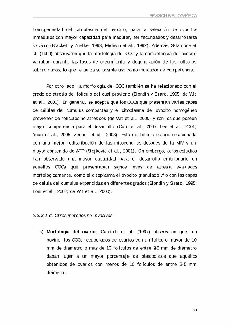
REVISIÓN BIBLIOGRÁFICA
35
homogeneidad del citoplasma del ovocito, para la selección de ovocitos
inmaduros con mayor capacidad para madurar, ser fecundados y desarrollarse
in vitro (Brackett y Zuelke, 1993; Madison et al., 1992). Además, Salamone et
al. (1999) observaron que la morfología del COC y la competencia del ovocito
variaban durante las fases de crecimiento y degeneración de los folículos
subordinados, lo que refuerza su posible uso como indicador de competencia.
Por otro lado, la morfología del COC también se ha relacionado con el
grado de atresia del folículo del cual proviene (Blondin y Sirard, 1995; de Wit
et al., 2000). En general, se acepta que los COCs que presentan varias capas
de células del cumulus compactas y el citoplasma del ovocito homogéneo
provienen de folículos no atrésicos (de Wit et al., 2000) y son los que poseen
mayor competencia para el desarrollo (Corn et al., 2005; Lee et al., 2001;
Yuan et al., 2005; Zeuner et al., 2003). Esta morfología estaría relacionada
con una mejor redistribución de las mitocondrias después de la MIV y un
mayor contenido de ATP (Stojkovic et al., 2001). Sin embargo, otros estudios
han observado una mayor capacidad para el desarrollo embrionario en
aquellos COCs que presentaban signos leves de atresia evaluados
morfológicamente, como el citoplasma el ovocito granulado y/o con las capas
de célula del cumulus expandidas en diferentes grados (Blondin y Sirard, 1995;
Boni et al., 2002; de Wit et al., 2000).
2.3.3.1.d. Otros métodos no invasivos
a) Morfología del ovario: Gandolfi et al. (1997) observaron que, en
bovino, los COCs recuperados de ovarios con un folículo mayor de 10
mm de diámetro o más de 10 folículos de entre 2-5 mm de diámetro
daban lugar a un mayor porcentaje de blastocistos que aquéllos
obtenidos de ovarios con menos de 10 folículos de entre 2-5 mm
diámetro.
REVISIÓN BIBLIOGRÁFICA
36
b) Concentración de diferentes componentes en el líquido folicular: La
concentración de estradiol (Anifandis et al., 2005; Wunder et al.,
2005), leptina (Anifandis et al., 2005), inhibina B (Chang et al., 2002) y
mio-inositol (Chiu et al., 2002) en el líquido folicular se ha relacionado
con una mayor capacidad ovocitaria para dar lugar a embriones de
mejor calidad, mayor tasa de implantación y gestaciones.
2.3.3.2. Métodos invasivos
Todas las técnicas invasivas se basan en métodos moleculares, como la
PCR, el radiomarcaje o la tinción con fluorocromos, que impiden que el
ovocito pueda seguir con su desarrollo. Por este motivo, es necesario
relacionar características moleculares del ovocito con características que
puedan ser evaluadas visualmente, como los métodos no invasivos
mencionados anteriormente. Algunas de las características moleculares que se
han relacionado con la competencia ovocitaria se describen a continuación.
2.3.3.2.a. Contenido mitocondrial del ovocito
Santos et al. (2006) observaron que el contenido mitocondrial de los
ovocitos, tras haber realizado un protocolo de FIV o ICSI (Intracytoplasmic
sperm injection), era superior en los ovocitos fecundados que en los que no
presentaban extrusión del segundo corpúsculo polar o formación de los
pronúcleos (considerados no fecundados), con lo que podría ser un buen
marcador para predecir el éxito de la fecundación in vitro.
2.3.3.2.b. Abundancia de transcritos específicos
Se ha observado una relación de la morfología del ovocito, y por tanto
de su competencia meiótica, con la abundancia relativa de determinados
transcritos en el ovocito (De Sousa et al., 1998). En embriones considerados
REVISIÓN BIBLIOGRÁFICA
37
de buena calidad se ha observado una mayor transcripción de genes
relacionados con el estrés, con el metabolismo (Gutiérrez-Adán et al., 2004) y
con la regulación de la expresión génica (Dode et al., 2006).
2.3.3.2.c. Grado de apoptosis
Las técnicas invasivas más utilizadas hasta ahora para evaluar el grado
de apoptosis, tanto en el ovocito y en las células del cumulus que lo rodean
como en los embriones resultantes, han sido la técnica de TUNEL (Corn et al.,
2005; Hao et al., 2004; Lee et al., 2001; Van Blerkom y Davis, 1998; Vandaele
et al., 2006; Warzych et al., 2006; Wu et al., 2000; Yuan et al., 2005; Zeuner
et al., 2003), la tinción con Annexin-V (Giampietro et al., 2006; Tseng et al.,
2006; Van Blerkom and Davis, 1998), la expresión relativa de Bax y Bcl-2 (Yang
y Rajamahendran, 2002), o la actividad de caspasas (Bilby et al., 2006; Bosco
et al., 2005; Ortiz et al., 2006).
Generalmente, se ha observado que la capacidad para el desarrollo
embrionario se incrementa en los complejos cumulus-ovocito que presentan
menor incidencia de apoptosis, evaluada mediante técnicas invasivas (Corn et
al., 2005; Lee et al., 2001; Yang y Rajamahendran, 2002; Yuan et al., 2005;
Zeuner et al., 2003). Curiosamente, estos resultados contradicen los estudios
que relacionaban la presencia de signos leves de atresia, evaluados según la
morfología de los COCs, con su mayor capacidad para desarrollarse hasta
blastocisto (Bilodeau-Goeseels y Panich, 2002; Blondin y Sirard, 1995).

OBJETIVOS
3. OBJETIVOS El objetivo principal de esta tesis es la búsqueda de marcadores
moleculares que nos ayuden a determinar la calidad del ovocito para poder
predecir su capacidad de desarrollarse hasta blastocisto después de un
proceso de maduración y fecundación in vitro. Para su consecución, nos
planteamos los siguientes objetivos:
• Estudiar la relación entre el diámetro del ovocito y su competencia
para dar lugar a un embrión viable y así poder utilizar este parámetro
como indicador no invasivo de competencia ovocitaria.
• Analizar la expresión de las dos subunidades del MPF, así como su
actividad y la acumulación de RNA y de proteína totales en ovocitos de
diferente diámetro, con la finalidad de valorar su posible implicación
en la adquisición de competencia del ovocito para el desarrollo
embrionario.
• Valorar el efecto de la incidencia de la apoptosis, tanto en ovocitos
como en células del cumulus, en la adquisición de competencia
ovocitaria para el desarrollo embrionario, así como su efecto en los
blastocistos resultantes.
41

www.theriojournal.com
Theriogenology 67 (2007) 526–536
Effect of oocyte diameter on meiotic competence,
embryo development, p34 (cdc2) expression and
MPF activity in prepubertal goat oocytes
Begona Anguita a, Ana R. Jimenez-Macedo a, Dolors Izquierdo a,Teresa Mogas b, Maria-Teresa Paramio a,*
a Departament de Ciencia Animal I dels Aliments, Universitat Autonoma de Barcelona, 08193 Bellaterra, Barcelona, Spainb Departament de Medicina I Cirurgia Animals, Universitat Autonoma de Barcelona, 08193 Bellaterra, Barcelona, Spain
Received 18 April 2006; received in revised form 1 September 2006; accepted 1 September 2006
Abstract
The aim of this study was to analyze the relationship between oocyte diameter, meiotic and embryo developmental competence
and the expression of the catalytic subunit of MPF, the p34cdc2, at mRNA, RNA and protein level, as well as its kinase activity, in
prepubertal (1–2 months old) goat oocytes. MPF is the main meiotic regulator and a possible regulator of cytoplasmic maturation;
therefore, it could be a key factor in understanding the differences between competent and incompetent oocytes. Oocytes were
classified according to oocyte diameter in four categories: <110, 110–125, 125–135 and >135 mm and matured, fertilized and
cultured in vitro. The p34cdc2 was analyzed in oocytes at the time of collection (0 h) and after 27 h of IVM (27 h) in each of the
oocyte diameter categories. The oocyte diameter was positively related to the percentage of oocytes at MII after IVM (0, 20.7, 58
and 78%, respectively) and the percentage of blastocysts obtained at 8 days postinsemination (0, 0, 1.95 and 12.5%, respectively).
The expression of RNA and mRNA p34cdc2 did not vary between oocyte diameters at 0 and 27 h. Protein expression of p34cdc2
increased in each oocyte category after 27 h of maturation. MPF activity among diameter groups did not vary at 0 h but after IVM
there was a clear and statistically significant increase of MPF activity in the biggest oocytes.
# 2006 Elsevier Inc. All rights reserved.
Keywords: Goat; Oocyte diameter; IVF; MPF
1. Introduction
Mammalian oocytes are arrested at the dictyate stage
of meiosis, also known as germinal vesicle (GV) stage,
during their growth. Fully grown oocytes are able to
resume meiosis in vivo after the LH surge, or
spontaneously after their release from the follicle and
subsequent in vitro culture [1]. In contrast, growing
oocytes are not able to resume or complete meiosis.
* Corresponding author. Tel.: +34 935811456; fax: +34 935811494.
E-mail address: [email protected] (M.-T. Paramio).
0093-691X/$ – see front matter # 2006 Elsevier Inc. All rights reserved.
doi:10.1016/j.theriogenology.2006.09.003
Oocyte competence is acquired during the growth
phase, when the synthesis and storage of proteins and
ribosomal and heterogeneous RNA take place [2] and
implies its ability to complete not only nuclear
maturation, but also cytoplasmic changes needed to
maintain embryo development. These cytoplasmic
changes include: (a) protein and RNA storage, (b)
development of calcium regulatory mechanisms, (c)
changes in the activity of MPF and MAPK, (d)
redistribution of cellular organelles, etc. The regulation
of cytoplasmic maturation is not as well known as
nuclear maturation regulation, however Naito et al. [3]
suggested that MPF plays an important part in this
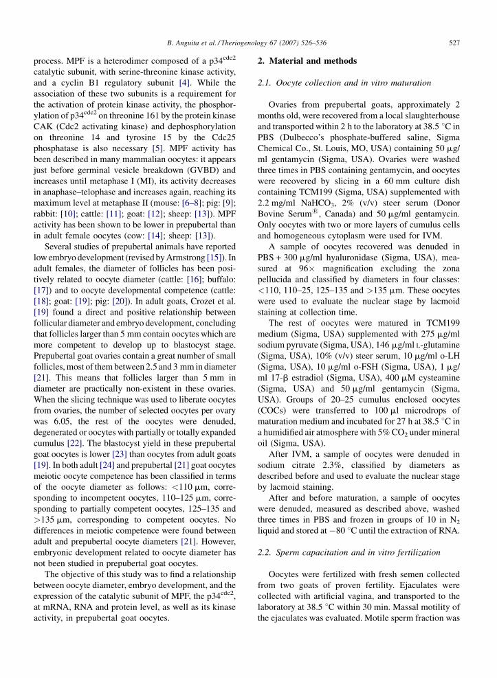
B. Anguita et al. / Theriogenology 67 (2007) 526–536 527
process. MPF is a heterodimer composed of a p34cdc2
catalytic subunit, with serine-threonine kinase activity,
and a cyclin B1 regulatory subunit [4]. While the
association of these two subunits is a requirement for
the activation of protein kinase activity, the phosphor-
ylation of p34cdc2 on threonine 161 by the protein kinase
CAK (Cdc2 activating kinase) and dephosphorylation
on threonine 14 and tyrosine 15 by the Cdc25
phosphatase is also necessary [5]. MPF activity has
been described in many mammalian oocytes: it appears
just before germinal vesicle breakdown (GVBD) and
increases until metaphase I (MI), its activity decreases
in anaphase–telophase and increases again, reaching its
maximum level at metaphase II (mouse: [6–8]; pig: [9];
rabbit: [10]; cattle: [11]; goat: [12]; sheep: [13]). MPF
activity has been shown to be lower in prepubertal than
in adult female oocytes (cow: [14]; sheep: [13]).
Several studies of prepubertal animals have reported
low embryo development (revised by Armstrong [15]). In
adult females, the diameter of follicles has been posi-
tively related to oocyte diameter (cattle: [16]; buffalo:
[17]) and to oocyte developmental competence (cattle:
[18]; goat: [19]; pig: [20]). In adult goats, Crozet et al.
[19] found a direct and positive relationship between
follicular diameter and embryo development, concluding
that follicles larger than 5 mm contain oocytes which are
more competent to develop up to blastocyst stage.
Prepubertal goat ovaries contain a great number of small
follicles, most of them between 2.5 and 3 mm in diameter
[21]. This means that follicles larger than 5 mm in
diameter are practically non-existent in these ovaries.
When the slicing technique was used to liberate oocytes
from ovaries, the number of selected oocytes per ovary
was 6.05, the rest of the oocytes were denuded,
degenerated or oocytes with partially or totally expanded
cumulus [22]. The blastocyst yield in these prepubertal
goat oocytes is lower [23] than oocytes from adult goats
[19]. In both adult [24] and prepubertal [21] goat oocytes
meiotic oocyte competence has been classified in terms
of the oocyte diameter as follows: <110 mm, corre-
sponding to incompetent oocytes, 110–125 mm, corre-
sponding to partially competent oocytes, 125–135 and
>135 mm, corresponding to competent oocytes. No
differences in meiotic competence were found between
adult and prepubertal oocyte diameters [21]. However,
embryonic development related to oocyte diameter has
not been studied in prepubertal goat oocytes.
The objective of this study was to find a relationship
between oocyte diameter, embryo development, and the
expression of the catalytic subunit of MPF, the p34cdc2,
at mRNA, RNA and protein level, as well as its kinase
activity, in prepubertal goat oocytes.
2. Material and methods
2.1. Oocyte collection and in vitro maturation
Ovaries from prepubertal goats, approximately 2
months old, were recovered from a local slaughterhouse
and transported within 2 h to the laboratory at 38.5 8C in
PBS (Dulbecco’s phosphate-buffered saline, Sigma
Chemical Co., St. Louis, MO, USA) containing 50 mg/
ml gentamycin (Sigma, USA). Ovaries were washed
three times in PBS containing gentamycin, and oocytes
were recovered by slicing in a 60 mm culture dish
containing TCM199 (Sigma, USA) supplemented with
2.2 mg/ml NaHCO3, 2% (v/v) steer serum (Donor
Bovine Serum1, Canada) and 50 mg/ml gentamycin.
Only oocytes with two or more layers of cumulus cells
and homogeneous cytoplasm were used for IVM.
A sample of oocytes recovered was denuded in
PBS + 300 mg/ml hyaluronidase (Sigma, USA), mea-
sured at 96� magnification excluding the zona
pellucida and classified by diameters in four classes:
<110, 110–25, 125–135 and >135 mm. These oocytes
were used to evaluate the nuclear stage by lacmoid
staining at collection time.
The rest of oocytes were matured in TCM199
medium (Sigma, USA) supplemented with 275 mg/ml
sodium pyruvate (Sigma, USA), 146 mg/ml L-glutamine
(Sigma, USA), 10% (v/v) steer serum, 10 mg/ml o-LH
(Sigma, USA), 10 mg/ml o-FSH (Sigma, USA), 1 mg/
ml 17-b estradiol (Sigma, USA), 400 mM cysteamine
(Sigma, USA) and 50 mg/ml gentamycin (Sigma,
USA). Groups of 20–25 cumulus enclosed oocytes
(COCs) were transferred to 100 ml microdrops of
maturation medium and incubated for 27 h at 38.5 8C in
a humidified air atmosphere with 5% CO2 under mineral
oil (Sigma, USA).
After IVM, a sample of oocytes were denuded in
sodium citrate 2.3%, classified by diameters as
described before and used to evaluate the nuclear stage
by lacmoid staining.
After and before maturation, a sample of oocytes
were denuded, measured as described above, washed
three times in PBS and frozen in groups of 10 in N2
liquid and stored at�80 8C until the extraction of RNA.
2.2. Sperm capacitation and in vitro fertilization
Oocytes were fertilized with fresh semen collected
from two goats of proven fertility. Ejaculates were
collected with artificial vagina, and transported to the
laboratory at 38.5 8C within 30 min. Massal motility of
the ejaculates was evaluated. Motile sperm fraction was

B. Anguita et al. / Theriogenology 67 (2007) 526–536528
obtained using the swim-up method [25] whereby 70 ml
semen were layered below 2 ml of defined medium ([26],
modified by Younis et al. [27]), referred to here as mDM,
and it was incubated for 45–60 min in a humidified air
atmosphere with 5% CO2 at 38.5 8C. After incubation,
600 ml of the supernatant of each tube was recovered and
centrifuged at 500 rpm for 10 min. After discarding the
supernatant, the pellet was resuspended in proportion 1:1
with mDM containing heparin (Sigma, USA) and
ionomycin (Sigma, USA) (final concentration: 10 mg/
ml heparin and 200 nM of ionomycin), and incubated for
15 min in a humidified air atmosphere with 5% CO2 at
38.5 8C [28]. Sperm concentration was determined using
a Neubauer chamber cell.
After maturation, we transferred groups of 20–25
oocytes to 100 ml microdrops of modified Tyrode’s
medium (TALP) as described by Parrish et al. [25],
supplemented with 1 mg/ml hypotaurine (Sigma, USA),
under mineral oil. Oocytes were co-cultured with
capacitated spermatozoa in a final concentration in the
microdrop of 4 � 106 cells/ml.
A sample of oocytes were recovered after 17 h of
IVF, denuded with sodium citrate 2.3% and classified by
diameters to evaluate fertilization rate.
2.3. Embryo in vitro culture
At 24 h after insemination, zygotes were washed to
remove both sperm cells and remnant cumulus cells,
and they were classified by diameters as described
above. Groups of 18–25 embryos of the same diameter
were placed in 25 ml microdrops of culture medium
(SOF, synthetic oviductal fluid [29]; modified by
Takahashi and First [30]) supplemented with aminoa-
cids and BSA [31] in 35 mm culture dishes under
mineral oil, and they were cultured in a humidified
atmosphere with 5% CO2, 5% O2 and 90% N2 at 38.5 8Cfor 7 days. At 48 h after insemination, we added 0.1 ml
foetal bovine serum (FBS, Life Technologies) per
embryo. At the end of the culture period, total cell
number of embryos was evaluated by Hoescht 33342
staining (Sigma, USA) under a fluorescence micro-
scope, and the percentage of total embryos (embryos
with two blastomeres or more), morulae (embryos with
16 or more cells and without blastocoele) and
blastocysts (embryos with 60 or more cells and with
blatocoele formation) was recorded.
2.4. Evaluation of nuclear stage
To evaluate the nuclear stage of oocytes at collection
time, after IVM and after IVF, oocytes were fixed in
ethanol: acid acetic (3:1, v/v) for 24 h at 4 8C. After that
time, oocytes were stained with 1% lacmoid (Sigma,
USA) in 45% acetic acid.
The classification of oocytes at collection time and
after IVM was the following: germinal vesicle (GV),
when the nuclear membrane and the nucleolus were
visible and chromatin was not condensed; germinal
vesicle breakdown (GVBD), when the nuclear mem-
brane and the nucleolus were not visible, and chromo-
somes had begun to condense; metaphase I (MI), when
the chromosomes were condensed and arranged in the
metaphase spindle; anaphase–telophase (A–T), when
homologous chromosomes begin to separate; and
metaphase II (MII), when the metaphase plate and the
first polar body were observed.
After IVF, oocytes with a sperm tail in the
cytoplasm were considered fertilized and they were
classified into one of the three groups: asynchrony,
when a condensed sperm head and the female
pronucleus were visible in the cytoplasm; 2PN
(normal fertilization): the two pronuclei (male and
female) and one sperm tail were visible in the
cytoplasm; and polyspermy, when there were two or
more sperm tails with condensed or descondensed
sperm heads in the cytoplasm. Oocytes without
pronuclei and sperm tails in the cytoplasm were
classified as no fertilized.
2.5. RNA extraction
Total RNA was extracted from groups of 10
oocytes using 100 ml of TriReagent1 (T-9424, Sigma,
USA). We added 20 mg of glycogen (Roche,
Germany) to the 100 ml as a carrier for the RNA
precipitation, and 1 pg of rabbit globin mRNA per
oocyte (Sigma, USA), that was used as an extrinsic
control of the whole process. After adding 20 ml of
chloroform, we centrifuged the sample at 12 000 � g
for 20 min at 4 8C. The aqueous phase was recovered
and the RNA was precipitated with 60 ml of
isopropanol at �20 8C overnight, while the resulting
organic phase was frozen at �80 8C until its use in
protein extraction. The next day, after centrifugation
at 12 000 � g at 4 8C for 20 min, the supernatant was
discarded and the pellet was washed sequentially with
70% ethanol and 100% ethanol. For the washing
steps, the sample was centrifuged at 12 000 � g at
4 8C for 15 min. The pellet was dried and resuspended
in 4 ml of H2O DEPC.
The RNA extracted was incubated for 30 min at
37 8C with 1U/ml DNase (Invitrogen Corporation,
USA), and stored at �80 8C.
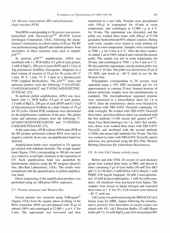
B. Anguita et al. / Theriogenology 67 (2007) 526–536 529
2.6. Reverse transcription (RT) and polymerase
chain reaction (PCR)
Total RNA corresponding to 10 oocytes was reverse-
transcribed with ThermoScriptTM RT-PCR System
(Invitrogen Corporation, USA), following the manu-
facturer’s instruction, in a final volume of 20 ml. The RT
was performed using oligodT and random primers. Four
microliters of these reactions were used to amplify
p34cdc2.
To perform p34cdc2 amplification, cDNA was
incubated with 1� PCR buffer, 0.5 mM of each primer,
2 mM of MgCl2, 200 mm of each dNTP and 0.1 U/ml of
Taq polymerase GoldStar (Eurogentec, Belgium) in a
final volume of reaction of 25 ml for 36 cycles (94 8C1 min, 56 8C 1 min, 72 8C 1 min) in a thermocycler
9700 (Applied BioSystems). The p34cdc2 sense and
antisense primers were the following: 50-GAATTAG-
CAACGAAGAAACC and 50-GTACAATATATCTGC-
TCTTGAC [32].
To perform globin amplification, cDNA was incu-
bated with 1� PCR buffer, 0.5 mm of each primer,
1.5 mM of MgCl2, 200 mm of each dNTP and 0.1 U/ml
of Taq polymerase GoldStar in a final volume of 25 ml
for 30 cycles. Globin PCR conditions were determined
by the amplification conditions of the gene. The globin
sense and antisense primers were the following: 50-GCAGCCACGGTGGCGAGTAT and 50-GTGGGA-
CAGGAGCTTGAAAT [33].
At the same time, a PCR without cDNA and a PCR of
the RT product performed without RNA were used as
negative controls. In no case, an amplification band was
detected.
Amplification bands were visualized in 2% agarose
gel stained with ethidium bromide. The weight marker
band (Sigma, USA) corresponding to 300 pb was used
as a control to avoid light variations in the exposition to
UV. Each amplification band was quantified by
densitometric analysis using the PC program Quantity
One (Bio-Rad Laboratories, USA). The results were
normalized with the quantification of globin amplifica-
tion band.
Direct sequencing of the amplification products was
performed using an ABI-prism DNA sequencer.
2.7. Protein extraction and Western blot
Oocyte protein was extracted using TriReagent1
(Sigma, USA) from the organic phase resulting of the
RNA extraction. DNA was precipitated with 30 ml of
ethanol 100% and centrifuged at 12 000 � g at 4 8C for
5 min. The supernatant was recovered and then
transferred to a new tube. Proteins were precipitated
with 150 ml of isopropanol for 10 min at room
temperature, and centrifuged at 12 000 � g at 4 8Cfor 10 min. The supernatant was discarded, and the
pellet was washed three times with 200 ml of 0.3 M
guanidine hydrochloride/95% ethanol solution. During
each wash, samples were stored in wash solution for
20 min at room temperature. Samples were centrifuged
at 7500 � g for 5 min at 4 8C. After the three washes,
we added 1 ml of 100% ethanol and vortered the protein
pellet. The sample was left at room temperature for
20 min, and centrifuged at 7500 � g for 5 min at 4 8C.
We discarded the supernatant, and the protein pellet was
dried for 5–10 min. The pellet was dissolved in 20 ml of
1% SDS, and stored at �80 8C until its use for the
Western blot.
Polypeptides corresponding to 20 oocytes were
separated using a 10% SDS acrylamide gel for 2 h
approximately at constant 25 mA. Stained proteins of
known molecular weights were run simultaneously as
standards. The electrophoretical separated proteins
were transferred to a nitrocellulose sheet for 1 h at
100 V. After the transference, sheets were blocked by
incubation with TBS 0.05% Tween20 containing 1%
milk overnight. We washed with TBS 0.05% Tween20
three times, and nitrocellulose sheet was incubated with
the first antibody (1:100, mouse IgG against p34cdc2,
Santa Cruz Biotechnology) for 1 h. After that incuba-
tion, sheets were washed three times with TBS 0.05%
Tween20, and incubated with the second antibody
(1:2000, anti-mouse IgG antibody) for 30 min. The blot
was washed six times with TBS 0.05% Tween20, and its
detection was performed using the ECL-Plus Western
Blotting Detection Kit (Amersham Biosciences).
2.8. In vitro Cdc2 kinase activity assay
Before and after IVM, 20 oocytes of each diameter
group were washed three times in PBS, and placed in
tubes containing 5 ml of lysis buffer (50 mM Tris–HCl
pH 7.5, 0.5 M NaCl, 5 mM EDTA, 0.01% Brij35, 1 mM
PMSF, 0.05 mg/ml leupeptin, 50 mM 2-mercaptoetha-
nol, 25 mM b-glycerophosphate, 1 mM Na-orthovana-
date). All chemicals were purchased from Sigma. The
samples were frozen in liquid nitrogen and sonicated
three times at 1 8C for 25 s. Cell extracts were stored at
�80 8C until use.
Cdc2 assay was performed using the MESACUP cdc2
kinase assay kit (MBL, Japan) following the manufac-
turer’s protocol. Five microliters of oocyte extract was
mixed with 10� cdc2 Reaction Buffer (25 mM Hepes
buffer pH 7.5, 10 mM MgCl2) and 10% biotynilated MV

B. Anguita et al. / Theriogenology 67 (2007) 526–536530
Table 1
Nuclear stage of different sizes of prepubertal goat oocytes at collection time
Diameter (mm) No. GV (%) GVBD (%) MI (%) A–T (%) MII (%) DEG (%)
<110 75 62 a (82.66) 4 a (5.33) 2 (2.66) 0 (0) 0 (0) 7 a, b (9.33)
110–125 105 51 b (48.57) 28 b (26.66) 5 (4.76) 0 (0) 0 (0) 21 a (20.00)
125–135 250 34 c (13.60) 161 c (64.40) 20 (8.00) 1 (0.40) 4 (1.60) 30 a, b (12.00)
>135 198 3 d (1.51) 159 d (80.30) 15 (7.57) 0 (0) 4 (2.02) 17 b (8.58)
GV: germinal vesicle; GVBD: germinal vesicle breakdown; MI: metaphase I; A–T: anaphase–telophase; MII: metaphase II; DEG: degenerated
oocytes. a, b, c, d: different letters in each column show statistically differences (P < 0.05).
Peptide (SLYSSPGGAYC). The phosphorylation reac-
tion was started adding 0.1 mM ATP (Sigma, USA), in a
final volume of 50 ml. The mixture was incubated at
30 8C for 30 min. The reaction was finished by adding
200 ml of phosphorylation Stop Reagent (PBS containing
50 mM EGTA). The phosphorylated MV peptide was
detected by ELISA at 492 nm.
2.9. Statistical analysis
Comparisons between groups were performed using
the statistical program Graph-Pad Instat 3.01 for
Windows 95 (Graph-Pad software, San Diego, CA,
USA). Fisher’s exact test was used to determine which
diameter groups differed in nuclear stage at collection
time, after IVM and after IVF.
PCR semi-quantification results of different diameter
groups were compared with a one-way ANOVA test
with Tukey’s post-test. Comparison between 0 and 27 h
IVM of the same diameter group was performed using
the t-test. For each diameter group, each experiment
was performed a minimum of three times.
In every test, values with P < 0.05 were considered
statistically significant.
3. Results
3.1. Nuclear stage of oocytes at collection time and
after in vitro maturation
Results on the nuclear stages of oocytes at the time of
collection are presented in Table 1. The percentages of
Table 2
Nuclear stage of different sizes of prepuberal goat oocytes at 27 h of IVM
Diameter (mm) No. GV (%) GVBD (%)
<110 40 38 a (95.00) 1 a, b (25.00)
110–125 145 38 b (26.21) 11 a (7.58)
125–135 327 3 c (0.92) 8 b (2.45)
>135 91 0 c (0) 1 b (1.10)
GV: germinal vesicle; GVBD: germinal vesicle breakdown; MI: metaphase
oocytes. a, b, c, d: different letters in each column show statistically differ
oocytes arrested at germinal vesicle stage (GV) differed
statistically (P < 0.05), being higher in the smallest
oocytes. As the diameter increased, the percentage of
oocytes in GVBD stage also increased (P < 0.05).
After 27 h of IVM, the percentage of oocytes that
reached MII was significantly higher when oocyte
diameter increased, as shown in Table 2.
3.2. Nuclear stage of oocytes after in vitro
fertilization
Table 3 represents pronuclear stage of prepubertal
goat oocytes at 17 h post-fertilization. The percentage
of total zygotes (asynchronics, with two pronuclei and
polyspermics) was higher when the oocyte diameter
increased (P < 0.05). In addition, the percentage of
zygotes that presented normal fertilization, that is, a
male and a female pronuclei, was lower in the smallest
oocytes when compared to the other three groups of size
(P < 0.05).
3.3. In vitro embryo development
Embryo development within oocyte size is shown in
Table 4. Total number of embryos obtained after the
culture period was statistically higher when oocyte
diameter increased (P < 0.05). Statistical differences in
the percentage of 2–7 cells embryos obtained were also
observed in oocytes smaller than 125 mm when
compared to oocytes larger than 125 mm. There were
no statistical differences in the percentage of 8–16 cells
and morulae obtained in the four groups of size.
MI (%) A–T (%) MII (%) DEG (%)
0 a (0) 0 (0) 0 a (0) 1 (2.50)
64 b (44.14) 0 (0) 30 b (20.69) 2 (1.38)
124 b (28.03) 0 (0) 189 c (57.97) 3 (0.92)
17 c (18.68) 1 (1.10) 71 d (78.02) 1 (1.10)
I; A–T: anaphase–telophase; MII: metaphase II; DEG: degenerated
ences (P < 0.05).
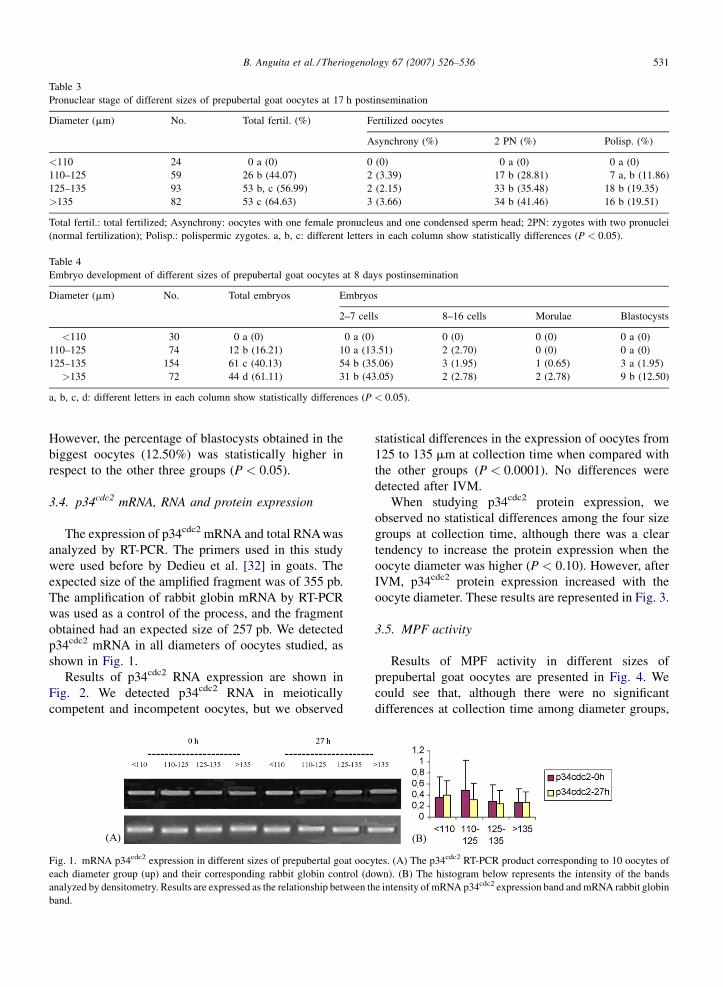
B. Anguita et al. / Theriogenology 67 (2007) 526–536 531
Table 3
Pronuclear stage of different sizes of prepubertal goat oocytes at 17 h postinsemination
Diameter (mm) No. Total fertil. (%) Fertilized oocytes
Asynchrony (%) 2 PN (%) Polisp. (%)
<110 24 0 a (0) 0 (0) 0 a (0) 0 a (0)
110–125 59 26 b (44.07) 2 (3.39) 17 b (28.81) 7 a, b (11.86)
125–135 93 53 b, c (56.99) 2 (2.15) 33 b (35.48) 18 b (19.35)
>135 82 53 c (64.63) 3 (3.66) 34 b (41.46) 16 b (19.51)
Total fertil.: total fertilized; Asynchrony: oocytes with one female pronucleus and one condensed sperm head; 2PN: zygotes with two pronuclei
(normal fertilization); Polisp.: polispermic zygotes. a, b, c: different letters in each column show statistically differences (P < 0.05).
Table 4
Embryo development of different sizes of prepubertal goat oocytes at 8 days postinsemination
Diameter (mm) No. Total embryos Embryos
2–7 cells 8–16 cells Morulae Blastocysts
<110 30 0 a (0) 0 a (0) 0 (0) 0 (0) 0 a (0)
110–125 74 12 b (16.21) 10 a (13.51) 2 (2.70) 0 (0) 0 a (0)
125–135 154 61 c (40.13) 54 b (35.06) 3 (1.95) 1 (0.65) 3 a (1.95)
>135 72 44 d (61.11) 31 b (43.05) 2 (2.78) 2 (2.78) 9 b (12.50)
a, b, c, d: different letters in each column show statistically differences (P < 0.05).
However, the percentage of blastocysts obtained in the
biggest oocytes (12.50%) was statistically higher in
respect to the other three groups (P < 0.05).
3.4. p34cdc2 mRNA, RNA and protein expression
The expression of p34cdc2 mRNA and total RNA was
analyzed by RT-PCR. The primers used in this study
were used before by Dedieu et al. [32] in goats. The
expected size of the amplified fragment was of 355 pb.
The amplification of rabbit globin mRNA by RT-PCR
was used as a control of the process, and the fragment
obtained had an expected size of 257 pb. We detected
p34cdc2 mRNA in all diameters of oocytes studied, as
shown in Fig. 1.
Results of p34cdc2 RNA expression are shown in
Fig. 2. We detected p34cdc2 RNA in meiotically
competent and incompetent oocytes, but we observed
Fig. 1. mRNA p34cdc2 expression in different sizes of prepubertal goat oocy
each diameter group (up) and their corresponding rabbit globin control (do
analyzed by densitometry. Results are expressed as the relationship between th
band.
statistical differences in the expression of oocytes from
125 to 135 mm at collection time when compared with
the other groups (P < 0.0001). No differences were
detected after IVM.
When studying p34cdc2 protein expression, we
observed no statistical differences among the four size
groups at collection time, although there was a clear
tendency to increase the protein expression when the
oocyte diameter was higher (P < 0.10). However, after
IVM, p34cdc2 protein expression increased with the
oocyte diameter. These results are represented in Fig. 3.
3.5. MPF activity
Results of MPF activity in different sizes of
prepubertal goat oocytes are presented in Fig. 4. We
could see that, although there were no significant
differences at collection time among diameter groups,
tes. (A) The p34cdc2 RT-PCR product corresponding to 10 oocytes of
wn). (B) The histogram below represents the intensity of the bands
e intensity of mRNA p34cdc2 expression band and mRNA rabbit globin

B. Anguita et al. / Theriogenology 67 (2007) 526–536532
Fig. 2. RNA p34cdc2 expression in different sizes of prepubertal goat oocytes. (A) The p34cdc2 RT-PCR product corresponding to 10 oocytes of each
diameter group (up) and their corresponding rabbit globin control (down). (B) The histogram represents the intensity of the bands analyzed by
densitometry. Results are expressed as the relationship between the intensity of RNA p34cdc2 expression band and mRNA rabbit globin band. §
indicates a significant difference in p34cdc2 RNA at collection time (P < 0.001).
Fig. 3. p34cdc2 protein expression in different sizes of prepubertal goat oocytes at collection time (0 h) and after IVM (27 h). (A) Western blot. Each
band has a size of 34 kDa and corresponds to 20 oocytes. (B) The histogram represents the intensity of the bands analyzed by densitometry. Results
are expressed as the relationship between the intensity of protein p34cdc2 expression in 20 oocytes and the p34cdc2 protein expression in granulose
cells (GC). a,bIndicate a significant difference in p34cdc2 protein expression among diameter groups (P < 0.05).
Fig. 4. MPF activity in different sizes of prepubertal goat oocytes at
collection time and after IVM. MPF activity is expressed as OD at
492 nm. a,bIndicate a significant difference in kinase activity among
diameter groups (P < 0.05).
MPF activity tended to increase in the biggest oocytes in
comparison to the smallest ones (P < 0.10). In addition,
after IVM, there is a clear and statistically significant
increase of MPF activity in the biggest oocytes.
4. Discussion
The results of this study show that the oocyte
diameter of prepubertal goat oocytes was positively
related to the percentage of oocytes reaching metaphase
II stage and to the percentage of oocytes developing up
to blastocyst stage. Oocyte diameter was also positively
related to the amount of p34cdc2 protein and MPF
activity in prepubertal females. Several studies have
examined the relationship between follicle diameter,
oocyte diameter and its meiotic and developmental
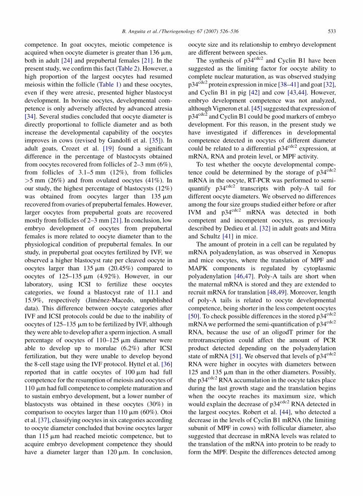
B. Anguita et al. / Theriogenology 67 (2007) 526–536 533
competence. In goat oocytes, meiotic competence is
acquired when oocyte diameter is greater than 136 mm,
both in adult [24] and prepubertal females [21]. In the
present study, we confirm this fact (Table 2). However, a
high proportion of the largest oocytes had resumed
meiosis within the follicle (Table 1) and these oocytes,
even if they were atresic, presented higher blastocyst
development. In bovine oocytes, developmental com-
petence is only adversely affected by advanced atresia
[34]. Several studies concluded that oocyte diameter is
directly proportional to follicle diameter and as both
increase the developmental capability of the oocytes
improves in cows (revised by Gandolfi et al. [35]). In
adult goats, Crozet et al. [19] found a significant
difference in the percentage of blastocysts obtained
from oocytes recovered from follicles of 2–3 mm (6%),
from follicles of 3.1–5 mm (12%), from follicles
>5 mm (26%) and from ovulated oocytes (41%). In
our study, the highest percentage of blastocysts (12%)
was obtained from oocytes larger than 135 mm
recovered from ovaries of prepubertal females. However,
larger oocytes from prepubertal goats are recovered
mostly from follicles of 2–3 mm [21]. In conclusion, low
embryo development of oocytes from prepubertal
females is more related to oocyte diameter than to the
physiological condition of prepubertal females. In our
study, in prepubertal goat oocytes fertilized by IVF, we
observed a higher blastocyst rate per cleaved oocyte in
oocytes larger than 135 mm (20.45%) compared to
oocytes of 125–135 mm (4.92%). However, in our
laboratory, using ICSI to fertilize these oocytes
categories, we found a blastocyst rate of 11.1 and
15.9%, respectively (Jimenez-Macedo, unpublished
data). This difference between oocyte categories after
IVF and ICSI protocols could be due to the inability of
oocytes of 125–135 mm to be fertilized by IVF, although
they were able to develop after a sperm injection. A small
percentage of oocytes of 110–125 mm diameter were
able to develop up to morulae (6.2%) after ICSI
fertilization, but they were unable to develop beyond
the 8-cell stage using the IVF protocol. Hyttel et al. [36]
reported that in cattle oocytes of 100 mm had full
competence for the resumption of meiosis and oocytes of
110 mm had full competence to complete maturation and
to sustain embryo development, but a lower number of
blastocysts was obtained in these oocytes (30%) in
comparison to oocytes larger than 110 mm (60%). Otoi
et al. [37], classifying oocytes in six categories according
to oocyte diameter concluded that bovine oocytes larger
than 115 mm had reached meiotic competence, but to
acquire embryo development competence they should
have a diameter larger than 120 mm. In conclusion,
oocyte size and its relationship to embryo development
are different between species.
The synthesis of p34cdc2 and Cyclin B1 have been
suggested as the limiting factor for oocyte ability to
complete nuclear maturation, as was observed studying
p34cdc2 protein expression in mice [38–41] and goat [32],
and Cyclin B1 in pig [42] and cow [43,44]. However,
embryo development competence was not analyzed,
although Vigneron et al. [45] suggested that expression of
p34cdc2 and Cyclin B1 could be good markers of embryo
development. For this reason, in the present study we
have investigated if differences in developmental
competence detected in oocytes of different diameter
could be related to a differential p34cdc2 expression, at
mRNA, RNA and protein level, or MPF activity.
To test whether the oocyte developmental compe-
tence could be determined by the storage of p34cdc2
mRNA in the oocyte, RT-PCR was performed to semi-
quantify p34cdc2 transcripts with poly-A tail for
different oocyte diameters. We observed no differences
among the four size groups studied either before or after
IVM and p34cdc2 mRNA was detected in both
competent and incompetent oocytes, as previously
described by Dedieu et al. [32] in adult goats and Mitra
and Schultz [41] in mice.
The amount of protein in a cell can be regulated by
mRNA polyadenylation, as was observed in Xenopus
and mice oocytes, where the translation of MPF and
MAPK components is regulated by cytoplasmic
polyadenylation [46,47]. Poly-A tails are short when
the maternal mRNA is stored and they are extended to
recruit mRNA for translation [48,49]. Moreover, length
of poly-A tails is related to oocyte developmental
competence, being shorter in the less competent oocytes
[50]. To check possible differences in the stored p34cdc2
mRNA we performed the semi-quantification of p34cdc2
RNA, because the use of an oligodT primer for the
retrotranscription could affect the amount of PCR
product detected depending on the polyadenylation
state of mRNA [51]. We observed that levels of p34cdc2
RNA were higher in oocytes with diameters between
125 and 135 mm than in the other diameters. Possibly,
the p34cdc2 RNA accumulation in the oocyte takes place
during the last growth stage and the translation begins
when the oocyte reaches its maximum size, which
would explain the decrease of p34cdc2 RNA detected in
the largest oocytes. Robert et al. [44], who detected a
decrease in the levels of Cyclin B1 mRNA (the limiting
subunit of MPF in cows) with follicular diameter, also
suggested that decrease in mRNA levels was related to
the translation of the mRNA into protein to be ready to
form the MPF. Despite the differences detected among

B. Anguita et al. / Theriogenology 67 (2007) 526–536534
diameter groups, the semi-quantification of p34cdc2
mRNA and RNA showed no relationship with oocyte
developmental competence.
p34cdc2 mRNA and RNA levels do not have to be
necessarily related to the p34cdc2 protein content in the
oocyte. For this reason, the study of the expression of the
p34cdc2 protein as a possible marker of oocyte develop-
mental competence was performed. At collection time,
although the corresponding RNA and mRNA were
already present in the smallest oocytes, p34cdc2 protein
was absent from these oocytes. These results were in
accordance to previous studies performed in mouse [52]
and adult goats [32], which did not detect p34cdc2 in
incompetent oocytes. However, in other species, p34cdc2
was detected in incompetent oocytes (equine: [53]; pig:
[54]; rat: [55]), showing that regulation of p34cdc2
expression differs between species. Accumulation of
p34cdc2 increases during oocyte growth (mouse: [52,39])
and during acquisition of meiotic competence (adult
goat: [32]; mouse: [39,56]). In contrast, our results did
not shown any statistical differences between diameter
groups at collection time although p34cdc2 was detected
in all groups but the smallest ones, probably due to the
high variability of our results. We detected the
appearance of the protein in the smallest oocytes after
27 h of culture, which clearly shows that the transcript
has been translated during the culture period. The
appearance of p34cdc2 has previously been observed in
incompetent oocytes from adult goats after in vitro
culture [32]. In our study, the amount of p34cdc2 protein
detected after in vitro maturation increased with oocyte
diameter and was associated with the ability to complete
meiosis and to develop into embryos. Dedieu et al. [32]
and Chesnel and Eppig [52] also observed an increase in
p34cdc2 according to the follicle diameter from which
oocytes were recovered, in adult goats and mice,
respectively. Nevertheless, it is important to note that
p34cdc2 accumulation during culture was observed for all
oocyte diameters, although the p34cdc2 levels achieved in
the smallest oocytes were lower than those detected in the
largest ones. Our results are in accordance to Dedieu et al.
[32], who also detected p34cdc2 in both competent and
incompetent oocytes after in vitro culture, although the
accumulation of p34cdc2 in the partially competent
oocytes was higher, reaching similar levels to fully
competent oocytes. However, accumulation of p34cdc2
during invitro culture does not promote the acquisition of
meiotic competence in oocytes [52] probably because
changes in the ability of p34cdc2 and Cyclin B1 to
dimerize [56] which depends on Cyclin B1 phosphor-
ylation [57], MPF coincidence in the time with its
regulators, such as cdc25 phosphatase or Wee kinases
([53]; reviewed by Jones [58]), or the accumulation of
MPF regulators. In fact, in adult goats, the presence of
active cdc25C, responsible for dephosphorylation of
p34cdc2 to activate MPF, is lower in meiotically
incompetent than competent oocytes [59]. Therefore,
not only the synthesis of p34cdc2 protein, but its regulation
to become active could also affect the acquisition of
oocyte competence. For this reason, it was decided to
study MPF activity in addition to p34cdc2 expression. In
our study, MPF activity was detected in oocytes just after
follicle release, when oocytes are supposed to be at the
GV stage and, as a consequence, MPF activity should be
non-existent in goats [12]. However, we found a high
percentage of oocytes at GVBD stage before culture,
especially in the largest oocytes, which would explain the
detection of MPF activity at collection time. After invitro
maturation, MPF activity increased with oocyte dia-
meter, and it was closely related to oocyte developmental
competence in prepubertal oocytes. Our results are in
accordance to previous studies that related MPF activity
in oocytes with their developmental competence. In fact,
Salamone et al. [14] and Ledda et al. [13] reported that
MPF activity in prepubertal females was substantially
lower than in adult females (cow and ewe, respectively),
while Bogliolo et al. [60] reported higher MPF activity in
in vivo than in vitro matured oocytes. In addition, higher
developmental competence in oocytes treated with
caffeine during nuclear transfer has also been related
to an increase in MPF activity promoted by caffeine [61].
Bringing together our results of maturation and MPF
activity, we observed that, after in vitro maturation, the
largest oocytes, which presented the highest MPF
activity, also had the highest percentage of oocytes at
the metaphase II stage, when the maximum level of
MPF activity is detected (mouse: [6–8]; pig: [9]; rabbit:
[10]; cattle: [11]; goat: [12]; sheep: [13]). Therefore, it
is difficult to establish whether differences in MPF
activity among our diameter groups are only caused by
differential developmental competence and not by
nuclear stage. Nevertheless, as we did not detect
differences in the rate of normal fertilization (two
pronuclei zygotes) in oocytes bigger than 110 mm, we
hypothesis that differences in MPF activity are not only
a consequence of different rates in MII oocytes, but to
different degrees of cytoplasmic maturation in those
oocytes, which is reflected in the differences in
blastocyst yield between oocyte diameters. However
we have to consider that other factors could affect MPF
activity, such as the oocyte’s ability to translocate MPF
from the cytoplasm to the nucleus (mouse: [62]; cattle:
[56]). In summary, we observed a positive relationship
between MPF activity (p34cdc2 kinase activity), oocyte
B. Anguita et al. / Theriogenology 67 (2007) 526–536 535
diameter and developmental competence, which indi-
cates that MPF activity not only takes part in nuclear
maturation, but could also play an important role in
cytoplasmic maturation in a direct or indirect way.
Nevertheless, more studies on the specific role of MPF
in cytoplasmic maturation and its regulation are needed.
In conclusion, our results indicate that the size of
oocytes from prepubertal goats is related to their
capability to undergo meiotic maturation, in vitro
fertilization and to reach the blastocyst stage. Oocyte
diameter and, therefore, oocyte developmental compe-
tence seems to be related to the amount of p34cdc2
protein and MPF activity found in these oocytes after in
vitro maturation. MPF activity seems to confer higher
developmental competence to oocytes through promo-
tion of cytoplasmic maturation.
Acknowledgment
Grant sponsor MEC: Grant number: AGL2004-
04737-CO3-01-GAN.
References
[1] Edwards R. Maturation in vitro of human oocytes. Lancet
1965;6:926–9.
[2] Crozet N, Motlik J, Szollosi D. Nucleolar fine structure and RNA
synthesis in porcine oocytes during early stages of antrum
formation. Biol Cell 1981;41:35–42.
[3] Naito K, Daen FP, Toyoda Y. Comparison of histone H1 kinase
activity during meiotic maturation between two types of porcine
oocytes matured in different media in vitro. Biol Reprod
1992;47:43–7.
[4] Labbe JC, Capony JP, Caput D, Cavadore JC, Derancourt J,
Kaghad M, et al. MPF from starfish oocytes at first meiotic
metaphase is a heterodimer containing one molecule of cdc2 and
one molecule of cyclin B. Embo J 1989;8:3053–8.
[5] Nebreda AR, Ferby I. Regulation of the meiotic cell cycle in
oocytes. Curr Opin Cell Biol 2000;12:666–75.
[6] Hashimoto N, Kishimoto T. Regulation of meiotic metaphase by
a cytoplasmic maturation-promoting factor during mouse oocyte
maturation. Dev Biol 1988;126:242–52.
[7] Fulka Jr J, Jung T, Moor RM. The fall of biological maturation
promoting factor (MPF) and histone H1 kinase activity during
anaphase and telophase in mouse oocytes. Mol Reprod Dev
1992;32:378–82.
[8] Verlhac MH, Kubiak JZ, Clarke HJ, Maro B. Microtubule and
chromatin behavior follow MAP kinase activity but not MPF
activity during meiosis in mouse oocytes. Development 1994;
120:1017–25.
[9] Naito K, Toyoda Y. Fluctuation of histone H1 kinase activity
during meiotic maturation in porcine oocytes. J Reprod Fertil
1991;93:467–73.
[10] Jelinkova L, Kubelka M, Motlik J, Guerrier P. Chromatin con-
densation and histone H1 kinase activity during growth and
maturation of rabbit oocytes. Mol Reprod Dev 1994;37:210–5.
[11] Collas P, Sullivan EJ, Barnes FL. Histone H1 kinase activity in
bovine oocytes following calcium stimulation. Mol Reprod Dev
1993;34:224–31.
[12] Dedieu T, Gall L, Crozet N, Sevellec C, Ruffini S. Mitogen-
activated protein kinase activity during goat oocyte maturation
and the acquisition of meiotic competence. Mol Reprod Dev
1996;45:351–8.
[13] Ledda S, Bogliolo L, Leoni G, Naitana S. Cell coupling and
maturation-promoting factor activity in in vitro-matured prepu-
bertal and adult sheep oocytes. Biol Reprod 2001;65:247–52.
[14] Salamone D, Damiani P, Fissore R, Robl J, Duby R. Biochemical
and developmental evidence that ooplasmic maturation of pre-
pubertal bovine oocytes is compromised. Biol Reprod 2001;64:
1761–8.
[15] Armstrong DT. Effects of maternal age on oocyte developmental
competence. Theriogenology 2001;55:1303–22.
[16] Fair T, Hyttel P, Greve T. Bovine oocyte diameter in relation to
maturational competence and transcriptional activity. Mol
Reprod Dev 1995;42:437–42.
[17] Raghu HM, Nandi S, Reddy SM. Follicle size and oocyte
diameter in relation to developmental competence of buffalo
oocytes in vitro. Reprod Fertil Dev 2002;14:55–61.
[18] Furher F, Matr B, Schellander K, Kalat M, Schleger W. Matura-
tion competence and chromatin behaviour in growing and fully
grown cattle oocytes. J Vet Med 1989;36:285–91.
[19] Crozet N, Ahmed-Ali M, Dubos MP. Developmental compe-
tence of goat oocytes from follicles of different size categories
following maturation, fertilization and culture in vitro. J Reprod
Fertil 1995;103:293–8.
[20] Marchal R, Vigneron C, Perreau C, Bali-Papp A, Mermillod P.
Effect of follicular size on meiotic and developmental compe-
tence of porcine oocytes. Theriogenology 2002;57:1523–32.
[21] Martino A, Mogas T, Palomo M, Paramio M. Meiotic compe-
tence of prepubertal goat oocytes. Theriogenology 1994;41:
969–80.
[22] Martino A, Mogas T, Palomo M, Paramio M. In vitro maturation
and fertilization of prepubertal goat oocytes. Theriogenology
1995;43:473–85.
[23] Izquierdo D, Villamediana P, Lopez-Bejar M, Paramio MT.
Effect of in vitro and in vivo culture on embryo development
from prepubertal goat IVM-IVF oocytes. Theriogenology
2002;57:1431–41.
[24] De Smedt V, Crozet N, Gall L. Morphological and functional
changes accompanying the acquisition of meiotic competence in
ovarian goat oocyte. J Exp Zoo 1994;269:128–39.
[25] Parrish J, Susko-Parrish J, Leibfried-Rutledge M, Crister E,
Eyeston W, First N. Bovine in vitro fertilization with frozen
thawed semen. Theriogenology 1986;25:591–600.
[26] Brackett BG, Oliphant G. Capacitation of rabbit spermatozoa in
vitro. Biol Reprod 1975;12:260–74.
[27] Younis AI, Zuelke KA, Harper KM, Oliveira MA, Brackett BG.
In vitro fertilization of goat oocytes. Biol Reprod 1991;44:
1177–82.
[28] Wang B, Baldassarre H, Tao T, Gauthier M, Neveu N, Zhou JF,
et al. Transgenic goats produced by DNA pronuclear micro-
injection of in vitro derived zygotes. Mol Reprod Dev 2002;63:
437–43.
[29] Tervit HR, Whittingham DG, Rowson LE. Successful culture in
vitro of sheep and cattle ova. J Reprod Fertil 1972;30:493–7.
[30] Takahashi Y, First L. In vitro development of bovine one-cell
embryos: influence of glucose, lactate, pyruvate, amino acids
and vitamins. Theriogenology 1992;37:963–78.

B. Anguita et al. / Theriogenology 67 (2007) 526–536536
[31] Gardner D, Lane M, Spitzer R, Batt P. Enhanced rates of
cleavage and development for sheep zygotes cultures to the
blastocyst stage in vitro in the absence of serum and somatic
cells: amino acids, vitamins, and culturind embryos in groups
stimulate development. Biol Reprod 1994;50:390–400.
[32] Dedieu T, Gall L, Hue I, Ledan E, Crozet N, Ruffini S, et al.
p34cdc2 expression and meiotic competence in growing goat
oocytes. Mol Reprod Dev 1998;50:251–62.
[33] Knijn HM, Wrenzycki C, Hendriksen PJ, Vos PL, Herrmann D,
van der Weijden GC, et al. Effects of oocyte maturation regimen on
the relative abundance of gene transcripts in bovine blastocysts
derived in vitro or in vivo. Reproduction 2002;124:365–75.
[34] Blondin P, Sirard MA. Oocyte and follicular morphology as
determining characteristics for developmental competence in
bovine oocytes. Mol Reprod Dev 1995;41:54–62.
[35] Gandolfi F, Brevini TA, Cillo F, Antonini S. Cellular and
molecular mechanisms regulating oocyte quality and the rele-
vance for farm animal reproductive efficiency. Rev Sci Tech
2005;24:413–23.
[36] Hyttel P, Fair T, Callesen H, Greve T. Oocyte growth, capacitation
and final maturation in cattle. Theriogenology 1997;47:23–32.
[37] Otoi T, Yamamoto K, Koyama N, Tachikawa S, Suzuki T. Bovine
oocyte diameter in relation to developmental competence. Ther-
iogenology 1997;48:769–74.
[38] Chesnel F, Eppig J. Synthesis and accumulation of p34cdc2 and
cyclin B in mouse oocytes during acquisition of competence to
resume meiosis. Mol Reprod Dev 1995;40:503–8.
[39] De Vantery C, Gavin A, Vassali J, Schorderet-Slatkine S. An
accumulation of p34cdc2 at the end of mouse oocyte growth
correlates with the acquisition of meiotic competence. Dev Biol
1996;174:335–44.
[40] De Vantery C, Stutz A, Vassalli J, Schorderet-Slatkine S. Acqui-
sition of meiotic competence in growing mouse oocytes is
controlled at both translational and posttranslational levels.
Dev Biol 1997;187:43–54.
[41] Mitra J, Schultz R. Regulation of the acquisition of meiotic
competence in the mouse: changes in the subcellular localization
of cdc2, cyclin B1, cdc25c and wee1, and in the concentration of
these proteins and their transcripts. J Cell Sci 1996;109:2407–15.
[42] Sun Q, Lai L, Bonk A, Prather R, Schatten H. Cytoplasmic
changes in relation to nuclear maturation and early embryo
developmental potential of porcine oocytes: effects of gonado-
tropins, cumulus cells, follicular size, and protein synthesis
inhibition. Mol Reprod Dev 2001;59:192–8.
[43] Levesque JT, Sirard MA. Resumption of meiosis is initiated by
the accumulation of cyclin B in bovine oocytes. Biol Reprod
1996;55:1427–36.
[44] Robert C, Hue I, McGraw S, Gagne D, Sirard MA. Quantifica-
tion of cyclin B1 and p34(cdc2) in bovine cumulus–oocyte
complexes and expression mapping of genes involved in the
cell cycle by complementary DNA macroarrays. Biol Reprod
2002;67:1456–64.
[45] Vigneron C, Perreau C, Dupont J, Uzbekova S, Prigent C,
Mermillod P. Several signaling pathways are involved in the
control of cattle oocyte maturation. Mol Reprod Dev 2004;69:
466–74.
[46] De Moor C, Richter J. Cytoplasmic polyadenilation elements
mediate masking and unmasking of Cyclin B1 mRNA. EMBO J
1999;18:2294–303.
[47] Charlesworth A, Welk J, MacNicol AM. The temporal control of
Wee1 mRNA translation during Xenopus oocyte maturation is
regulated by cytoplasmic polyadenylation elements within the
30-untranslated region. Dev Biol 2000;227:706–19.
[48] Huarte J, Stutz A, O’Connell ML, Gubler P, Belin D, Darrow AL,
et al. Transient translational silencing by reversible mRNA
deadenylation. Cell 1992;69:1021–30.
[49] Temeles GL, Schultz RM. Transient polyadenylation of a mater-
nal mRNA following fertilization of mouse eggs. J Reprod Fertil
1997;109:223–8.
[50] Gandolfi TA, Gandolfi F. The maternal legacy to the embryo:
cytoplasmic components and their effects on early development.
Theriogenology 2001;55:1255–76.
[51] Lequarre AS, Grisart B, Moreau B, Schuurbiers N, Massip A,
Dessy F. Glucose metabolism during bovine preimplantation
development: analysis of gene expression in single oocytes and
embryos. Mol Reprod Dev 1997;48:216–26.
[52] Chesnel F, Eppig JJ. Synthesis and accumulation of p34cdc2 and
cyclin B in mouse oocytes during acquisition of competence to
resume meiosis. Mol Reprod Dev 1995;40:503–8.
[53] Goudet G, Belin F, Bezard J, Gerard N. Maturation-promoting
factor (MPF) and mitogen activated protein kinase (MAPK)
expression in relation to oocyte competence for in-vitro matura-
tion in the mare. Mol Hum Reprod 1998;4:563–70.
[54] Christmann L, Jung T, Moor RM. MPF components and meiotic
competence in growing pig oocytes. Mol Reprod Dev 1994;38:
85–90.
[55] Goren S, Dekel N. Maintenance of meiotic arrest by a phos-
phorylated p34cdc2 is independent of cyclic adenosine 30,50-monophosphate. Biol Reprod 1994;51:956–62.
[56] Kanatsu-Shinohara M, Schultz RM, Kopf GS. Acquisition of
meiotic competence in mouse oocytes: absolute amounts of
p34(cdc2), cyclin B1, cdc25C, and wee1 in meiotically incom-
petent and competent oocytes. Biol Reprod 2000;63:1610–6.
[57] Yang J, Bardes ESG, Moore JD, Brennan J, Powers MA,
Kornbluth S. Control of cyclin B1 localization through regulated
binding of the nuclear export factor CRM1. Genes Dev 1998;12:
2131–43.
[58] Jones KT. Turning it on and off: M-phase promoting factor
during meiotic maturation and fertilization. Mol Hum Reprod
2004;10:1–5.
[59] Gall L, Ruffini S, Le Bourhis D, Boulesteix C. Cdc25C expres-
sion in meiotically competent and incompetent goat oocytes.
Mol Reprod Dev 2002;62:4–12.
[60] Bogliolo L, Leoni G, Ledda S, Zedda MT, Bonelli P, Madau L,
et al. M-phase promoting factor (MPF) and mitogen activated
protein kinases (MAPK) activities of domestic cat oocytes
matured in vitro and in vivo. Cloning Stem Cells 2004;6:15–23.
[61] Kawahara M, Wakai T, Yamanaka K, Kobayashi J, Sugimura S,
Shimizu T, et al. Caffeine promotes premature chromosome
condensation formation and in vitro development in porcine
reconstructed embryos via a high level of maturation promoting
factor activity during nuclear transfer. Reproduction 2005;130:
351–7.
[62] Mitra J, Schultz RM. Regulation of the acquisition of meiotic
competence in the mouse: changes in the subcellular localization
of cdc2, cyclin B1, cdc25C and wee1, and in the concentration
of these proteins and their transcripts. J Cell Sci 1996;109
(Pt 9):2407–15.

CICLINA B1, RNA Y PROTEÍNA TOTAL
Total RNA and protein content, Cyclin B1 expression and developmental
competence in prepubertal goat oocytes
Begoña Anguita1, Maria-Teresa Paramio1, Ana R Jiménez-Macedo1, Roser
Morató2, Teresa Mogas2, Dolors Izquierdo1a
1 Departament de Ciència Animal i dels Aliments. Universitat Autònoma de
Barcelona. 08193 Bellaterra. Barcelona. Spain 2 Departament de Medicina i Cirurgia Animals. Universitat Autònoma de Barcelona.
08193 Bellaterra. Barcelona. Spain a: Correspondence and reprint requests. Dolors Izquierdo. Departament de Ciència
Animal i dels Aliments. 08193 Bellaterra. Barcelona. Spain. E-mail:
[email protected], Fax: 34935811494, Phone: 34935811495.
59
CICLINA B1, RNA Y PROTEÍNA TOTAL
Abstract
The aim of this study was to examine the relationship between the
developmental competence of oocytes and their total RNA and protein contents,
and the level of Cyclin B1 transcription. Ovaries from prepubertal goats were
collected from a slaughterhouse. Oocytes were recovered by slicing and those with
two or more layers of cumulus cells and homogenous cytoplasm were matured in
vitro (20 to 25 oocytes per drop) for 27 h. Both before and after IVM, samples of
oocytes were denuded and categorised into four group treatments by diameter (<
110 µm, 110 to 125 µm, 125 to 135 µm; > 135 µm), separated into sub-groups of 10
oocytes per treatment-replicate and stored in liquid nitrogen until total RNA
content analysis by spectophotometry, total protein content analysis by a
colorimetric assay and Cyclin B1 transcription analysis by RT-PCR. For the study of
developmental competence, the rest of the matured oocytes were fertilised in
vitro in groups of 20 to 25 for 24 h. Presumptive zygotes were denuded, sorted into
the four categories of diameter noted above, and placed into culture drops in
groups of 18 to 25 for in vitro culture. Cleavage rate was evaluated at 48 hpi and
embryo development at 8 d post-insemination. There were four replicates of each
treatment for each assay or evaluation point of the experiment. There were no
significant differences between the size categories of oocytes at collection in total
RNA content, total protein content and Cyclin B1 mRNA. There were significant
differences (P < 0.05) in the expression of Cyclin B1 before IVM with oocytes in the
> 135 mm diameter category having the highest value for this variant. There were
no significant differences in these characteristics between the categories of oocyte
diameter after IVM except in respect of total RNA content, which was lower for the
largest size of oocytes (>135 µm; mean ± SD = 12.3 ± 1.84 ng/oocyte) than the
other three size groups (19.2 ± 1.38 to 22.1 ± 4.44 ng/oocyte; P < 0.05). Significant
differences (P < 0.05) in cleavage rate were observed between the different oocyte
size categories (<110 µm, 3.0 %; 110 to 125 µm, 32 %; 125 to 135 µm, 50 %; >135
µm, 73 %). Only oocytes >125 µm diameter developed to the blastocyst stage (125
to 135 µm, 7 %; >135 µm, 10 %). This study showed that the RNA content and the
Cyclin B1 RNA expression of prepubertal goat oocytes, and their development to
embryos varied between the different size categories of the oocytes.
Keywords: Cyclin B1, MPF, goat, oocyte, diameter
60

CICLINA B1, RNA Y PROTEÍNA TOTAL
1. Introduction
Some studies have demonstrated that the low blastocyst rate obtained in
vitro is mainly due to insufficient cytoplasmic maturation in the oocytes, which is
reflected in the inability of the fertilized oocyte to decondense the sperm head, to
block polyspermy, or inability to maintain embryo development beyond the
transition of embryonic genome activation (Sirard et al., 2006). In addition,
problems related to insufficient cytoplasmic maturation seem to be higher in
oocytes obtained from prepubertal than adult females (Armstrong, 2001). Results
from previous studies in our laboratory also indicate a deficient cytoplasmic
maturation in oocytes recovered from prepubertal goats at the slaughterhouse, a
low penetration rate (Urdaneta et al., 2004), different distributions of cortical
granules (Velilla et al., 2004) and mitochondria (Velilla et al., 2006) when
compared to adult goats, and a low blastocyst rate (Jimenez-Macedo et al., 2005;
Urdaneta et al., 2004). The small follicular diameter of prepubertal goat ovaries,
most of them between 2.5 to 3 mm (Martino et al., 1994), makes it necessary to
recover the oocytes by slicing the ovarian surface, which thus gives a high number
of oocytes. However, the oocyte population recovered is highly heterogeneous and
the oocytes have different maturational status and different degrees of atresia.
Oocyte selection made so far in our laboratory, based on cumulus-oocyte complex
(COC) morphology, seemed not to be good enough to distinguish between
developmentally competent and incompetent oocytes due to the low blastocyst
numbers obtained. In adult goats, Crozet et al. (1995) found a direct and positive
relationship between follicular diameter and embryo development, concluding that
follicles bigger than 5 mm contain oocytes which have a better competence to
develop up to blastocyst stage. In prepubertal goat ovaries, follicles larger than 5
mm in diameter are practically non-existent (Martino et al., 1995). In both adult
(De Smedt et al., 1994) and prepubertal (Martino et al., 1994) goat oocytes meiotic
oocyte competence has been classified in terms of the oocyte diameter as: <110
µm, corresponding to incompetent oocytes, 110 to 125 µm, corresponding to
partially competent oocytes, 125 to 135 µm and > 135 µm, corresponding to
competent oocytes. A study performed in our laboratory comparing embryo
development between different diameters of prepubertal goat oocytes showed a
61

CICLINA B1, RNA Y PROTEÍNA TOTAL
higher blastocyst rate in oocytes with a diameter higher than 135 µm (12.5 %;
Anguita et al., 2006).
During the oocyte growth phase, synthesis and storage of proteins and
heterogeneous RNA take place (Crozet et al., 1981). This storage is necessary to
maintain early embryonic development until the activation of the embryonic
genome (Bachvarova, 1992). It is possible that the low rate of in vitro development
could be explained by an insufficient storage of proteins and RNA in the oocyte
cytoplasm, maybe because they are recovered for IVM when they have not yet
finished the growth phase.
Oocyte meiotic competence is described as its ability to resume meiosis and
progress to metaphase II (MII) stage, where the oocyte remains in arrest until
fertilization. Meiotic maturation is regulated by several protein kinases, and MPF is
considered the most important. MPF is a heterodimer composed of a p34cdc2
catalytic subunit, with serine-threonine kinase activity, and a Cyclin B1 regulatory
subunit (Labbe et al., 1989). MPF activity has been described in many mammalian
oocytes: it appears just before GVBD and increase until metaphase I stage, then its
activity decreases in anaphase-telophase and increases again, reaching its
maximum level in metaphase II (mouse: Fulka et al., 1992; Hashimoto and
Kishimoto, 1986; Verlhac et al., 1994; pig: Naito and Toyoda, 1991; rabbit:
Jelinkova et al., 1994; cattle: Collas et al., 1993; goat: Dedieu et al., 1996; sheep:
Ledda et al., 2001). The limiting factor for MPF activation has been studied in
several species and species-specific differences have been identified (mouse:
Chesnel and Eppig, 1995; Mitra and Schultz, 1996; pigs: Naito et al., 1995; Sun et
al., 2001; goat: Dedieu et al., 1998; cattle: Levesque and Sirard, 1996; Robert et
al., 2002). Moreover, MPF activity has been shown to be lower in prepubertal than
in adult female oocytes (cow: Salamone et al., 2001; sheep: Ledda et al., 2001).
Therefore, low developmental competence could be explained by a deficiency of
one of the MPF subunits.
The objective of this study was to analyze the possible relationship between
total RNA and protein content, Cyclin B1 mRNA and RNA expression in prepubertal
62

CICLINA B1, RNA Y PROTEÍNA TOTAL
goat oocytes with oocyte developmental competence. To this purpose, analyses
were performed in oocytes of different diameter.
2. Material and methods
Unless otherwise indicated, all chemicals were purchased from Sigma
Chemical Co. (St. Louis, MO, USA).
2.1. Oocyte collection and IVM
Ovaries from prepubertal goats, approximately 2 months old, were
recovered from a local slaughterhouse and transported within 2 hours to the
laboratory at 38.5 ºC in PBS (Solution Dulbecco’s phosphate-buffered saline, P-4417)
which contained 50 µg/mL gentamycin (G-3632). Ovaries were washed three times
in PBS containing gentamycin, and COCs (cumulus-oocyte complexes) were
recovered by slicing in a 60 mm culture dish containing TCM199 (M-2520)
supplemented with 2.2 mg/mL NaHCO3, 2% (v/v) steer serum (Donor Bovine
Serum®, CanSera, Canada) and 50 μg/mL gentamycin. Only oocytes with two or
more layers of cumulus cells and homogeneous cytoplasm were used for IVM.
COCs were matured in TCM199 medium (M-7528) supplemented with 275
μg/mL sodium pyruvate (P-3662), 146 µg/mL L-glutamine (G-5763), 10% (v/v) steer
serum, 10 µg/mL o-LH (L-5269), 10 µg/mL o-FSH (F-4520), 1 µg/mL 17-β estradiol
(E-2257), 400 µM cysteamine (M-9768) and 50 µg/mL gentamycin (G-3632). Groups
of 20-25 COCs were transferred to 100 µl microdrops of maturation medium under
mineral oil (M-3516) and incubated for 27 hours at 38.5 ºC in a humidified air
atmosphere with 5% CO2.
2.2. Sperm capacitation and IVF
Matured oocytes were fertilized with fresh semen collected from two goats
of proven fertility. Ejaculates were collected with artificial vaginas and
63
CICLINA B1, RNA Y PROTEÍNA TOTAL
transported to the laboratory at 38.5 ºC within 30 minutes. Ejaculates were mixed
and the motile sperm fraction was separated by the swim-up method (Parrish et
al., 1986). Briefly, 70 µL of sperm was placed at the bottom of three conic tubes
containing 2 mL of mDM (Defined medium (Brackett and Oliphant, 1975), modified
by Younis et al. (1991)), and it was incubated for 45 min at 38.5 ºC in a humidified
air atmosphere with 5% CO2. After incubation, 600 µL of the supernatant of each
tube was recovered and centrifuged at 170 X g for 3 minutes. After discarding the
supernatant, the pellet was resuspended in mDM (1:1; v/v) containing heparin (H-
3149) and ionomycin (I-0634) (Final concentration: 10 µg/mL heparin and 200 nM of
ionomycin) and incubated for 15 min in a humidified air atmosphere with 5%CO2
and 38.5 ºC (Wang et al., 2002). Sperm concentration was determined using a
Neubauer chamber cell.
After maturation, groups of 20 to 25 oocytes were transferred to 100 µL
microdrops of modified Tyrode’s medium (TALP) as described by Parrish et al.
(Parrish et al., 1986), supplemented with 1 µg/mL hypotaurine (H-1384) and 0.3
mg/mL glutathione (G-6013) under mineral oil. Oocytes were co-cultured with
capacitated spermatozoa in a final concentration in the microdrop of 4 x 10⁶ sperm
cells/mL.
2.3. In vitro embryo culture
At 24 hours after insemination (hpi), presumptive zygotes were washed in
SOF medium (Synthetic Oviductal Fluid, (Holm et al., 1999)) and denuded by
pipetting to separate them from sperm cells. Groups of 18-25 embryos were placed
to 25 µL microdrops of SOF medium in 35 mm culture dishes under mineral oil, and
they were cultured in a humidified atmosphere with 5% CO2, 5% O2 and 90% N2 at
38.5 ºC for 7 days. At 48 hpi cleavage rate was evaluated and 0.1 µL fetal bovine
serum (F-7524) per embryo was added to the medium. At the end of the culture
period (8 days post-insemination), total cell number of embryos was evaluated by
Hoechst 33342 staining (B-2261) under a fluorescence microscope.
64
CICLINA B1, RNA Y PROTEÍNA TOTAL
2.4. RNA extraction
Total RNA from 10 oocytes was extracted using 100 µL of TriReagent® (T-
9424). We added 20 µg of glycogen (92760323, Roche, Germany) to the 100 µL of
TriReagent® as a carrier for the RNA precipitation and 1 pg of rabbit globin mRNA
(R-1253) per oocyte that was used as an extrinsic control of the whole process.
After adding 20 µL of chloroform, we centrifuged the sample at 12000 X g for 20
min at 4 ºC. The aqueous phase was recovered and the RNA was precipitated with
60 µL of isopropanol at -20 ºC overnight, while the resulting organic phase was
frozen at -80 ºC until its use in protein extraction. The next day, after
centrifugation at 12000 X g at 4 ºC for 20 min, the supernatant was discarded and
the pellet was washed sequentially with 70% ethanol and 100% ethanol. For the
washing steps, the sample was centrifuged at 12000 X g at 4 ºC for 15 min. The
pellet was dried and resuspended in 4 µL of H2O DEPC.
The RNA extracted was incubated for 30 min at 37 ºC with 1U/µL DNase
(18047-019, Invitrogen Corporation, USA), and stored at -80 ºC if the RT-PCR or the
RNA quantification could not be done immediately.
2.5. Total RNA quantification
Total RNA obtained after RNA extraction and DNA digestion was quantified
by spectrophotometry, in a wavelength of 260 nm. A known amount of goat ovary
RNA was used as an efficiency control of the process.
2.6. Protein extraction and protein quantification
Protein from 40 oocytes was extracted using TriReagent® (T-9424). DNA was
precipitated with 30 µL of 100% ethanol and centrifuged at 12000 X g at 4 ºC for 5
min. The supernatant was recovered and then transferred to a new tube. Proteins
were precipitated with 150 µL of isopropanol for 10 min at room temperature, and
centrifuged at 12000 X g at 4 ºC for 10 min. The supernatant was discarded, and
the pellet was washed three times with 200 µL of 0.3 M guanidine
hydrochloride/95% ethanol solution. During each wash, samples were stored in
65

CICLINA B1, RNA Y PROTEÍNA TOTAL
wash solution for 20 min at room temperature. Samples were centrifuged at 7500 X
g for 5 min at 4ºC. After the three washes, we added 1 mL of 100% ethanol and
vortered the protein pellet. The sample was left at room temperature for 20 min,
and centrifuged at 7500 X g for 5 min at 4 ºC. We discarded the supernatant, and
the protein pellet was dried for 5-10 min. The pellet was dissolved in 20 µL of 1%
SDS, and stored at -80 ºC until its use for quantification.
Total protein quantification of 5 µL of the protein extract (corresponding to
10 oocytes) was performed with DC Protein Assay (BioRad, USA), following the
manufacturer’s instructions. This is a colorimetric assay, and the color developed
in each reaction is proportional to the protein content of the sample. Different
concentrations of BSA (0, 50, 100, 200, 300, 400, 500, 600, 700, 1000 and 1410
µg/mL) diluted in SDS 1% were used as the control. The developed color of each
reaction was measured in a microplate ELISA reader (iEMS Reader, LabSystems) at
690 nm.
2.7. Reverse Transcription (RT) and Polymerase Chain Reaction (PCR)
In order to know Cyclin B1 mRNA and RNA expression in prepubertal goat
oocytes according to their diameter, for each size group total RNA corresponding to
10 oocytes was reverse-transcribed with ThermoScript™ RT-PCR System (Invitrogen
Corporation, USA), following the manufacturer’s instruction, in a final volume of 20
µL. The RT was performed using oligodT and random primers. Four µL of these
reactions were used to amplify Cyclin B1.
To perform Cyclin B1 amplification, cDNA was incubated with 1 X PCR
buffer, 1 µM of each primer, 2 mM of MgCl2, 200 µM of each dNTP and 0.1 U/ µL of
Taq polymerase GoldStar (ME-0064-01, Eurogentec, Belgium) in a final volume of
reaction of 25 µL for 34 cycles (94 ºC 1 min, 58 ºC 1 min, 72 ºC 1 min) in a
thermocycler 9700 (Applied BioSystems, USA). The Cyclin B1 sense and antisense
primers were the following: 5’-GAGCCATCCTCATTGACTGGC and 5’-
CTTAGATGCTCTCCGAAGG (Hue et al., 1997).
66

CICLINA B1, RNA Y PROTEÍNA TOTAL
To perform globin amplification, cDNA was incubated with 1 X PCR buffer, 0.5 µM
of each primer, 1.5 mM of MgCl2, 200 µM of each dNTP and 0.1 U/µL of Taq
polymerase GoldStar in a final volume of 25 µL for 30 cycles. Globin PCR conditions
were determined by the amplification conditions of Cyclin B1. The globin sense and
antisense primers were the following: 5’-GCAGCCACGGTGGCGAGTAT and 5’-
GTGGGACAGGAGCTTGAAAT (Knijn et al., 2002) .
At the same time, a PCR without cDNA and a PCR of the RT product
performed without RNA were used as negative controls. In no case was an
amplification band detected.
Amplification bands were visualized in a 2% agarose gel stained with
ethidium bromide. The weight marker band (P-9577) corresponding to 300 pb was
used as a control to avoid light variations in the exposition to UV. Each
amplification band was quantified by densitometric analysis using the PC program
Quantity One (Bio-Rad Laboratories, USA). The results were normalized with the
quantification of globin amplification band. Direct sequencing of the amplifications
products was performed using an ABI-prism DNA sequencer.
2.8. Experimental design
All the oocytes used for each replicate and assay were collected at the same
day. Four replicates of each assay were performed. Each replicate was started at a
separate day.
Before IVM, a sample of oocytes selected at random was denuded in PBS plus
300 µg hyaluronidase/mL (H-4272), measured at X 96 excluding the zona pellucida
and classified by diameters in four categories: <110 µm, 110 to 125 µm, 125 to 135
µm and >135 µm according to the criteria described by Anguita et al. (2006) in
prepubertal goats. From each diameter category, at least four sub-samples each of
10 oocytes were washed, frozen in liquid nitrogen and stored at -80 ºC until its use
in one of the four analyses. The remainder of the oocytes, still not separated by
diameter categories, were matured in vitro. After 27 h of in vitro maturation, a
67

CICLINA B1, RNA Y PROTEÍNA TOTAL
sample of oocytes were denuded, measured and classified by diameters as
described above. At least four sub-samples of 10 oocytes of each category were
washed and stored as described above until its use in one of the four analyses. The
remainder of oocytes was fertilized in vitro. At 24 hpi, the presumptive zygotes
were measured and classified in the four categories and culture in vitro for
additional 7 days. Each drop contained 18-25 embryos of the same diameter
category.
RNA extraction was performed in four sub-samples of 10 oocytes of each
replicate and size category. The RNA corresponding to one sub-sample per size
category and replicate was used to perform total RNA content and Cyclin B1
transcription analyses. The organic phase obtained in the RNA extraction of the
four sub-samples of each size category (40 oocytes per category) and replicate was
stored at -80ºC until its use in protein extraction and quantification.
2.9. Statistical analysis
Embryo development, PCR semi-quantification, total RNA quantification and
total protein quantification results of different diameter groups were compared
with a One-Way ANOVA test with Tukey’s post-test by the PROC MIXED procedure of
SAS (Version 9.2, Inst., Inc, Cary, NC). Data from embryo development were
transformed using arcsine square root before statistical analysis. Each oocyte size
group was represented by four replicates for each of the variates studied. A value
of P < 0.05 was accepted as the minimum level of significance in all the statistical
tests. Data are presented as percentage ± SD.
3. Results
3.1. Embryonic development
Results of embryonic development of different size oocytes from prepubertal
goats are shown in table 1. Total number of embryos obtained increased with
oocyte diameter, and there were significant differences among the four diameter
68

CICLINA B1, RNA Y PROTEÍNA TOTAL
groups (<110 µm: 3%; 110 to 125 µm: 32%; 125 to 135 µm: 50%; >135 µm: 73%).
Blastocyst rate in 125-135 µm oocytes (7%) and > 135 µm oocytes (10%) was
significantly higher than in <125 µm oocytes (0%) (P < 0.05).
3.2. Total RNA quantification
For each diameter group, total RNA corresponding to 10 oocytes, before and
after IVM, was quantified by spectrophometry at 260 nm. Results, expressed as
ng/oocyte, are shown in table 2. As we can observe, we detected no statistically
significant differences among the four categories of diameters before IVM (<110
µm: 22.9 ± 6.5; 110 to 125 µm: 21.9 ± 6.8; 125 to 135 µm: 19.7 ± 6.5; >135 µm:
26.5 ± 11.1) (P>0.05). However, after IVM, the total RNA detected was lower in the
biggest oocytes (>135 μm: 12.3 ± 1.8) when compared to oocytes 110-125 μm
oocytes (22.1 ± 4.4), and oocytes from 125 to 135 μm (19.2 ± 1.4) (P=0.08).
3.3. Total protein quantification
Total protein oocyte quantification from each size group was performed
using the DC Protein Assay. As we can observe in table 3, we detected no
statistically significant differences between the four groups of size at any of the
hours analyzed.
3.4. Cyclin B1 mRNA and RNA expression
Results of Cyclin B1 mRNA expression at collection time and after IVM are
represented in figure 1. No statistically significant differences in Cyclin B1 mRNA
expression were detected among the four groups of oocyte size before and after
IVM. However, the expression of Cyclin B1 mRNA at collection increased as oocyte
diameter increased from 0.50 ± 0.07 for oocytes < 110 µm to 0.96 ± 0.33 for 110 to
125 µm oocytes, but declined with further increase in diameter (125 to 135 µm:
0.86 ± 0.33; >135 µm: 0.58 ± 0.37).
69
CICLINA B1, RNA Y PROTEÍNA TOTAL
Results of Cyclin B1 RNA expression are shown in figure 2. Before IVM, Cyclin
B1 RNA expression was highest in the largest oocytes (<110 µm: 0.23 ± 0.13; 110 to
125 µm: 0.59 ± 0.11; 125 to 135 µm: 0.53 ± 0.54; >135 µm: 1.27 ± 0.78) (P=0.04).
However, after IVM no statistically significant differences were detected among
size categories. The expression of Cyclin B1 RNA increased from collection to the
completion of IVM for oocytes < 125 µm (P < 0.05), 110 to 125 µm (P < 0.05) and
125 to 135 µm (P < 0.10), but decreased for oocytes > 135 µm diameter (NS).
4. Discussion
The characteristics that determine whether an oocyte is competent to
develop into an embryo are still unknown. It seems that cytoplasmic oocyte
maturation is the key to a better understanding of oocyte developmental
competence. However, processes that take place during that maturation and, most
importantly, their regulation, are still poorly understood. The aim of our study was
to try to clarify how the storage and expression of certain molecules that could be
related to cytoplasmic oocyte maturation differ between oocytes with different
developmental competence.
We have observed that oocyte developmental competence in prepubertal
goat oocytes is related to the diameter; oocytes smaller than 125 µm were not able
to develop to blastocyst stage and a maximum blastocyst rate was reached in
oocytes with a diameter larger than 135 µm (10%), but there were no differences
when compared to oocytes from 125 to 135 µm. The capacity of prepubertal goat
oocytes to reach blastocyst stage was previously study in our laboratory, and we
observed for the first time that prepubertal goat oocytes with a diameter higher
than 125 µm were able to develop to blastocyst stage (Anguita et al., 2006).
Moreover, we observed that the inability of oocytes smaller than 125 µm to
maintain embryonic development was due to a deficient nuclear maturation in
these oocytes, and only 0 and 20.7% of oocytes reached metaphase II stage after
IVM in oocytes smaller than 110 µm and oocytes form 110- to 125 µm, respectively
(Anguita et al., 2006). In this earlier study, we also observed that oocytes from 110
to 125 µm which completed nuclear maturation were able to be normally fertilized
70
CICLINA B1, RNA Y PROTEÍNA TOTAL
(2PN = 28.8%), showing no differences compared to oocytes higher than 125 µm,
but they were unable to surpass the 2-8 cell stage in embryonic development,
which may indicate that these oocytes have not stored enough RNA and proteins to
reach blastocyst stage. The relationship between follicular size, oocyte diameter
and developmental competence has been observed in many species (goat: Crozet
et al., 1995; cow: Kauffold et al., 2005; Lequarre et al., 2005; Otoi et al., 1997;
pig: Marchal et al., 2002; buffalo: Raghu et al., 2002). In adult goats, Crozet et al.
(1995) observed a significant difference in the percentage of blastocysts obtained
from oocytes recovered from follicles of 2 to 3 mm (6%), from follicles of 3.1 to 5
mm (12%), from follicles bigger than 5 mm (26%) and from ovulated oocytes (41%).
Crozet et al. (2000) described the relationship between goat oocyte diameter and
follicle diameter in adult goats. Thus, oocytes with a mean diameter of 96 μm were
recovered from follicles smaller than 0.5 mm; oocytes of 120 μm were recovered
from follicles of 0.5 to 0.8 mm; oocytes of 125 μm from follicles of 1 to 1.8 mm;
oocytes of 136 μm (range 125 to 146 μm) from follicles of 2 to 3 mm and oocytes
ranging from 130 to 146 μm from follicles larger than 3 mm. In the present study,
we obtained a higher blastocyst rate in 125 to 135 µm oocytes (7%) than in a
previous study (1.95%; Anguita et al., 2006). This improvement could be due to the
use of glutathione (GSH) during fertilization in vitro. Urdaneta et al. (2004)
observed an increase in normal fertilization (zygotes with two pronuclei) and
cleavage when GSH was used in in vitro fertilization of prepubertal goat oocytes.
Also, an increase in blastocyst rate has been reported when glutathione was used in
the IVF medium (pigs: Boquest et al., 1999; cattle: Van Soom et al., 1998). Thus,
GSH plays an important role as cellular protection during oxidative stress and
helping cell proliferation during embryonic events (Del Corso et al., 1994; Lafleur
et al., 1994; Yu, 1994). These results seem to indicate that the improvement in
blastocyst rate detected in oocytes with 125-135 µm diameters in comparison to
the results of Anguita et al. (2006) could be related to a beneficial effect of the
GSH during fertilization.
During oocyte growth in vivo, RNA and protein storage take place (Gosden et
al., 1997). RNA synthesis ceases when oocytes resume meiosis (Hyttel et al., 2001).
Meiotic progression up to MII, fertilization and early embryonic development must
71

CICLINA B1, RNA Y PROTEÍNA TOTAL
be maintained with the stored RNA and proteins (Bachvarova, 1992). Deficient
storage of RNA and proteins, therefore, could lead to a decrease in the
developmental competence of the oocyte. To check if the differences in
developmental competence between different diameters of prepubertal goat
oocytes could be explained by a differential storage of RNA or proteins, we
performed total RNA and protein quantification before and after IVM. We observed
that the total RNA stored before IVM in the oocytes was not related to oocyte size.
After maturation, total RNA decreased in the largest oocytes, perhaps due to a
higher capability of these oocytes to mobilize the stored RNA and to synthesize
new proteins, which could explain their high developmental competence. However,
no significant differences in total protein content were observed between the
different oocyte diameters before or after maturation. Therefore, we could not
determine whether the lower RNA content detected in the biggest oocytes is due to
a mobilization of stored RNA or not.
MPF is the main meiotic regulator, and it is thought to be a possible
cytoplasmic maturation regulator too. It is composed of two subunits, the catalytic
subunit p34cdc2 and the regulatory subunit Cyclin B1 (Labbe et al., 1989). It has
been suggested that the oocytes inability to resume or complete meiosis could be
related to deficient MPF activation or the lack of one of the MPF subunits, but the
limiting factor varies among species: in the mouse, meiotic competence acquisition
is related to p34cdc2 protein synthesis (Chesnel and Eppig, 1995; De Vantéry et al.,
1996; de Vantery et al., 1997; Mitra and Schultz, 1996); in adult goats p34cdc2 is
considered the key factor for meiotic resumption (Crozet et al., 2000; Dedieu et
al., 1998); and in pigs (Sun et al., 2001) and cattle (Levesque and Sirard, 1996) the
limiting factor for meiotic resumption is the Cyclin B1 subunit. As observed, many
studies have tried to establish a relationship between MPF subunit expression and
oocyte meiotic competence, but we have not found any study of MPF expression
related to developmental competence, although Vigneron et al. (2004) suggested
that the expression of p34cdc2 and Cyclin B1 could be good markers of embryo
development. In our study, we have analyzed Cyclin B1 expression at a
transcriptional level by RT-PCR. We observed no differences in Cyclin B1 mRNA
expression in the four groups of size before or after IVM. It is important to notice
72
CICLINA B1, RNA Y PROTEÍNA TOTAL
that Cyclin B1 mRNA was detected in all the diameters evaluated, what is in
accordance with the results obtained by Hue et al. (1997) who detected Cyclin B1
mRNA in both meiotically competent and incompetent oocytes. Although it is not
statistically different, before IVM Cyclin B1 mRNA expression seemed to increase in
oocytes with diameters from 110 to 135 µm, possibly due to the accumulation of
the mRNA that takes place during the last growth phase in the oocytes (Gosden et
al., 1997). The decrease of Cyclin B1 mRNA observed in the biggest oocytes could
indicate the use of that mRNA to synthesize the Cyclin B1 protein needed to
associate with p34cdc2 to form pre-MPF. In fact, in an earlier study we observed
that most oocytes had resumed meiosis at collection time, and the percentage of
GVBD-oocytes increased with oocyte diameter (<110 µm: 5.33 %; 110 to 125 µm:
26.66 %; 125 to 135 µm: 64.40 %; >135 µm: 80.30 %; Anguita et al., 2006). The
association of Cyclin B1 and p34cdc2 and its subsequent activation is a requirement
for meiosis resumption in many mammalian oocytes (mouse: Chesnel and Eppig,
1995; Choi et al., 1991; pig: Christmann et al., 1994; cattle: Wu et al., 1997;
rabbit: Jelinkova et al., 1994; goat: Dedieu et al., 1996). Our results concur with
our previous study which detected a positive relationship between the
developmental competence of prepubertal goat oocytes and diameters, higher
p34cdc2 protein content and MPF activity (Anguita et al., 2006). As MPF requires not
only the synthesis of p34cdc2, but also of Cyclin B1, the present results suggest that
the decrease of Cyclin B1 mRNA in the biggest oocytes and the supposed Cyclin B1
synthesis could lead to an increase in MPF formation and activity, which could also
be related to the higher developmental competence of these oocytes.
In addition to Cyclin B1 mRNA expression, we also evaluated Cyclin B1 RNA
expression by RT-PCR using random primers instead of oligo-dT primers. The
protein amount in a cell can be regulated by mRNA polyadenylation, as was
observed in Xenopus and mice oocytes, where translation of MPF and MAPK
components is regulated by cytoplasmic polyadenylation (Xenopus: Charlesworth et
al., 2000; de Moor and Richter, 1999; mouse: Krischek and Meinecke, 2002). Poli-A
tails are short when the maternal mRNA is stored and they are extended to recruit
mRNA for translation (Huarte et al., 1992; Temeles and Schultz, 1997). As a
73

CICLINA B1, RNA Y PROTEÍNA TOTAL
consequence, the use of oligo-dT primers could affect the PCR product detected
depending on the polyadenylation state of mRNA (Lequarre et al., 1997).
Cyclin B1 RNA expression increased with increasing oocyte diameter before
IVM, but not after maturation. After IVM, there were no significant differences
between the treatments, probably due to the Cyclin B1 RNA storage that takes
place during maturation in oocytes smaller than 135 µm. In the largest oocytes,
however, Cyclin B1 expression during maturation did not increase, probably due to
the higher RNA consumption rate of the larger oocytes in comparison to the other
size groups. Although the rest of size groups reach the levels of the biggest group,
they did not reach the blastocyst rate achieved by the oocytes bigger than 135 µm.
These results could suggest that the amount of stored Cyclin B1 RNA at oocyte
collection time is related to oocyte size as well as to meiotic and developmental
competence. These results also suggest the possible role that Cyclin B1 plays not
only in oocyte nuclear maturation but, also in cytoplasmic maturation.
5. Conclusion
In conclusion, under the conditions of the present study, development to the
blastocyst stage of prepubertal goat oocytes depended upon the denuded oocytes
having a minimum diameter of 125 µm. This study showed that the RNA content
and the Cyclin B1 RNA expression of prepubertal goat oocytes, and their
development to embryos, varied between the different size categories of the
oocytes.
Acknowledgements
Financial support: Ministry of Education and Science of Spain, grant number:
AGL2004-04737-C03-01/GAN; Generalitat de Catalunya, grant number: 2006FIC
00187. The authors would like to thank Dr. Paul W. Cardozo for his valued help in
statistical analysis.
74
CICLINA B1, RNA Y PROTEÍNA TOTAL
Referentes
Anguita B., Jiménez-Macedo A., Izquierdo D., Paramio M., 2006. Effect of oocyte
diameter on meiotic competence, embryo development, P34 (cdc2) expression and
MPF activity in prepubertal goat oocytes. Theriogenology,
doi:10.1016/j.theriogenology.2006.09.003
Armstrong D.T., 2001. Effects of maternal age on oocyte developmental
competence. Theriogenology. 55, 1303-1322.
Bachvarova R.F., 1992. A maternal tail of poly(A): the long and the short of it. Cell.
69, 895-897.
Boquest A.C., Abeydeera L.R., Wang W.H., Day B.N., 1999. Effect of adding
reduced glutathione during insemination on the development of porcine embryos in
vitro. Theriogenology. 51, 1311-1319.
Brackett B.G., Oliphant G., 1975. Capacitation of rabbit spermatozoa in vitro. Biol.
Reprod.12, 260-274.
Charlesworth A., Welk J., MacNicol A.M., 2000. The temporal control of Wee1
mRNA translation during Xenopus oocyte maturation is regulated by cytoplasmic
polyadenylation elements within the 3'-untranslated region. Dev. Biol. 227, 706-
719.
Chesnel F., Eppig J.J., 1995. Synthesis and accumulation of p34cdc2 and Cyclin B in
mouse oocytes during acquisition of competence to resume meiosis. Mol. Reprod.
Dev. 40, 503-508.
Choi T., Aoki F., Mori M., Yamashita M., Nagahama Y., Kohmoto K., 1991.
Activation of p34cdc2 protein kinase activity in meiotic and mitotic cell cycles in
mouse oocytes and embryos. Development. 113, 789-795.
Christmann L., Jung T., Moor R.M., 1994. MPF components and meiotic competence
in growing pig oocytes. Mol. Reprod. Dev. 38, 85-90.
Collas P., Sullivan E.J., Barnes F.L., 1993. Histone H1 kinase activity in bovine
oocytes following calcium stimulation. Mol. Reprod. Dev. 34, 224-231.
Crozet N., Ahmed-Ali M., Dubos M.P., 1995. Developmental competence of goat
oocytes from follicles of different size categories following maturation,
fertilization and culture in vitro. J. Reprod. Fertil. 103, 293-298.
75

CICLINA B1, RNA Y PROTEÍNA TOTAL
Crozet N., Dahirel M., Gall L., 2000. Meiotic competence of in vitro grown goat
oocytes. J. Reprod. Fertil. 118, 367-373.
Crozet N., Motlik J., Szöllösi D., 1981. Nucleolar fine structure and RNA synthesis in
porcine oocytes during early stages of antrum formation. Biol. Cell. 41, 35-42.
De Moor C.H., Richter J.D., 1999. Cytoplasmic polyadenylation elements mediate
masking and unmasking of cyclin B1 mRNA. Embo. J. 18, 2294-2303.
De Smedt V., Crozet N., Gall L., 1994. Morphological and functional changes
accompanying the acquisition of meiotic competence in ovarian goat oocyte. J.
Exp. Zoo. 269, 128-139.
De Vantéry C., Gavin A., Vassali J., Schorderet-Slatkine S., 1996. An accumulation
of p34cdc2 at the end of mouse oocyte growth correlates with the acquisition of
meiotic competence. Dev. Biol. 174, 335-344.
De Vantéry C., Stutz A., Vassalli J.D., Schorderet-Slatkine S., 1997. Acquisition of
meiotic competence in growing mouse oocytes is controlled at both translational
and posttranslational levels. Dev. Biol. 187, 43-54.
Dedieu T., Gall L., Crozet N., Sevellec C., Ruffini S., 1996. Mitogen-activated
protein kinase activity during goat oocyte maturation and the acquisition of meiotic
competence. Mol. Reprod. Dev. 45, 351-358.
Dedieu T., Gall L., Hue I., Ledan E., Crozet N., Ruffini S., Sevellec C., 1998.
p34cdc2 expression and meiotic competence in growing goat oocytes. Mol. Reprod.
Dev. 50, 251-262.
Del Corso A., Cappiello M., Mura U., 1994. Thiol dependent oxidation of enzymes:
the last chance against oxidative stress. Int. J. Biochem. 26, 745-750.
Fulka J., Jr., Jung T., Moor R.M., 1992. The fall of biological maturation promoting
factor (MPF) and histone H1 kinase activity during anaphase and telophase in mouse
oocytes. Mol. Reprod. Dev. 32, 378-382.
Gosden R., Krapez J., Briggs D., 1997. Growth and development of the mammalian
oocyte. Bioessays. 19, 875-882.
Hashimoto N., Kishimoto T., 1986. Cell cycle dynamics of maturation-promoting
factor during mouse oocyte maturation. Tokai. J. Exp. Clin. Med. 11, 471-477.
Holm P., Booth P.J., Schmidt M.H., Greve T., Callesen H., 1999. High bovine
blastocyst development in a static in vitro production system using SOFaa medium
76
CICLINA B1, RNA Y PROTEÍNA TOTAL
supplemented with sodium citrate and myo-inositol with or without serum-
proteins. Theriogenology. 52, 683-700.
Huarte J., Stutz A., O'Connell M.L., Gubler P., Belin D., Darrow A.L., Strickland S.,
Vassalli J.D., 1992. Transient translational silencing by reversible mRNA
deadenylation. Cell. 69, 1021-1030.
Hue I., Dedieu T., Huneau D., Ruffini S., Gall L., Crozet N., 1997. Cyclin B1
expression in meiotically competent and incompetent goat oocytes. Mol. Reprod.
Dev. 47, 222-228.
Hyttel P., Viuff D., Fair T., Laurincik J., Thomsen P.D., Callesen H., Vos P.L.,
Hendriksen P.J., Dieleman S.J., Schellander K., Besenfelder U., Greve T., 2001.
Ribosomal RNA gene expression and chromosome aberrations in bovine oocytes and
preimplantation embryos. Reproduction. 122, 21-30.
Jelinkova L., Kubelka M., Motlik J., Guerrier P., 1994. Chromatin condensation and
histone H1 kinase activity during growth and maturation of rabbit oocytes. Mol.
Reprod. Dev. 37, 210-215.
Jimenez-Macedo A.R., Izquierdo D., Urdaneta A., Anguita B., Paramio M.T., 2006.
Effect of roscovitine on nuclear maturation, MPF and MAP kinase activity and
embryo development of prepubertal goat oocytes. Theriogenology. 65, 1769-1782.
Kauffold J., Amer H.A., Bergfeld U., Weber W., Sobiraj A., 2005. The in vitro
developmental competence of oocytes from juvenile calves is related to follicular
diameter. J. Reprod. Dev. 51, 325-332.
Knijn H.M., Wrenzycki C., Hendriksen P.J., Vos P.L., Herrmann D., van der Weijden
G.C., Niemann H., Dieleman S.J., 2002. Effects of oocyte maturation regimen on
the relative abundance of gene transcripts in bovine blastocysts derived in vitro or
in vivo. Reproduction. 124, 365-375.
Krischek C., Meinecke B., 2002. In vitro maturation of bovine oocytes requires
polyadenylation of mRNAs coding proteins for chromatin condensation, spindle
assembly, MPF and MAP kinase activation. Anim. Reprod. Sci. 73, 129-140.
Labbe J.C., Picard A., Peaucellier G., Cavadore J.C., Nurse P., Doree M., 1989.
Purification of MPF from starfish: identification as the H1 histone kinase p34cdc2
and a possible mechanism for its periodic activation. Cell. 57, 253-263.
77

CICLINA B1, RNA Y PROTEÍNA TOTAL
Lafleur M.V., Hoorweg J.J., Joenje H., Westmijze E.J., Retel J., 1994. The
ambivalent role of glutathione in the protection of DNA against singlet oxygen.
Free Radic. Res. 21, 9-17.
Ledda S., Bogliolo L., Leoni G., Naitana S., 2001. Cell coupling and maturation-
promoting factor activity in in vitro-matured prepubertal and adult sheep oocytes.
Biol. Reprod. 65, 247-252.
Lequarre A.S., Grisart B., Moreau B., Schuurbiers N., Massip A., Dessy F., 1997.
Glucose metabolism during bovine preimplantation development: analysis of gene
expression in single oocytes and embryos. Mol. Reprod. Dev. 48, 216-226.
Lequarre A.S., Vigneron C., Ribaucour F., Holm P., Donnay I., Dalbies-Tran R.,
Callesen H., Mermillod P., 2005. Influence of antral follicle size on oocyte
characteristics and embryo development in the bovine. Theriogenology. 63, 841-
859.
Levesque J.T., Sirard M.A., 1996. Resumption of meiosis is initiated by the
accumulation of cyclin B in bovine oocytes. Biol. Reprod. 55, 1427-1436.
Marchal R., Vigneron C., Perreau C., Bali-Papp A., Mermillod P., 2002. Effect of
follicular size on meiotic and developmental competence of porcine oocytes.
Theriogenology. 57, 1523-1532.
Martino A., Mogas T., Palomo M., Paramio M., 1994. Meiotic competence of
prepubertal goat oocytes. Theriogenology. 41, 969-980.
Martino A., Mogas T., Palomo M., Paramio M., 1995. In vitro maturation and
fertilization of prepubertal goat oocytes. Theriogenology. 43, 473-485.
Mitra J., Schultz R.M., 1996. Regulation of the acquisition of meiotic competence
in the mouse: changes in the subcellular localization of cdc2, cyclin B1, cdc25C and
wee1, and in the concentration of these proteins and their transcripts. J. Cell. Sci.
109, 2407-2415.
Naito K., Hawkins C., Yamashita M., Nagahama Y., Aoki F., Kohmoto K., Toyoda Y.,
Moor R.M., 1995. Association of p34cdc2 and cyclin B1 during meiotic maturation in
porcine oocytes. Dev. Biol. 168, 627-634.
Naito K., Toyoda Y., 1991. Fluctuation of histone H1 kinase activity during meiotic
maturation in porcine oocytes. J. Reprod. Fertil. 93, 467-473.
78

CICLINA B1, RNA Y PROTEÍNA TOTAL
Otoi T., Yamamoto K., Koyama N., Tachikawa S., Murakami M., Kikkawa Y., Suzuki
T., 1997. Cryopreservation of mature bovine oocytes following centrifugation
treatment. Cryobiology. 34, 36-41.
Parrish J., Susko-Parrish J., Leibfried-Rutledge M., Crister E., Eyeston W., First N.,
1986. Bovine in vitro fertilization with frozen thawed semen. Theriogenology. 25,
591-600.
Raghu H.M., Nandi S., Reddy S.M., 2002. Follicle size and oocyte diameter in
relation to developmental competence of buffalo oocytes in vitro. Reprod. Fertil.
Dev. 14, 55-61.
Robert C., Hue I., McGraw S., Gagne D., Sirard M.A., 2002. Quantification of cyclin
B1 and p34(cdc2) in bovine cumulus-oocyte complexes and expression mapping of
genes involved in the cell cycle by complementary DNA macroarrays. Biol. Reprod.
67, 1456-1464.
Salamone D.F., Damiani P., Fissore R.A., Robl J.M., Duby R.T., 2001. Biochemical
and developmental evidence that ooplasmic maturation of prepubertal bovine
oocytes is compromised. Biol. Reprod. 64, 1761-1768.
Sirard M.A., Richard F., Blondin P., Robert C., 2006. Contribution of the oocyte to
embryo quality. Theriogenology. 65, 126-136.
Sun Q., Lai L., Bonk A., Prather R., Schatten H., 2001. Cytoplasmic changes in
relation to nuclear maturation and early embryo developmental potential of
porcine oocytes: Effects of gonadotropins, cumulus cells, follicular size, and
protein synthesis inhibition. Mol. Reprod. Dev. 59, 192-198.
Temeles G.L., Schultz R.M., 1997. Transient polyadenylation of a maternal mRNA
following fertilization of mouse eggs. J. Reprod. Fertil. 109, 223-228.
Urdaneta A., Jimenez A.R., Paramio M.T., Izquierdo D., 2004. Cysteamine,
glutathione and ionomycin treatments improve in vitro fertilization of prepubertal
goat oocytes. Zygote. 12, 277-284.
Van Soom A., Vanroose G., de Kruif A., 1998. Glutathione addition during
fertilisation doubles embryo production but has no effect upon embryo quality in
cattle. Theriogenology. 49, 301.
Velilla E., Izquierdo D., Rodriguez-Gonzalez E., Lopez-Bejar M., Vidal F., Paramio
M.T., 2004. Distribution of prepubertal and adult goat oocyte cortical granules
79
CICLINA B1, RNA Y PROTEÍNA TOTAL
during meiotic maturation and fertilisation: ultrastructural and cytochemical study.
Mol. Reprod. Dev. 68, 507-514.
Velilla E., Rodriguez-Gonzalez E., Vidal F., Izquierdo D., Paramio M.T., 2006.
Mitochondrial organization in prepubertal goat oocytes during in vitro maturation
and fertilization. Mol. Reprod. Dev. 73, 617-626.
Verlhac M.H., Kubiak J.Z., Clarke H.J., Maro B., 1994. Microtubule and chromatin
behavior follow MAP kinase activity but not MPF activity during meiosis in mouse
oocytes. Development. 120, 1017-1025.
Vigneron C., Perreau C., Dupont J., Uzbekova S., Prigent C., Mermillod P., 2004.
Several signaling pathways are involved in the control of cattle oocyte maturation.
Mol. Reprod. Dev. 69, 466-474.
Wang B., Baldassarre H., Tao T., Gauthier M., Neveu N., Zhou J.F., Leduc M.,
Duguay F., Bilodeau A.S., Lazaris A., Keefer C., Karatzas C.N., 2002. Transgenic
goats produced by DNA pronuclear microinjection of in vitro derived zygotes. Mol.
Reprod. Dev. 63, 437-443.
Wu B., Ignotz G., Currie W.B., Yang X., 1997. Dynamics of maturation-promoting
factor and its constituent proteins during in vitro maturation of bovine oocytes.
Biol. Reprod. 56, 253-259.
Younis A.I., Zuelke K.A., Harper K.M., Oliveira M.A., Brackett B.G., 1991. In vitro
fertilization of goat oocytes. Biol. Reprod. 44, 1177-1182.
Yu B.P., 1994. Cellular defenses against damage from reactive oxygen species.
Physiol. Rev. 74, 139-162.
80

CICLINA B1, RNA Y PROTEÍNA TOTAL
Table 1: In vitro embryo development up to 8 days post-insemination of
prepubertal goat oocytes of different diameters.
<110 μm 110 to 125
μm
125 to 135 μm >135 μm
Oocytes
inseminated
25 52 108 61
Oocytes cleaved*
(% ± SD)
3 ± 6 c 32 ± 16 b 50 ± 12 ab 73 ± 15 a
2-7 cells*
(% ± SD)
3 ± 6 b 18 ± 6 a 33 ± 8 a 47 ± 24 a
8-16 cells*
(% ± SD)
0 b 9 ± 8 a 8 ± 3 a 10 ± 4 a
Morulae*
(% ± SD)
0 5 ± 6 2 ± 3 5 ± 3
Blastocysts*
(% ± SD)
0 b 0 b 7 ± 3 a 10 ± 9 a
a, b, c, d Values within a line with different letters are significantly different (P <
0.05)
* Results are expressed as the mean percentage of embryos obtained in each
classification in relation to the total number of embryos analyzed. These data were
transformed using arcsine square root to perform statistical analysis.
81
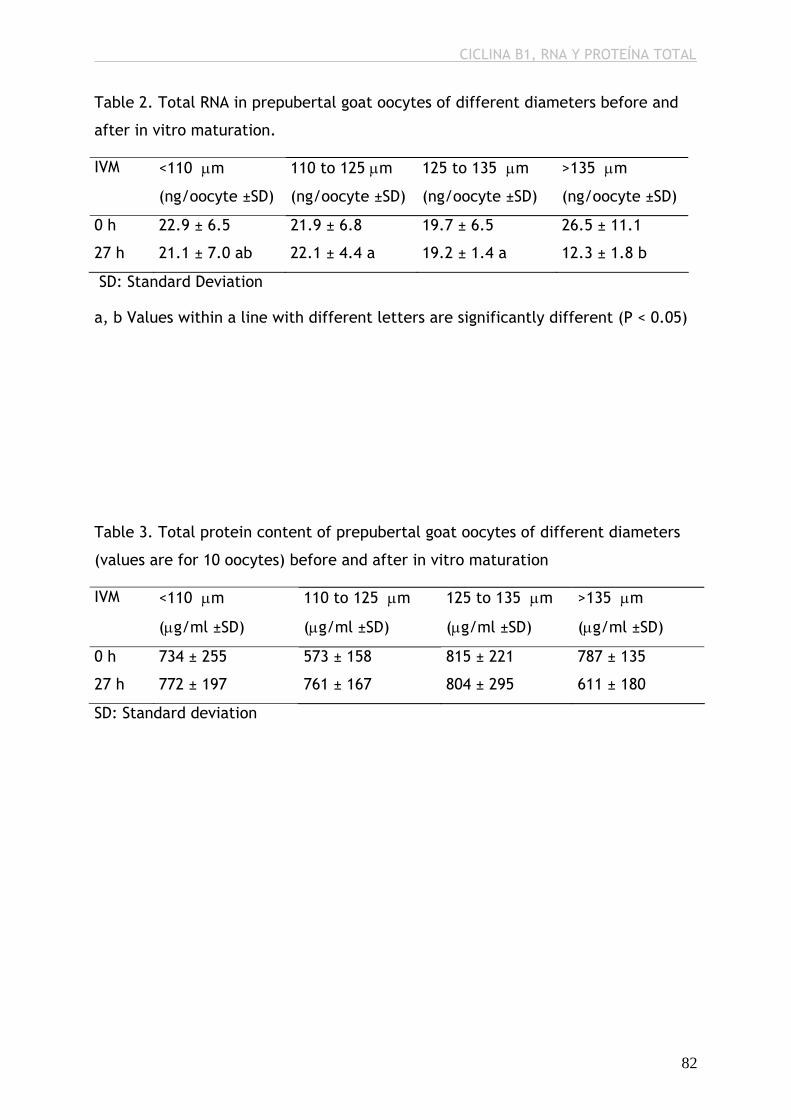
CICLINA B1, RNA Y PROTEÍNA TOTAL
Table 2. Total RNA in prepubertal goat oocytes of different diameters before and
after in vitro maturation.
IVM <110 μm
(ng/oocyte ±SD)
110 to 125 μm
(ng/oocyte ±SD)
125 to 135 μm
(ng/oocyte ±SD)
>135 μm
(ng/oocyte ±SD)
0 h 22.9 ± 6.5 21.9 ± 6.8 19.7 ± 6.5 26.5 ± 11.1
27 h 21.1 ± 7.0 ab 22.1 ± 4.4 a 19.2 ± 1.4 a 12.3 ± 1.8 b
SD: Standard Deviation
a, b Values within a line with different letters are significantly different (P < 0.05)
Table 3. Total protein content of prepubertal goat oocytes of different diameters
(values are for 10 oocytes) before and after in vitro maturation
IVM <110 μm
(μg/ml ±SD)
110 to 125 μm
(μg/ml ±SD)
125 to 135 μm
(μg/ml ±SD)
>135 μm
(μg/ml ±SD)
0 h 734 ± 255 573 ± 158 815 ± 221 787 ± 135
27 h 772 ± 197 761 ± 167 804 ± 295 611 ± 180
SD: Standard deviation
82
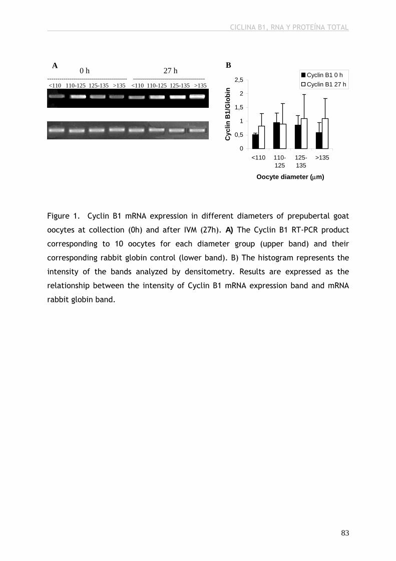
CICLINA B1, RNA Y PROTEÍNA TOTAL
B
0
0,5
1
1,5
2
2,5
<110 110-125
125-135
>135
Oocyte diameter (μm)
Cyc
lin B
1/G
lobi
n
Cyclin B1 0 hCyclin B1 27 h
0 h 27 h ---------------------------------------- ------------------------------------ <110 110-125 125-135 >135 <110 110-125 125-135 >135
A
Figure 1. Cyclin B1 mRNA expression in different diameters of prepubertal goat
oocytes at collection (0h) and after IVM (27h). A) The Cyclin B1 RT-PCR product
corresponding to 10 oocytes for each diameter group (upper band) and their
corresponding rabbit globin control (lower band). B) The histogram represents the
intensity of the bands analyzed by densitometry. Results are expressed as the
relationship between the intensity of Cyclin B1 mRNA expression band and mRNA
rabbit globin band.
83
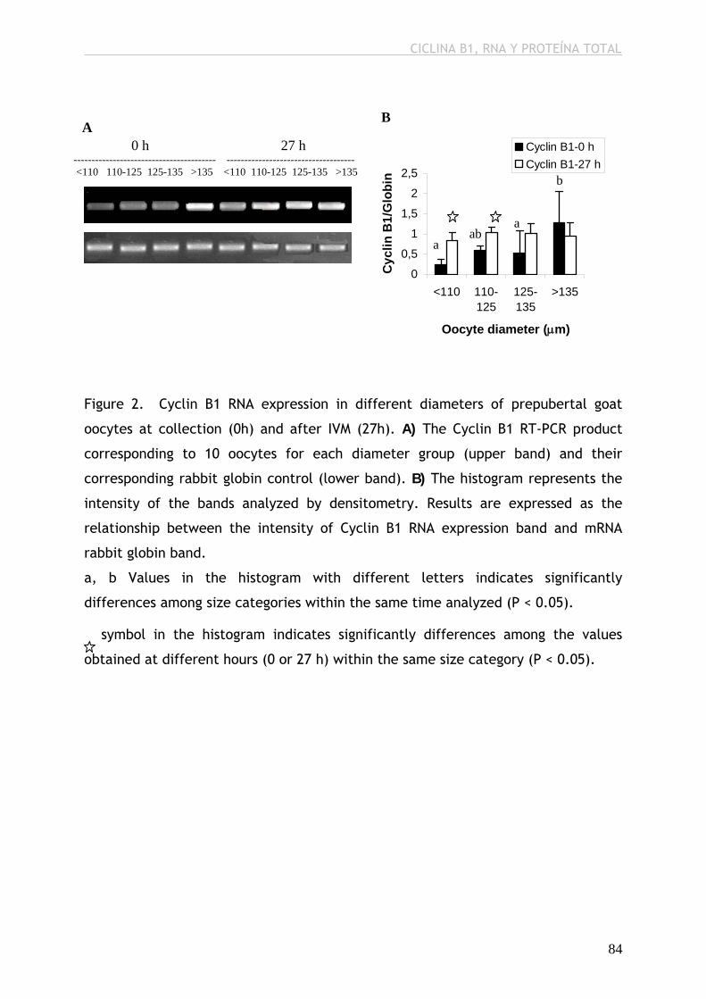
CICLINA B1, RNA Y PROTEÍNA TOTAL
B
A 0 h 27 h ---------------------------------------- ------------------------------------ <110 110-125 125-135 >135 <110 110-125 125-135 >135
0
0,5
1
1,5
2
2,5
<110 110-125
125-135
>135
Oocyte diameter (μm)
Cyc
lin B
1/G
lobi
n
Cyclin B1-0 hCyclin B1-27 h
aba
b
a
Figure 2. Cyclin B1 RNA expression in different diameters of prepubertal goat
oocytes at collection (0h) and after IVM (27h). A) The Cyclin B1 RT-PCR product
corresponding to 10 oocytes for each diameter group (upper band) and their
corresponding rabbit globin control (lower band). B) The histogram represents the
intensity of the bands analyzed by densitometry. Results are expressed as the
relationship between the intensity of Cyclin B1 RNA expression band and mRNA
rabbit globin band.
a, b Values in the histogram with different letters indicates significantly
differences among size categories within the same time analyzed (P < 0.05).
symbol in the histogram indicates significantly differences among the values
obtained at different hours (0 or 27 h) within the same size category (P < 0.05).
84
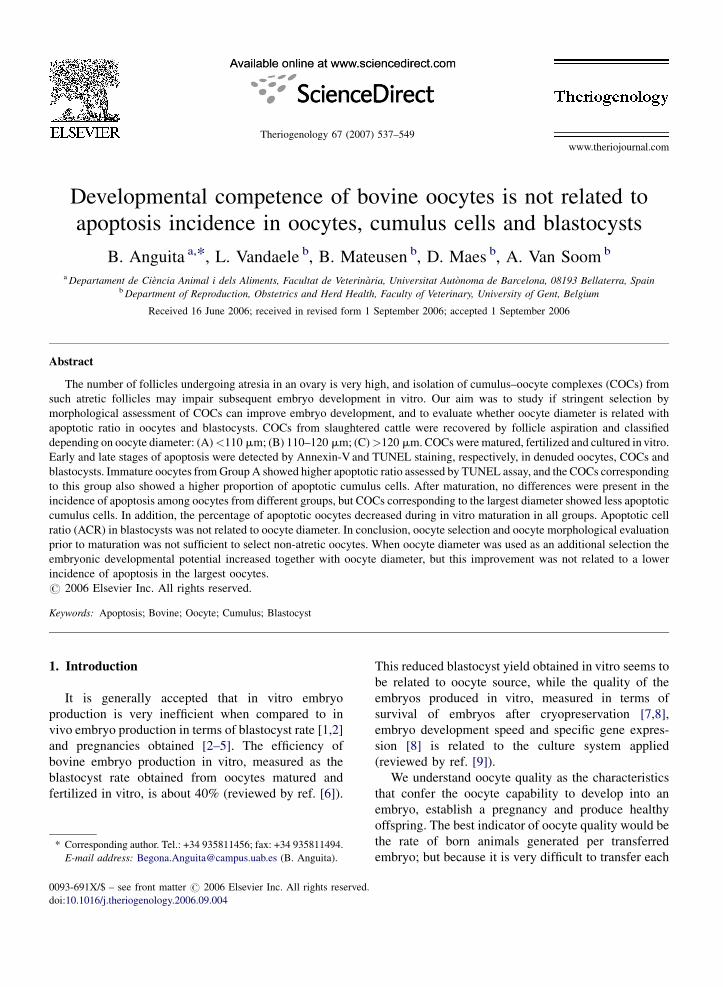
www.theriojournal.com
Theriogenology 67 (2007) 537–549
Developmental competence of bovine oocytes is not related to
apoptosis incidence in oocytes, cumulus cells and blastocysts
B. Anguita a,*, L. Vandaele b, B. Mateusen b, D. Maes b, A. Van Soom b
a Departament de Ciencia Animal i dels Aliments, Facultat de Veterinaria, Universitat Autonoma de Barcelona, 08193 Bellaterra, Spainb Department of Reproduction, Obstetrics and Herd Health, Faculty of Veterinary, University of Gent, Belgium
Received 16 June 2006; received in revised form 1 September 2006; accepted 1 September 2006
Abstract
The number of follicles undergoing atresia in an ovary is very high, and isolation of cumulus–oocyte complexes (COCs) from
such atretic follicles may impair subsequent embryo development in vitro. Our aim was to study if stringent selection by
morphological assessment of COCs can improve embryo development, and to evaluate whether oocyte diameter is related with
apoptotic ratio in oocytes and blastocysts. COCs from slaughtered cattle were recovered by follicle aspiration and classified
depending on oocyte diameter: (A)<110 mm; (B) 110–120 mm; (C)>120 mm. COCs were matured, fertilized and cultured in vitro.
Early and late stages of apoptosis were detected by Annexin-V and TUNEL staining, respectively, in denuded oocytes, COCs and
blastocysts. Immature oocytes from Group A showed higher apoptotic ratio assessed by TUNEL assay, and the COCs corresponding
to this group also showed a higher proportion of apoptotic cumulus cells. After maturation, no differences were present in the
incidence of apoptosis among oocytes from different groups, but COCs corresponding to the largest diameter showed less apoptotic
cumulus cells. In addition, the percentage of apoptotic oocytes decreased during in vitro maturation in all groups. Apoptotic cell
ratio (ACR) in blastocysts was not related to oocyte diameter. In conclusion, oocyte selection and oocyte morphological evaluation
prior to maturation was not sufficient to select non-atretic oocytes. When oocyte diameter was used as an additional selection the
embryonic developmental potential increased together with oocyte diameter, but this improvement was not related to a lower
incidence of apoptosis in the largest oocytes.
# 2006 Elsevier Inc. All rights reserved.
Keywords: Apoptosis; Bovine; Oocyte; Cumulus; Blastocyst
1. Introduction
It is generally accepted that in vitro embryo
production is very inefficient when compared to in
vivo embryo production in terms of blastocyst rate [1,2]
and pregnancies obtained [2–5]. The efficiency of
bovine embryo production in vitro, measured as the
blastocyst rate obtained from oocytes matured and
fertilized in vitro, is about 40% (reviewed by ref. [6]).
* Corresponding author. Tel.: +34 935811456; fax: +34 935811494.
E-mail address: [email protected] (B. Anguita).
0093-691X/$ – see front matter # 2006 Elsevier Inc. All rights reserved.
doi:10.1016/j.theriogenology.2006.09.004
This reduced blastocyst yield obtained in vitro seems to
be related to oocyte source, while the quality of the
embryos produced in vitro, measured in terms of
survival of embryos after cryopreservation [7,8],
embryo development speed and specific gene expres-
sion [8] is related to the culture system applied
(reviewed by ref. [9]).
We understand oocyte quality as the characteristics
that confer the oocyte capability to develop into an
embryo, establish a pregnancy and produce healthy
offspring. The best indicator of oocyte quality would be
the rate of born animals generated per transferred
embryo; but because it is very difficult to transfer each
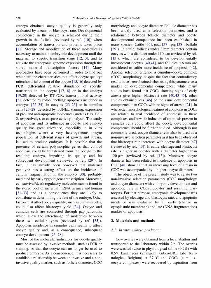
B. Anguita et al. / Theriogenology 67 (2007) 537–549538
embryo obtained, oocyte quality is generally only
evaluated by means of blastocyst rate. Developmental
competence in the oocyte is achieved during their
growth in the follicle (reviewed by ref. [10]) when
accumulation of transcripts and proteins takes place
[11]. Storage and mobilization of these molecules is
necessary to maintain embryonic development until the
maternal to zygotic transition stage [12,13], and to
activate the embryonic genome expression through the
stored maternal transcription factors [14]. Many
approaches have been performed in order to find out
which are the characteristics that affect oocyte quality:
mitochondrial content of the oocyte [15,16] detected by
PCR; differential relative abundance of specific
transcripts in the oocyte [17,18] or in the embryo
[19,20] detected by RT-PCR; transcriptional activity
[21] detected by radio-labelling; apoptosis incidence in
embryos [22–24], in oocytes [23–25] or in cumulus
cells [25–28] detected by TUNEL staining, expression
of pro- and anti-apoptotic molecules (such as Bax, Bcl-
2, respectively), or caspase activity analysis. The study
of apoptosis and its influence in oocyte and embryo
quality has great relevance, especially in in vitro
technologies where a very heterogeneous oocyte
population, at different stages of growth and atresia,
is used to produce embryos. It is possible that the
presence of certain polymorphic genes that control
apoptosis could be transferred from the oocyte to the
resulting embryo, impairing its quality and its
subsequent development (reviewed by ref. [29]). In
fact, it has already been observed that maternal
genotype has a strong effect on the incidence of
cellular fragmentation in the embryo [30], probably
mediated by early zygotic gene transcription. Moreover,
cell survival/death regulatory molecules can be found in
the stored pool of maternal mRNA in mice and human
[31–33] and as a consequence they are likely to
contribute in determining the fate of the embryo. Other
factors that affect oocyte quality, such as cumulus cells,
could also affect blastocyst yield [34]. Oocyte and
cumulus cells are connected through gap junctions,
which allow the interchange of molecules between
these two cellular types (reviewed by ref. [35]).
Apoptosis incidence in cumulus cells seems to affect
oocyte quality and, as a consequence, subsequent
embryo development [25–28].
Most of the molecular indicators for oocyte quality
must be assessed by invasive methods, such as PCR or
staining, so that the oocyte can no longer be used to
produce embryos. As a consequence, it is necessary to
establish a relationship between an invasive and a non-
invasive quality marker, such as follicle diameter, COC
morphology and oocyte diameter. Follicle diameter has
been widely used as a selection parameter, and a
relationship between follicle diameter and oocyte
developmental competence has been established in
many species (Cattle [36]; goat [37]; pig [38]; buffalo
[39]). In cattle, follicles under 3 mm diameter contain
oocytes with a diameter under 110 mm (reviewed by ref.
[13]), which are considered to be developmentally
incompetent oocytes [40,41], and follicles >6 mm are
considered to suffer more incidence of apoptosis [42].
Another selection criterion is cumulus–oocyte complex
(COC) morphology, despite the fact that contradictory
results have been obtained when using this parameter as a
marker of developmental competence: while many
studies have found that COCs showing signs of early
atresia give higher blastocyst rate [40,43–45], other
studies obtained less [46] or the same developmental
competence than COCs with no signs of atresia [21]. In
what extent morphological features of apoptosis in COCs
are related to real incidence of apoptosis in these
complexes, and how the inductors of apoptosis present in
cumulus cells could affect the oocyte developmental
competence should be further studied. Although is not
commonly used, oocyte diameter can also be used as a
non-invasive selection parameter as it has been observed
that blastocyst rate increases with oocyte diameter [47]
(reviewed by ref. [13]). In cattle, cleavage and blastocyst
rate is higher in oocytes with a diameter higher than
120 mm (reviewed by ref. [13]). Moreover, oocyte
diameter has been related to incidence of apoptosis in
COC [48] showing that an increasing level of atresia in
COC was accompanied by a higher oocyte diameter.
The objective of the present study was to relate two
non-invasive selection parameters (COC morphology
and oocyte diameter) with embryonic development and
apoptotic rate in COCs, oocytes and resulting blas-
tocysts. For that purpose, embryonic development was
assessed by cleavage and blastocyst rate, and apoptotic
incidence was evaluated by an early (change in
cytoplasmic membrane) and late (DNA fragmentation)
marker of apoptosis.
2. Materials and methods
2.1. In vitro embryo production
Cow ovaries were obtained from a local abattoir and
transported to the laboratory within 2 h. The ovaries
were washed twice in physiological saline (0.9%) with
0.5% kanamycin (25 mg/ml, Gibco-BRL Life Tech-
nologies, Belgium) at 37 8C and COCs (cumulus–
oocyte complexes) were recovered by aspiration from
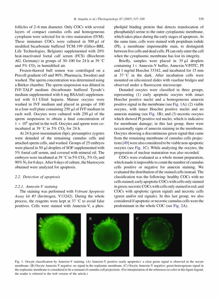
B. Anguita et al. / Theriogenology 67 (2007) 537–549 539
follicles of 2–6 mm diameter. Only COCs with several
layers of compact cumulus cells and homogeneous
cytoplasm were selected for in vitro maturation (IVM).
These immature COCs were cultured in 500 ml of
modified bicarbonate buffered TCM-199 (Gibco-BRL
Life Technologies, Belgium) supplemented with 20%
heat-inactivated foetal calf serum (FCS) (Biochrom
AG, Germany) in groups of 30–100 for 24 h at 39 8Cand 5% CO2 in humidified air.
Frozen-thawed bull semen was centrifuged on a
Percoll gradient (45 and 90%; Pharmacia, Sweden) and
washed. The sperm concentration was determined using
a Burker chamber. The sperm suspension was diluted in
IVF-TALP medium (bicarbonate buffered Tyrode’s
medium supplemented with 6 mg BSA/ml) supplemen-
ted with 0.1 UI/ml heparin. Mature oocytes were
washed in IVF medium and placed in groups of 100
in a four-well plate containing 250 ml of IVF medium in
each well. Oocytes were cultured with 250 ml of the
sperm suspension to obtain a final concentration of
1 � 106 spz/ml in the well. Oocytes and sperm were co-
incubated at 39 8C in 5% CO2 for 24 h.
At 24 h post-insemination (hpi), presumptive zygotes
were denuded of the remaining cumulus cells and
attached sperm cells, and washed. Groups of 25 embryos
were placed in 50 ml droplets of SOF supplemented with
5% foetal calf serum, and covered with mineral oil. The
embryos were incubated at 39 8C in 5% CO2, 5% O2 and
90% N2 for 6 days. After 6 days of culture, the blastocysts
obtained were analyzed for apoptosis.
2.2. Detection of apoptosis
2.2.1. Annexin-V staining
The staining was performed with Vybrant Apoptosis
Assay kit #3 (Invitrogen, V13242). During the whole
process, the reagents were kept at 37 8C to avoid false
positives. Cells were stained with Annexin-V, a phos-
Fig. 1. Oocyte classification by Annexin-V staining: (A) Annexin-V posit
membrane. (B) Oocyte Annexin-V negative: no signal in the ooplasmic mem
the ooplasmic membrane is considered to be a remnant of cumulus cell projec
the reader is referred to the web version of the article.)
pholipid binding protein that detects translocation of
phosphatidyl-serine to the outer cytoplasmic membrane,
which takes place during the early stages of apoptosis. At
the same time, cells were stained with propidium iodide
(PI), a membrane impermeable stain, to distinguish
between live cells and dead cells. PI can only enter the cell
when the cytoplasmic membrane has lost its integrity.
Briefly, samples were placed in 35 ml droplets
containing 1� Annexin-V buffer, Annexin-V/FITC, PI
and 1 mg/ml Hoechst 33342, and incubated for 15 min
at 37 8C in the dark. After incubation cells were
mounted on siliconized slides with vaseline bridges and
observed under a fluorescent microscope.
Denuded oocytes were classified in three groups,
representing (1) early apoptotic oocytes with intact
Hoechst positive nuclei and a homogeneous annexin
positive signal in the membrane (see Fig. 1A); (2) viable
oocytes, with intact Hoechst positive nuclei and no
annexin staining (see Fig. 1B); and (3) necrotic oocytes
which showed PI positive red nuclei, which is indicative
for membrane damage; in this last group, there were
occasionally signs of annexin staining in the membrane.
Oocytes showing a discontinuous green signal that came
from the remaining membrane of cumulus cells projec-
tions [49] were also considered to beviable non-apoptotic
oocytes (see Fig. 1C). While analyzing the oocytes, the
progression of nuclear maturation was also recorded.
COCs were evaluated as a whole mount preparation,
which made it impossible to count the number of cumulus
cells positive or negative for annexin staining. We
evaluated the distribution of the stained cells instead. The
classification was the following: healthy COCs with no
cells stained;earlyapoptotic COCs with cellsonly stained
in green; necrotic COCs with cells only stained in red; and
COCs with apoptotic (green signal) and necrotic cells
(green and/or red signals). In this last group, we also
considered if apoptotic or necrotic cumulus cells were the
predominant in the whole COC (see Fig. 2A).
ive (early apoptotic): a clear green signal is observed in the oocyte
brane. (C) Oocyte Annexin-V negative: green heterogenous signal in
tions. (For interpretation of the references to color in this figure legend,

B. Anguita et al. / Theriogenology 67 (2007) 537–549540
Fig. 2. (A) Annexin-V staining in COC and (B) COC with some TUNEL-positive cells in the outer cumulus layers.
Blastocyst cell number was determined by Hoechst
staining, which let us to divide the blastocyst of each
diameter group in three additional classes: blastocysts
with less than 35 blastomeres; blastocysts containing
35–60 blastomeres; and blastocysts with more than 60
blastomeres. Classification of annexin-stained blasto-
meres was the same as used for oocytes (see Fig. 3).
Results were represented as: EACR (early apoptotic cell
ratio); LACR (late apoptotic cell ratio); and NCR
(necrotic cell ratio).
After evaluation of oocytes and COCs, cells were
washed four times in PVP (1 mg/ml polyvinyl-pyrroli-
done in PBS), fixed in 4% paraformaldehyde overnight
and used for TUNEL staining the day after. Blastocysts to
be used for TUNEL assay were fixed in paraformalde-
hyde without performing Annexin-V staining first to
avoid misinterpretation of the fluorescent signal.
2.2.2. TUNEL assay
Fixed samples were washed twice in 1 mg/ml PVP
and incubated in 0.5% Triton X-100 (in PBS) for 1 h at
room temperature. Afterwards, positive and negative
Fig. 3. Annexin-V staining in blastocysts: (A) Blastocyst with two apopotic
Hoechst.
control samples were incubated with 0.1 U/ml DNAse
for 1 h at 37 8C. After DNAse treatment, the TUNEL
staining was performed with In Situ Cell Death
Detection Kit (Roche, 1684795) following the manu-
facturer’s instruction. The negative control was incu-
bated in the same conditions as the rest of the samples
but without enzyme Terminal deoxynucleotidyl trans-
ferase. Subsequently, samples were treated with 50 mg
RNAse/ml for 1 h at room temperature in the dark, and
stained with 6.25 mg/ml propidium iodide for 15 min
before mounting them on slides. Samples were
evaluated by means of fluorescence microscopy.
Denuded oocytes were classified as: apoptotic, cells
with fragmented nuclei (TUNEL-positive, green) (see
Fig. 4A and B); and non-apoptotic, cells with intact
nuclei (TUNEL-negative, red) (see Fig. 4A and C). At
the same time, the nuclear maturation stage was
recorded.
COCs were evaluated as a whole mount, and the
COCs were classified depending on the distribution of
the TUNEL-positive cells: viable, in COCs with no
apoptotic cells; outer cumulus layers, when apoptotic
and PI positive blastomeres and (B) the same blastocyst stained with
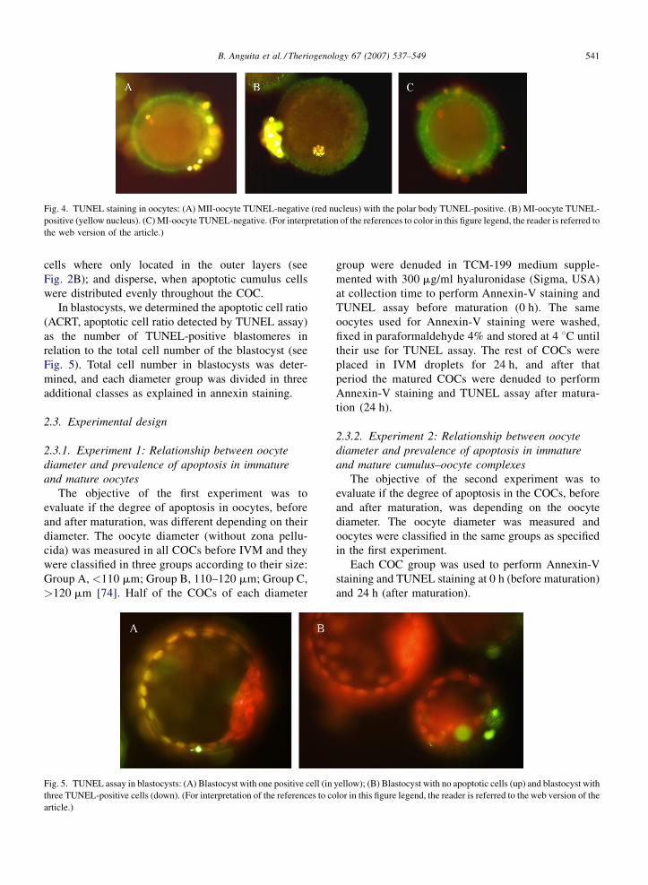
B. Anguita et al. / Theriogenology 67 (2007) 537–549 541
Fig. 4. TUNEL staining in oocytes: (A) MII-oocyte TUNEL-negative (red nucleus) with the polar body TUNEL-positive. (B) MI-oocyte TUNEL-
positive (yellow nucleus). (C) MI-oocyte TUNEL-negative. (For interpretation of the references to color in this figure legend, the reader is referred to
the web version of the article.)
cells where only located in the outer layers (see
Fig. 2B); and disperse, when apoptotic cumulus cells
were distributed evenly throughout the COC.
In blastocysts, we determined the apoptotic cell ratio
(ACRT, apoptotic cell ratio detected by TUNEL assay)
as the number of TUNEL-positive blastomeres in
relation to the total cell number of the blastocyst (see
Fig. 5). Total cell number in blastocysts was deter-
mined, and each diameter group was divided in three
additional classes as explained in annexin staining.
2.3. Experimental design
2.3.1. Experiment 1: Relationship between oocyte
diameter and prevalence of apoptosis in immature
and mature oocytes
The objective of the first experiment was to
evaluate if the degree of apoptosis in oocytes, before
and after maturation, was different depending on their
diameter. The oocyte diameter (without zona pellu-
cida) was measured in all COCs before IVM and they
were classified in three groups according to their size:
Group A, <110 mm; Group B, 110–120 mm; Group C,
>120 mm [74]. Half of the COCs of each diameter
Fig. 5. TUNEL assay in blastocysts: (A) Blastocyst with one positive cell (in
three TUNEL-positive cells (down). (For interpretation of the references to co
article.)
group were denuded in TCM-199 medium supple-
mented with 300 mg/ml hyaluronidase (Sigma, USA)
at collection time to perform Annexin-V staining and
TUNEL assay before maturation (0 h). The same
oocytes used for Annexin-V staining were washed,
fixed in paraformaldehyde 4% and stored at 4 8C until
their use for TUNEL assay. The rest of COCs were
placed in IVM droplets for 24 h, and after that
period the matured COCs were denuded to perform
Annexin-V staining and TUNEL assay after matura-
tion (24 h).
2.3.2. Experiment 2: Relationship between oocyte
diameter and prevalence of apoptosis in immature
and mature cumulus–oocyte complexes
The objective of the second experiment was to
evaluate if the degree of apoptosis in the COCs, before
and after maturation, was depending on the oocyte
diameter. The oocyte diameter was measured and
oocytes were classified in the same groups as specified
in the first experiment.
Each COC group was used to perform Annexin-V
staining and TUNEL staining at 0 h (before maturation)
and 24 h (after maturation).
yellow); (B) Blastocyst with no apoptotic cells (up) and blastocyst with
lor in this figure legend, the reader is referred to the web version of the

B. Anguita et al. / Theriogenology 67 (2007) 537–549542
Fig. 6. Distribution of nuclear stage and TUNEL outcome depending
on oocyte diameter before maturation. GV+ or GVBD+: oocytes at
GV or GVBD nuclear stage, respectively, that were positive for
TUNEL staining. GV� or GVBD�: viable oocytes at GV or GVBD
nuclear stage, respectively.
2.3.3. Experiment 3: Relationship between oocyte
diameter and prevalence of apoptosis in blastocysts
derived from differently sized oocytes
The objective of the third experiment was to study
the blastocyst rate and the degree of apoptosis in
blastocysts obtained from oocytes of different size.
Again, the diameters groups were the same as described
in experiments 1 and 2, but the size was measured
before in vitro culture (IVC) in denuded zygotes.
Cleavage rates were determined at 48 hpi for each
group. At 7 days post-insemination (dpi) half of the
blastocyst of each group were analyzed by annexin
staining, and the other half were fixed in paraformal-
dehyde 4% overnight and used for TUNEL assay the
day after.
2.4. Statistical analysis
Logistic regression analyses, including the effect of
replicate, were performed to compare the proportion of
TUNEL and Annexin-V positive cells in the different
groups of oocytes and COC, and to assess the
developmental competence among diameter groups.
The apoptotic cell ratio in blastocysts, detected by
Annexin-V staining and TUNEL assay in different
diameter groups, was analyzed using analyses of
variance, also including the effect of replicate.
Differences were considered significant when
P < 0.05. All analyses were performed using SPSS
12 for Windows (Analytical Software, Chicago, IL).
3. Results
3.1. Experiment 1: Relationship between oocyte
diameter and prevalence of apoptosis in immature
and mature oocytes
The results of Annexin-V and TUNEL staining in
bovine oocytes are represented in Table 1. When
Table 1
The incidence of early and late apoptosis, as assessed by Annexin-Vand TUN
according to oocyte diameter
Oocyte diameter <110 mm 110–12
Total Apoptotic % Total
Immature Annexin 23 5 21.7 53
Immature TUNEL 11 9 81.8a1 53
Mature Annexin 15 2 13.3 102
Mature TUNEL 23 2 8.72 152
Values in the same row with different superscripts (a,b) differ significantly (P
(1,2) differ significantly (P < 0.05).* One oocyte stained with PI, and thus it can also be considered as necr
Annexin-V staining was performed, we could not detect
any statistical differences in the rate of early apoptotic
oocytes in the three oocyte groups either before or after
maturation. Less apoptosis was observed in mature than
in immature oocytes in Groups B and C when annexin
staining was used. Oocyte nuclear stage had no effect on
the outcome of Annexin-V staining.
When late apoptotic events were assessed, we
observed that the percentage of TUNEL-positive
oocytes was statistically higher in immature oocytes
with a diameter below 110 mm in comparison with
larger oocytes. The apoptotic rate decreased signifi-
cantly during maturation, with only 10.5% or less of
mature oocytes showing signs of late apoptosis and no
statistical differences in rate of apoptotic oocytes
between groups.
The relationship between nuclear stage and late
apoptotic events is represented in Figs. 6 and 7. Most of
the TUNEL-positive immature oocytes were at germ-
inal vesicle breakdown stage (GVBD), and this
proportion was statistically higher in oocytes with a
diameter below 110 mm. In mature oocytes the nuclear
stage had no effect on the TUNEL staining outcome,
and we observed that the proportion of oocytes reaching
metaphase II stage increased with oocyte diameter.
EL staining in immature and in vitro matured bovine oocytes grouped
0 mm >120 mm
Apoptotic % Total Apoptotic %
19 35.91 64 13* 18.81
11 20.8b1 53 9 17.0b
5 4.92 81 2 2.52
11 7.22 124 13 10.5
< 0.05). Values in the same column and assay with different numbers
otic.

B. Anguita et al. / Theriogenology 67 (2007) 537–549 543
Fig. 7. Distribution of nuclear stage and TUNEL outcome depending
on oocyte diameter after maturation. MI+ or MII++: oocytes at MI or
MII nuclear stage, respectively, that were positive for TUNEL stain-
ing. DEG+: degenerate oocytes that were positive for TUNEL stain-
ing. GV�or GVBD�: viable oocytes at GV or GVBD nuclear stage,
respectively.
3.2. Experiment 2: Relationship between oocyte
diameter and prevalence of apoptosis in immature
and mature cumulus–oocyte complexes
The aim of the second experiment was to study if the
apoptosis degree of cumulus–oocytes complexes was
related to oocyte diameter.
When Annexin-V staining was performed, we did
not detect any difference among diameter groups before
maturation. It is important to notice that we observed
positive cells in the COCs of the three groups before and
after maturation. After maturation, a higher percentage
of COCs with early apoptotic cells was detected in
Group A in comparison to the other two groups. At the
same time, after maturation the proportion of COCs
with late apoptotic and/or necrotic cumulus cells was
significantly higher in Groups B and C (Table 2).
When TUNEL assay was performed in immature
COCs, we observed that the percentage of COCs with
apoptotic cells was statistically lower when the oocyte
diameter was 110–120 mm in comparison to the other
groups. Apoptotic cumulus cells were located exclu-
sively in the outer layers (Table 3).
Table 2
Annexin staining in COCs before and after maturation depending on oocy
Annexin
staining
<110 mm 110–120 mm
N Viable EA
(%)
LA/Necrotic
(%)
N Viable
Immature COC 15 0 0 15 (100) 40 0
Mature COC 15 0 3a (20) 12a (80) 61 0
Values in the same row and different diameter and classification group, with d
complexes evaluated; EA, early atretic; LA, late atretic.
During IVM, there was a significant increase in the
rate of COCs with apoptotic cells in Group B. We could
also observe that the distribution of the apoptotic cells in
the COCs varied during IVM: before IVM they were
almost only located in the outer cumulus cells (see
Fig. 2B), while after IVM, we observed a significant
increase in positive cells distributed in a dispersed way
in the COC in the two biggest groups (Table 3).
After maturation, there were no differences detected
among diameter groups. The percentage of COCs
showing no apoptotic cumulus cells was higher in the
largest group, but it was not statistically different
(Table 3).
3.3. Experiment 3: Relationship between oocyte
diameter and prevalence of apoptosis in blastocysts
derived from differently sized oocytes
Cleavage rate in embryos originating from oocytes
with the smallest diameter was 50%, which was
significantly lower than for embryos in the other
groups (Table 4). Furthermore, blastocyst rate was
significantly higher in Groups B and C when compared
to the smallest group (<110 mm: 9.18%; 110–120:
21.20%; >120: 23.97%) (Table 4). Relation blastocyst
rate/cleavage was also higher in the embryos coming
from oocytes with the largest diameters (18.36% in
Group A versus 36.63% and 40.81% in Groups B and C,
respectively) (Table 4). Moreover, none of the
blastocysts in Group A were hatched at 7 dpi.
Blastocysts from the Group C showed significantly
less late apoptotic and necrotic cells than Group B when
annexin staining was performed (late apoptotic: Group
B, 3.75%; Group C, 1.57%; Necrotic: Group B, 1.33%;
Group C, 0.22%) (Table 5). We detected no differences
in ACRT among diameter groups when TUNEL assay
was performed (Group A, 12.37%; Group B, 12.28%;
Group C, 12.54%).
We also evaluated the apoptosis rate among the
different groups depending on blastocyst cell number
(Table 6). We detected significant differences among
te diameter
>120 mm
EA
(%)
LA/Necrotic
(%)
N Viable EA
(%)
LA/Necrotic
(%)
2 (5) 38 (95) 43 0 3 (6.98) 40 (93.02)
2b (3.28) 59b (96.72) 60 0 0b 60b (100)
ifferent superscripts (a,b or a,b) differ significantly (P < 0.05). N, total
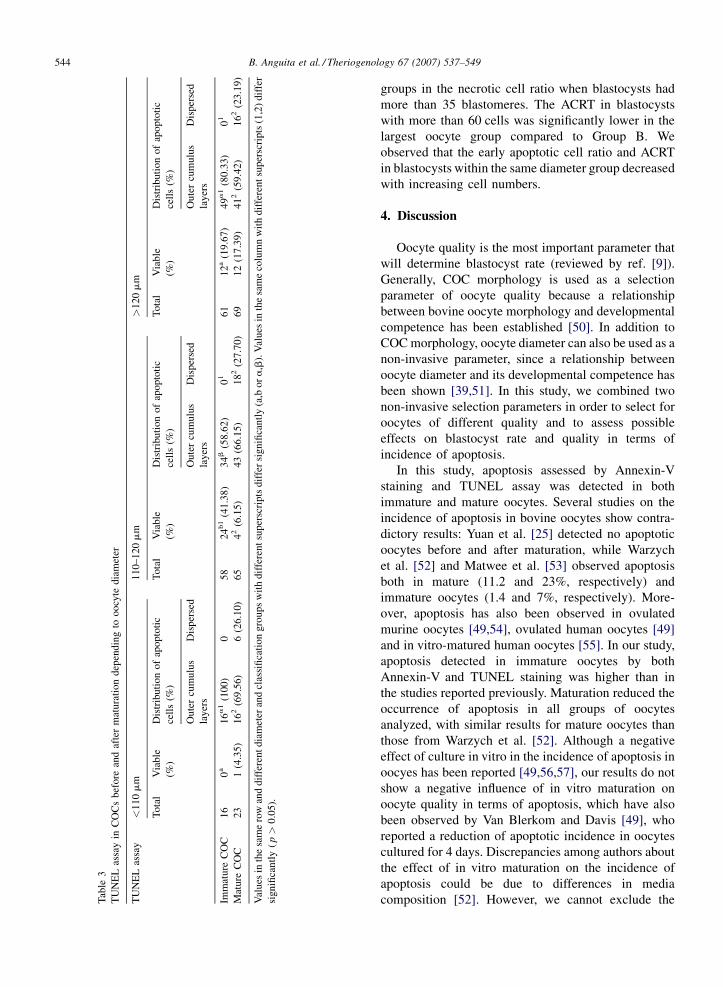
B. Anguita et al. / Theriogenology 67 (2007) 537–549544
Tab
le3
TU
NE
Las
say
inC
OC
sbef
ore
and
afte
rm
atura
tion
dep
endin
gto
oocy
tedia
met
er
TU
NE
Las
say
<1
10
mm
11
0–
12
0m
m>
12
0m
m
To
tal
Via
ble
(%)
Dis
trib
uti
on
of
apo
pto
tic
cell
s(%
)
To
tal
Via
ble
(%)
Dis
trib
uti
on
of
apo
pto
tic
cell
s(%
)
To
tal
Via
ble
(%)
Dis
trib
uti
on
of
apo
pto
tic
cell
s(%
)
Ou
ter
cum
ulu
s
lay
ers
Dis
per
sed
Ou
ter
cum
ulu
s
lay
ers
Dis
per
sed
Oute
rcu
mulu
s
lay
ers
Dis
per
sed
Imm
atu
reC
OC
16
0a
16
a1
(10
0)
05
82
4b1
(41
.38
)3
4b
(58
.62
)0
16
11
2a
(19
.67)
49
a1
(80
.33)
01
Mat
ure
CO
C2
31
(4.3
5)
16
2(6
9.5
6)
6(2
6.1
0)
65
42
(6.1
5)
43
(66
.15)
18
2(2
7.7
0)
69
12
(17
.39
)4
12
(59
.42
)1
62
(23
.19
)
Val
ues
inth
esa
me
row
and
dif
fere
nt
dia
met
eran
dcl
assi
fica
tio
ng
rou
ps
wit
hd
iffe
ren
tsu
per
scri
pts
dif
fer
sig
nifi
can
tly
(a,b
or
a,b
).V
alu
esin
the
sam
eco
lum
nw
ith
dif
fere
nt
sup
ersc
rip
ts(1
,2)
dif
fer
sign
ifica
ntl
y(p>
0.0
5).
groups in the necrotic cell ratio when blastocysts had
more than 35 blastomeres. The ACRT in blastocysts
with more than 60 cells was significantly lower in the
largest oocyte group compared to Group B. We
observed that the early apoptotic cell ratio and ACRT
in blastocysts within the same diameter group decreased
with increasing cell numbers.
4. Discussion
Oocyte quality is the most important parameter that
will determine blastocyst rate (reviewed by ref. [9]).
Generally, COC morphology is used as a selection
parameter of oocyte quality because a relationship
between bovine oocyte morphology and developmental
competence has been established [50]. In addition to
COC morphology, oocyte diameter can also be used as a
non-invasive parameter, since a relationship between
oocyte diameter and its developmental competence has
been shown [39,51]. In this study, we combined two
non-invasive selection parameters in order to select for
oocytes of different quality and to assess possible
effects on blastocyst rate and quality in terms of
incidence of apoptosis.
In this study, apoptosis assessed by Annexin-V
staining and TUNEL assay was detected in both
immature and mature oocytes. Several studies on the
incidence of apoptosis in bovine oocytes show contra-
dictory results: Yuan et al. [25] detected no apoptotic
oocytes before and after maturation, while Warzych
et al. [52] and Matwee et al. [53] observed apoptosis
both in mature (11.2 and 23%, respectively) and
immature oocytes (1.4 and 7%, respectively). More-
over, apoptosis has also been observed in ovulated
murine oocytes [49,54], ovulated human oocytes [49]
and in vitro-matured human oocytes [55]. In our study,
apoptosis detected in immature oocytes by both
Annexin-V and TUNEL staining was higher than in
the studies reported previously. Maturation reduced the
occurrence of apoptosis in all groups of oocytes
analyzed, with similar results for mature oocytes than
those from Warzych et al. [52]. Although a negative
effect of culture in vitro in the incidence of apoptosis in
oocyes has been reported [49,56,57], our results do not
show a negative influence of in vitro maturation on
oocyte quality in terms of apoptosis, which have also
been observed by Van Blerkom and Davis [49], who
reported a reduction of apoptotic incidence in oocytes
cultured for 4 days. Discrepancies among authors about
the effect of in vitro maturation on the incidence of
apoptosis could be due to differences in media
composition [52]. However, we cannot exclude the

B. Anguita et al. / Theriogenology 67 (2007) 537–549 545
Table 4
Embryos developing from oocytes with different diameters
Oocyte diameter N Cleavage at
4 dpi (%)
Hatched blastocysts
at 7 dpi (%)
Total blastocyst
rate (%)
Blastocyst/cleavage
(%)
<110 mm 294 147 (50)a 0 27 (9.18)a 18.36a
110–120 mm 1047 606 (57.88)b 3 (0.29) 222 (21.20)b 36.63b
>120 mm 584 343 (58.73)b 4 (0.68) 140 (23.97)b 40.81b
Values in the same column with different superscripts (a,b) differ significantly (P < 0.05).
Table 5
Detection of apoptosis in day 6 blastocysts from oocytes of different diameter
Oocyte, Ø (mm) Annexin-V staining TUNEL assay
Blastocysts
evaluated
EACR
(mean � S.D.)
LACR
(mean � S.D.)
NCR
(mean � S.D.)
Blastocysts
evaluated
Apoptotic cell ratio
(mean � S.D.)
<110 mm 13 6.49 � 3.84 3.36 � 6.34a,b 1.65 � 2.82a,b 11 12.37 � 7.23
110–120 mm 75 9.74 � 8.00 3.75 � 5.66a 1.33 � 2.37a 67 12.28 � 16.35
>120 mm 45 8.56 � 7.87 1.57 � 2.61b 0.22 � 0.86b 56 12.54 � 8.59
Values in the same column with different superscripts (a,b) differ significantly (P < 0.05). EACR, early apoptotic cell ratio; LACR, late apoptotic
cell ratio; NCR, necrotic cell ratio.
possibility that manipulation may have induced an early
stage of atresia in our immature oocytes, which would
be reverted by our in vitro culture conditions.
Differences in the incidence of apoptosis in oocytes
among the diameter groups studied was only observed
in immature oocytes, and the smallest oocytes showed
the highest rates of late apoptosis. After in vitro
maturation, no differences were detected among groups.
These results could indicate: (1) the quality of the
smallest oocytes, as assessed by apoptosis detection, is
Table 6
Detection of apoptosis in day 6 blastocysts from oocytes of different diam
Oocyte, Ø Annexin-V staining
Blastocysts
evaluated
Early apoptotic cell
ratio (mean � S.D.)
Late apoptotic
ratio (mean � S
<35 cells
<110 mm 0 0 0
110–120 mm 9 19.95 � 6.311 8.86 � 9.131
>120 mm 7 18.79 � 8.382 2.99 � 4.54
35–60 cells
<110 mm 7 7.09 � 3.60 5.09 � 8.45
110–120 mm 31 11.40 � 8.112 4.34 � 5.931,2
>120 mm 13 10.90 � 8.651 1.92 � 2.67
>60 cells
<110 mm 6 5.78 � 4.33 1.34 � 1.26
110–120 mm 35 5.65 � 4.953 1.91 � 2.892
>120 mm 25 4.49 � 2.812 1.00 � 1.67
Values in the same column and blastocyst cell class (<35 cells, 35–60 cells
(P < 0.05). Values in the same column and diameter group (<110 mm, 1
significantly (P < 0.05).
lower than the largest oocytes, which would be reflected
in the low blastocyst yield obtained from these oocytes;
or (2) the smallest oocytes have not yet finished their
growth period, and may be they are more sensitive than
the largest oocytes to manipulation.
We detected an effect of nuclear stage on the
incidence of late apoptotic events in immature oocytes:
most of the apoptotic oocytes had already resumed
meiosis (<110 mm: 88.88%; 110–120 mm: 90.91%;
>120 mm: 77.77%), while the proportion of oocytes at
eter depending on blastocyst cell number
TUNEL assay
cell
.D.)
Necrotic cell
ratio (mean � S.D.)
Blastocysts
evaluated
Apoptotic cell
ratio (mean � S.D.)
0 0
2.01 � 4.00 8 37.34 � 31.101
0.71 � 1.88 6 19.68 � 14.581
2.45 � 3.62a 4 19.68 � 5.531
1.43 � 2.35a,b 12 16.27 � 16.992
0.18 � 0.66b 11 14.62 � 9.381,2
0.72 � 1.21a,b 7 8.20 � 3.99a,b2
1.07 � 1.83a 47 7.00 � 5.04a2
0.11 � 0.40b 39 10.68 � 6.60b2
or >60 cells) and with different superscripts (a,b) differ significantly
10–120 mm, >120 mm) and with different superscripts (1,2) differ
B. Anguita et al. / Theriogenology 67 (2007) 537–549546
GVBD stage decreased in the non-apoptotic ones
(<110 mm: 50%; 110–120 mm: 57.14%; >120 mm:
72.72%). No immature oocyte beyond GVBD was
detected. These results would be in accordance with
Warzych et al [52] who detected that the apoptotic rate
was higher in ‘‘immature’’ oocytes that had reached MI
or MII (50%), than those that still were at GV (0.70%). It
has been suggested that apoptosis is a mechanism to
eliminate oocytes that have resumed meiosis inside the
follicle. Therefore, apoptosis in our immature oocytes
could be a signal that they come from follicles that could
not maintain meiotic arrest in those oocytes. In which
way this premature meiotic resumption could affect
blastocyst rate should be further studied, although it has
been suggested that oocytes showing early signs of
atresia suffer a phenomenon similar to maturation [58],
which confer higher embryonic developmental compe-
tence [40]. de Wit et al. [45] also found that COC showing
morphological characteristics of early stage of atresia
seemed to mature in vitro faster than healthy COCs,
because of a premature oocyte-cumulus uncoupling.
Several studies have concluded that the detection of
the incidence of apoptosis in COCs could be a good
indicator of oocyte developmental competence
[46,59,60]. Cumulus cells and oocytes communicate
in a bidirectional way through gap junctions [61].
Cumulus cells play an important role in maintaining
oocyte meiotic arrest, support oocyte cytoplasmic
maturation and participate in the induction of meiotic
resumption (reviewed by ref. [35]). In addition,
presence of cumulus cells during in vitro maturation
is essential to improve blastocyst rate [34], maybe due
to the protective effect of the cumulus cells against
oxidative stress that could induce apoptosis in the
oocytes [62]. Our second experiment tried to establish if
differences in blastocyst rate among oocyte diameters
could be related to the incidence of apoptosis in the
corresponding cumulus cells. At the time of collection,
we observed that most of the apoptotic cumulus cells
were located in the outer layers, which is in accordance
with the results of Yuan et al. [46]. The absence of
apoptotic cumulus cells in the corona of the COC is
explained by the anti-apoptotic effect of a gradient of
paracrine factors secreted by the oocyte, probably
BMP15 (Bone morphogenetic protein-15) and BMP6
[63]. A differential distribution of the late apoptotic
cumulus cells detected by TUNEL before and after IVM
was observed, indicating that they distribute in a more
dispersed way when in vitro maturation progresses.
Moreover, after maturation there was an increase in the
percentage of COCs showing late apoptotic cumulus
cells in Group B. Yuan et al. [46] and Ikeda et al. [64]
obtained similar results and they detected an increase of
apoptotic cumulus cells after maturation, detected by
TUNEL and ligation mediated-PCR, respectively.
Annexin-V assay in COCs revealed that, after
maturation, the percentage of COCs showing late
apoptotic cells was higher in Groups B and C than in
Group A, which had more COCs with early apoptotic
cumulus cells. These results could also indicate a
deficiency in cumulus expansion and maturation in
COCs containing the smallest oocytes. In fact, it was also
observed that only 43.47% of the smallest oocytes were
able to reach metaphase II after in vitro maturation, while
the percentage of MII oocytes increased up to 78.94 and
81.45% in Groups B and C, respectively.
Apoptosis incidence is related to embryo quality [65].
Apoptosis plays a very important role during pre- and
post-implantation development, removing abnormal
cells or cells that are no longer required and controlling
the embryo cell number. The protective role of apoptosis
in the pre-implantation period can turn into damage if the
incidence in the embryo is very high. However, a
detrimental threshold of apoptosis incidence has not been
established yet, and therefore consequences of apoptotic
incidence in blastocysts are difficult to assess. Our results
of apoptosis rate in bovine blastocysts show no
differences in ACR among diameter groups when
TUNEL assay was performed (average about 12%).
Other studies using TUNEL assay in in vitro produced
blastocysts showed an average apototic index of either
10.3% [66], 4–9% [67], 12% [68] and 9.8–14.2% [69],
which is very similar to our results. In addition to TUNEL
assay, Annexin-V staining was also performed to analyze
apoptosis in blastocysts, and we observed a reduction of
late apoptotic cell ratio and necrotic cell ratio in
blastocysts of Group C. Apoptosis incidence, although
lower than in blastocysts produced in vitro, has been also
observed in in vivo bovine, porcine, rodent [67], primate
and human blastocysts [70]. Therefore, it is difficult to
determine in what extent the apoptosis detected in our
blastocysts can affect their quality. However, as we did
not detect any difference in developmental competence
among Groups B and C, we can establish that the
differential incidence of late stage of apoptosis and
necrosis detected in blastocysts derived from each
diameter group seems to have no effect on the blastocyst
rate.
The incidence of apoptosis is variable during embryo
development: it has been reported that it appears after
compaction in human embryos, and varies during
progression of blastocyst stage [71], while in murine
blastocysts there is a wave of death cell during blastocyst
expansion and minimal levels of apoptosis incidence are

B. Anguita et al. / Theriogenology 67 (2007) 537–549 547
found in the late blastocyst stage, before implantation
[67,72]. We separated our blastocysts in three additional
groups depending on their cell number to study if
apoptosis in bovine blastocysts varies depending on the
stage too. In most cases, we observed a decrease in ACR
as the cell number increased, both with Annexin-V and
TUNEL assay, in all diameter groups. This decrease in
apoptosis incidence is explained by the increase of
blastocyst cell number rather than a reduction of the total
apoptotic blastomeres in the blastocyst.
Our results of developmental competence indicate
that cleavage and blastocyst rate increased with oocyte
diameter, as shown in previous studies [39,51].
Considering our results on the incidence of apoptosis
in oocytes, cumulus cells and blastocysts, it is difficult
to find a clear relationship between the parameters
analyzed and developmental competence of the oocyte.
Although we found a higher apoptosis rate in the
smallest oocytes at the time of collection time, the
reduction found after maturation may indicate that it is
not the main cause of the reduced developmental
competence of these oocytes. On the other hand, we
observed more apoptosis in cumulus cells of the
smallest oocytes at collection time, which could be
indicative for the low developmental potential of these
oocytes. However, it is difficult to affirm that it is the
only cause of the reduced blastocyst rate achieved in
these oocytes. It is accepted that small oocytes have less
developmental competence due to an insufficient
cytoplasmic maturation, and they normally do not
reach the blastocyst stage because of a lack of
accumulation of RNA and proteins, necessary to
maintain embryonic development beyond the embryo-
nic genome activation [12], which takes place in bovine
embryos at the 8–16 cell stage [73]. Therefore, it seems
more probable that the reduced developmental compe-
tence of the smallest oocytes is due to the fact that they
have not yet finished their growth rather than a higher
incidence of apoptosis.
In conclusion, oocyte selection by COC morphology
is not enough to select only non-apoptotic COCs. The use
of oocyte diameter as an additional selection parameter
helps to select the most competent oocytes, but this
developmental competence is not related to a difference
in incidence of apoptosis among oocyte diameters.
Acknowledgements
This work was supported by a grant from Generalitat
de Catalunya (2005BE00053). The authors are very
grateful to Johanna Mestach and Griet Spaepen for their
valuable technical assistance.
References
[1] Rizos D, Ward F, Duffy P, Boland MP, Lonergan P. Conse-
quences of bovine oocyte maturation, fertilization or early
embryo development in vitro versus in vivo: implications for
blastocyst yield and blastocyst quality. Mol Reprod Dev 2002;
61:234–48.
[2] Cognie Y, Baril G, Poulin N, Mermillod P. Current status of
embryo technologies in sheep and goat. Theriogenology 2003;
59:171–88.
[3] Galli C, Lazzari G. Practical aspects of IVM/IVF in cattle. Anim
Reprod Sci 1996;42:371–9.
[4] Van Wagtendonk-de Leeuw AM, Mullaart E, De Roos APW,
Merton JS, Den Daas JHG, Kemp B, et al. Effects of different
reproduction techniques: AI, MOET or IVP, on health
and welfare of bovine offspring. Theriogenology 2000;53:
575–97.
[5] Peterson AJ, Lee RS. Improving successful pregnancies after
embryo transfer. Theriogenology 2003;59:687–97.
[6] Lonergan P, Rizos D, Gutierrez-Adan A, Fair T, Boland MP.
Oocyte and embryo quality: effect of origin, culture conditions
and gene expression patterns. Reprod Domest Anim 2003;38:
259–67.
[7] Enright BP, Lonergan P, Dinnyes A, Fair T, Ward FA, Yang X,
et al. Culture of in vitro produced bovine zygotes in vitro vs. in
vivo: implications for early embryo development and quality.
Theriogenology 2000;54:659–73.
[8] Rizos D, Gutierrez-Adan A, Perez-Garnelo S, De La Fuente J,
Boland MP, Lonergan P. Bovine embryo culture in the presence
or absence of serum: implications for blastocyst development,
cryotolerance, and messenger RNA expression. Biol Reprod
2003;68:236–43.
[9] Lonergan P, Rizos D, Ward F, Boland MP. Factors influencing
oocyte and embryo quality in cattle. Reprod Nutr Dev 2001;41:
427–37.
[10] Sirard MA. Resumption of meiosis: mechanism involved in
meiotic progression and its relation with developmental compe-
tence. Theriogenology 2001;55:1241–54.
[11] Crozet N, Motlik J, Szollosi D. Nucleolar fine structure and RNA
synthesis in porcine oocytes during early stages of antrum
formation. Biol Cell 1981;41:35–42.
[12] Bachvarova RF. A maternal tail of poly(A): the long and the short
of it. Cell 1992;69:895–7.
[13] Gandolfi TA, Gandolfi F. The maternal legacy to the embryo:
cytoplasmic components and their effects on early development.
Theriogenology 2001;55:1255–76.
[14] Vigneault C, McGraw S, Massicotte L, Sirard MA. Transcription
factor expression patterns in bovine in vitro-derived embryos
prior to maternal-zygotic transition. Biol Reprod 2004;70:
1701–9.
[15] Tamassia M, Nuttinck F, May-Panloup P, Reynier P, Heyman Y,
Charpigny G, et al. In vitro embryo production efficiency in
cattle and its association with oocyte adenosine triphosphate
content, quantity of mitochondrial DNA, and mitochondrial
DNA haplogroup. Biol Reprod 2004;71:697–704.
[16] Santos TA, El Shourbagy S, St John JC. Mitochondrial content
reflects oocyte variability and fertilization outcome. Fertil Steril
2006;85:584–91.
[17] De Sousa PA, Westhusin ME, Watson AJ. Analysis of variation
in relative mRNA abundance for specific gene transcripts in
single bovine oocytes and early embryos. Mol Reprod Dev 1998;
49:119–30.
B. Anguita et al. / Theriogenology 67 (2007) 537–549548
[18] Watson AJ, De Sousa P, Caveney A, Barcroft LC, Natale D,
Urquhart J, et al. Impact of bovine oocyte maturation media on
oocyte transcript levels, blastocyst development, cell number,
and apoptosis. Biol Reprod 2000;62:355–64.
[19] Gutierrez-Adan A, Rizos D, Fair T, Moreira PN, Pintado B, de la
Fuente J, et al. Effect of speed of development on mRNA
expression pattern in early bovine embryos cultured in vivo or
in vitro. Mol Reprod Dev 2004;68:441–8.
[20] Dode MA, Dufort I, Massicotte L, Sirard MA. Quantitative
expression of candidate genes for developmental competence
in bovine two-cell embryos. Mol Reprod Dev 2006;73:288–97.
[21] Bilodeau-Goeseels S, Panich P. Effects of oocyte quality on
development and transcriptional activity in early bovine
embryos. Anim Reprod Sci 2002;71:143–55.
[22] Pomar FJ Rubio, Teerds KJ, Kidson A, Colenbrander B, Thar-
asanit T, Aguilar B, et al. Differences in the incidence of
apoptosis between in vivo and in vitro produced blastocysts
of farm animal species: a comparative study. Theriogenology
2005;63:2254–68.
[23] Yang MY, Rajamahendran R. Expression of Bcl-2 and Bax
proteins in relation to quality of bovine oocytes and embryos
produced in vitro. Anim Reprod Sci 2002;70:159–69.
[24] Liu HC, He ZY, Mele CA, Veeck LL, Davis O, Rosenwaks Z.
Expression of apoptosis-related genes in human oocytes and
embryos. J Assist Reprod Genet 2000;17:521–33.
[25] Yuan YQ, Van Soom A, Leroy JL, Dewulf J, Van Zeveren A, de
Kruif A, et al. Apoptosis in cumulus cells, but not in oocytes,
may influence bovine embryonic developmental competence.
Theriogenology 2005;63:2147–63.
[26] Lee KS, Joo BS, Na YJ, Yoon MS, Choi OH, Kim WW. Cumulus
cells apoptosis as an indicator to predict the quality of oocytes
and the outcome of IVF-ET. J Assist Reprod Genet 2001;18:
490–8.
[27] Zeuner A, Muller K, Reguszynski K, Jewgenow K. Apoptosis
within bovine follicular cells and its effect on oocyte develop-
ment during in vitro maturation. Theriogenology 2003;59:
1421–33.
[28] Corn CM, Hauser-Kronberger C, Moser M, Tews G, Ebner T.
Predictive value of cumulus cell apoptosis with regard to blas-
tocyst development of corresponding gametes. Fertil Steril
2005;84:627–33.
[29] Hansen PJ. Realizing the promise of IVF in cattle—an overview.
Theriogenology 2006;65:119–25.
[30] Han Z, Chung YG, Gao S, Latham KE. Maternal factors con-
trolling blastomere fragmentation in early mouse embryos. Biol
Reprod 2005;72:612–8.
[31] Jurisicova A, Latham KE, Casper RF, Varmuza SL. Expression
and regulation of genes associated with cell death during murine
preimplantation embryo development. Mol Reprod Dev
1998;51:243–53.
[32] Exley GE, Tang C, McElhinny AS, Warner CM. Expression of
caspase and BCL-2 apoptotic family members in mouse pre-
implantation embryos. Biol Reprod 1999;61:231–9.
[33] Metcalfe AD, Hunter HR, Bloor DJ, Lieberman BA, Picton HM,
Leese HJ, et al. Expression of 11 members of the BCL-2 family
of apoptosis regulatory molecules during human preimplantation
embryo development and fragmentation. Mol Reprod Dev
2004;68:35–50.
[34] Hashimoto S, Saeki K, Nagao Y, Minami N, Yamada M, Utsumi
K. Effects of cumulus cell density during in vitro maturation of
the developmental competence of bovine oocytes. Theriogenol-
ogy 1998;49:1451–63.
[35] Tanghe S, Van Soom A, Nauwynck H, Coryn M, de Kruif A.
Minireview: functions of the cumulus oophorus during oocyte
maturation, ovulation, and fertilization. Mol Reprod Dev
2002;61:414–24.
[36] Furher F, Matr B, Schellander K, Kalat M, Schleger W. Matura-
tion competence and chromatin behaviour in growing and fully
grown cattle oocytes. J Vet Med 1989;36:285–91.
[37] Crozet N, Ahmed-Ali M, Dubos MP. Developmental compe-
tence of goat oocytes from follicles of different size categories
following maturation, fertilization and culture in vitro. J Reprod
Fertil 1995;103:293–8.
[38] Marchal R, Vigneron C, Perreau C, Bali-Papp A, Mermillod P.
Effect of follicular size on meiotic and developmental compe-
tence of porcine oocytes. Theriogenology 2002;57:1523–32.
[39] Raghu HM, Nandi S, Reddy SM. Follicle size and oocyte
diameter in relation to developmental competence of buffalo
oocytes in vitro. Reprod Fertil Dev 2002;14:55–61.
[40] Blondin P, Sirard MA. Oocyte and follicular morphology as
determining characteristics for developmental competence in
bovine oocytes. Mol Reprod Dev 1995;41:54–62.
[41] Hendriksen PJ, Vos PL, Steenweg WN, Bevers MM, Dieleman
SJ. Bovine follicular development and its effect on the in vitro
competence of oocytes. Theriogenology 2000;53:11–20.
[42] Jewgenow K, Heerdegen B, Muller K. In vitro development of
individually matured bovine oocytes in relation to follicular wall
atresia. Theriogenology 1999;51:745–56.
[43] Boni R, Cuomo A, Tosti E. Developmental potential in bovine
oocytes is related to cumulus–oocyte complex grade, calcium
current activity, and calcium stores. Biol Reprod 2002;66:
836–42.
[44] Nicholas B, Alberio R, Fouladi-Nashta AA, Webb R. Relation-
ship between low-molecular-weight insulin-like growth factor-
binding proteins, caspase-3 activity, and oocyte quality. Biol
Reprod 2005;72:796–804.
[45] de Wit AA, Wurth YA, Kruip TA. Effect of ovarian phase and
follicle quality on morphology and developmental capacity of
the bovine cumulus–oocyte complex. J Anim Sci 2000;78:
1277–83.
[46] Yuan Y, Van Soom A, Leroy JLMN, Dewulf J, Van Zeveren A, de
Kruif A, et al. Apoptosis in cumulus cells, but not in oocytes,
may influence bovine embryonic developmental competence.
Theriogenology 2005;63:2147–63.
[47] Pujol M, Lopez-Bejar M, Paramio MT. Developmental compe-
tence of heifer oocytes selected using the brilliant cresyl blue
(BCB) test. Theriogenology 2004;61:735–44.
[48] De Wit AAC, Kruip TAM. Bovine cumulus–oocyte-complex-
quality is reflected in sensitivity for a-amanitin, oocyte-diameter
and developmental capacity. Anim Reprod Sci 2001;65:51–65.
[49] Van Blerkom J, Davis PW. DNA strand breaks and phosphati-
dylserine redistribution in newly ovulated and cultured mouse
and human oocytes: occurrence and relationship to apoptosis.
Hum Reprod 1998;13:1317–24.
[50] Khurana NK, Niemann H. Effects of oocyte quality, oxigen
tension, embryo density, cumulus cells and energy substrates on
cleavage and morula/blastocyst formation of bovine embryos.
Theriogenology 2000;54:741–56.
[51] Arlotto T, Schwartz JL, First NL, Leibfried-Rutledge ML.
Aspects of follicle and oocyte stage that affect in vitro maturation
and development of bovine oocytes. Theriogenology 1996;45:
943–56.
[52] Warzych E, Peippo J, Szydlowski M, Lechniak D. Supplements
to in vitro maturation media affect the production of bovine

B. Anguita et al. / Theriogenology 67 (2007) 537–549 549
blastocysts and their apoptotic index but not the proportions of
matured and apoptotic oocytes. Anim Reprod Sci 2006.
[53] Matwee C, Betts DH, King WA. Apoptosis in the early bovine
embryo. Zygote 2000;8:57–68.
[54] Perez GI, Tao XJ, Tilly JL. Fragmentation and death (a.k.a.
apoptosis) of ovulated oocytes. Mol Hum Reprod 1999;5:
414–20.
[55] Wu J, Zhang L, Wang X. Maturation and apoptosis of human
oocytes in vitro are age-related. Fertil Steril 2000;74:1137–41.
[56] Takase K, Ishikawa M, Hoshiai H. Apoptosis in the degeneration
process of unfertilized mouse ova. Tohoku J Exp Med 1995;175:
69–76.
[57] Fujino Y, Ozaki K, Yamamasu S, Ito F, Matsuoka I, Hayashi E,
et al. DNA fragmentation of oocytes in aged mice. Hum Reprod
1996;11:1480–3.
[58] Assey RJ, Hyttel P, Greve T, Purwantara B. Oocyte morphology
in dominant and subordinate follicles. Mol Reprod Dev 1994;37:
335–44.
[59] Lee KS, Joo BS, Na YJ, Yoon MS, Choi OH, Kim WW. Cumulus
cell apoptosis as an indicator to predict the quality of oocytes and
the outcome of IVF-ET. J Assist Reprod Genet 2001;18:490–8.
[60] Corn CM, Hauser-Kronberger C, Moser M, Tews G, Ebner T.
Predictive value of cumulus cell apoptosis with regard to blas-
tocyst development of correspoding gametes. Fertil Steril
2005;84:627–33.
[61] de Loos F, Kastrop P, Van Maurik P, Van Beneden TH, Kruip
TAM. Heterologous cell contacts and metabolic coupling in
bovine cumulus–oocyte complexes. Mol Reprod Dev 1991;28:
255–9.
[62] Tatemoto H, Sakurai N, Muto N. Protection of porcine oocytes
against apoptotic cell death caused by oxidative stress during In
vitro maturation: role of cumulus cells. Biol Reprod 2000;63:
805–10.
[63] Hussein TS, Froiland DA, Amato F, Thompson JG, Gilchrist RB.
Oocytes prevent cumulus cell apoptosis by maintaining a mor-
phogenic paracrine gradient of bone morphogenetic proteins. J
Cell Sci 2005;118:5257–68.
[64] Ikeda S, Imai H, Yamada M. Apoptosis in cumulus cells during
in vitro maturation of bovine cumulus-enclosed oocytes. Repro-
duction 2003;125:369–76.
[65] Hardy K, Handyside AH, Winston RM. The human blastocyst
cell number, death and allocation during late preimplantation
development in vitro. Development 1989;107:597–604.
[66] Pomar FJ, Teerds KJ, Kidson A, Colenbrander B, Tharasanit T,
Aguilar B, et al. Differences in the incidence of apoptosis
between in vivo and in vitro produced blastocysts of farm animal
species: a comparative study. Theriogenology 2005;63:2254–68.
[67] Fabian D, Koppel J, Maddox-Hyttel P. Apoptotic processes
during mammalian preimplantation development. Theriogenol-
ogy 2005;64:221–31.
[68] Yuan YQ, Van Soom A, Coopman FO, Mintiens K, Boerjan ML,
Van Zeveren A, et al. Influence of oxygen tension on apoptosis
and hatching in bovine embryos cultured in vitro. Theriogenol-
ogy 2003;59:1585–96.
[69] Vandaele L, Mateusen B, Maes D, de Kruif A, Van Soom A. Is
apoptosis in bovine in vitro produced embryos related to early
developmental kinetics and in vivo bull fertility? Theriogenol-
ogy 2005.
[70] Hardy K. Cell death in the mammalian blastocyst. Mol Hum
Reprod 1997;3:919–25.
[71] Hardy K. Apoptosis in the human embryo. Rev Reprod 1999;4:
125–34.
[72] Handyside AH, Hunter S. Cell division and death in the mouse
blastocyst before implantation. Dev Biol 1986;195:519–26.
[73] Telford NA, Watson AJ, Schultz GA. Transition from maternal to
embryonic control in early mammalian development: a compar-
ison of several species. Mol Reprod Dev 1990;26:90–100.
[74] Fair T, Hyttel P, Greve T. Bovine oocyte diameter in relation to
maturational competence and transcriptional activity. Mol
Reprod Dev 1995;42(4):437–42.
TUNEL EN COCs CAPRINOS
103
EFFECT OF PREVALENCE OF APOPTOSIS IN OOCYTES AND CUMULUS CELLS,
ASSESSED BY TUNEL ASSAY, ON EMBRYO DEVELOPMENT IN PREPUBERTAL
GOATS
Anguita B1, Paramio MT1, Romaguera R1, Jiménez-Macedo AR1, Morató R2,
Mogas T2, Izquierdo D1a. 1Department of Animal and Food Sciences, Universitat Autònoma de
Barcelona, Bellaterra, Spain 2Department of Medicine and Surgery, Universitat Autònoma de
Barcelona, Bellaterra, Spain
aCorresponding author: Dolors Izquierdo. Departament de Ciència Animal i
dels Aliments. Facultat de Veterinària. UAB. 08193 Bellaterra. Barcelona.
Spain. Tel: 34 935811495; Fax: 34 935811494 E-mail address:
TUNEL EN COCs CAPRINOS
104
ABSTRACT
Oocyte population recovered from prepubertal goats is very
heterogeneous, and strict selection criteria are needed to recover the
healthiest and most competent oocytes. The aim was to study apoptosis,
assessed by TUNEL assay, in prepubertal goat COCs from different diameters
and morphology, and its effect in embryo development. Oocytes were divided
in three categories depending on the diameter: A: 110-125 µm, B: 125-135 µm
and C: >135 µm. Each diameter category was classified in two groups
depending on COC morphology: Healthy (H: compact cumulus layers and
homogeneous cytoplasm) or Early atretic (EA: granulated cytoplasm and/or
dispersed cumulus layers). TUNEL assay was performed in COCs before and
after maturation, and in blastocysts. Before maturation, apoptosis in oocytes
and cumulus cells (CCs) was related to oocyte diameter, only in H group
(oocytes, A: 51. 6 %, B: 43.3 %, C: 12.1 %; CCs, A: 91.5 %, B: 65.8 %, C: 64.9
%). After IVM, the oocyte apoptosis decreased in the largest oocytes, in both
morphology groups; on the contrary, the apoptosis increased in CCs. The
blastocyst rate increased with increasing oocyte diameter, and it was higher
in H than in EA oocytes (HA: 0%, HB: 5.3 %, HC: 14.4 %; EA A: 0.3 %, EA B: 4.1
%, EA C: 5.1 %). COCs diameter and morphology had no effect on the
percentage of apoptosis in blastocysts cells. In conclusion oocyte
developmental competence in prepubertal goats is influenced by oocyte
diameter and COC morphology. Apoptosis in oocytes and cumulus cells could
affect embryo development, but not prevalence of apoptotic blastocyst cells.
Keywords: apoptosis, oocyte, goat, TUNEL assay

TUNEL EN COCs CAPRINOS
105
1. INTRODUCTION
Apoptosis is a type of programmed cellular death that allows the
regulation of biological processes, such as morphogenesis and tissular
homeostasis, through the removing of abnormal cells or cells that are no
longer required (Steller, 1995). In the ovary, apoptosis is the responsible of
follicular atresia, by which most of the follicles present at birth are lost
during further development. Follicular atresia can occur at any stage of
follicular development, but the follicular compartment where atresia initiates
differs: in antral follicles, atresia is initiated with degeneration of granulosa
cells (Irving-Rodgers et al., 2001) and the oocyte is only affected during the
last stage of follicular atresia (Driancourt et al., 1991; Yang and
Rajamahendran, 2000; Yang and Rajamahendran, 2002); however, in preantral
follicles, cell death is often initially observed within the oocyte (Marion et al.,
1968; Reynaud and Driancourt, 2000). Not only the compartment initially
affected, but also the mechanism of granulosa cell death varies depending on
follicle size (Alonso-Pozos et al., 2003). The process of atresia occurs
continuously from birth until the complete depletion of the follicle pool
(Hirshfield, 1991; Hsueh et al., 1994; Kaipia and Hsueh, 1997), and more than
half of follicles present in an ovary in a certain moment are undergoing
atresia at different stages (Kruip and Dieleman, 1982). This is especially
important when slaughtered female ovaries are used as source of oocytes for
in vitro embryo production, which means that most of these oocytes may
come from atretic follicles and, therefore, blastocyst production might be
impaired.
Many approaches have tried to find out the parameters that confer
oocyte developmental competence, such as stage of the estrous cycle
(Hagemann et al., 1999), hormonal patterns (Kruip and Dieleman, 1982),
composition of follicular fluid (Anifandis et al., 2005; Chang et al., 2002; Chiu
et al., 2002; Wunder et al., 2005), follicular diameter (Blondin and Sirard,
1995; Crozet et al., 1995; Hagemann et al., 1999; Lonergan et al., 1994) and
follicular atresia (Blondin and Sirard, 1995). However, the recovery of oocytes

TUNEL EN COCs CAPRINOS
106
from slaughtered female ovaries for in vitro embryo production makes
difficult the use of some of those parameters to select the most competent
oocytes. In addition, when prepubertal goats are used as oocyte donors,
oocyte recovery must be done by slicing the ovarian surface, and not by
follicular aspiration as in adults, which let us the obtaining of a high number
of oocytes but without considering diameter of follicles or their degree of
atresia. Consequently, the oocyte population recovered from prepubertal
goats is very heterogeneous, and therefore a strict selection must be done in
order to select the most competent oocytes.
Cumulus-oocyte complex (COC) morphology is a good selection
parameter since it is related to the grade of atresia of the follicle that
comprises it (Blondin and Sirard, 1995; de Wit et al., 2000). The strong
relationship between cumulus cells and the oocyte through gap junctions
allows the interchange of molecules in a bidirectional way (Tanghe et al.,
2002); therefore, any factor that could affect cumulus cells, such as
apoptosis, can also be reflected in a lower oocyte quality and, as a
consequence, in vitro embryo production can be impaired. In general, it is
accepted that COCs that come from non-atretic follicles present a compact
cumulus cell layers and homogeneous oocyte cytoplasm (de Wit et al., 2000)
and have higher developmental competence (Corn et al., 2005; Lee et al.,
2001; Yuan et al., 2005; Zeuner et al., 2003). However, contradictory results
have been obtained in other studies, concluding that COCs showing mild signs
of atresia yield higher blastocyst rate (Blondin and Sirard, 1995; Boni et al.,
2002; de Wit et al., 2000; Hazeleger and Stubbings, 1992).
The study of apoptosis in oocytes can be also a good marker of oocyte
quality and its capability to develop into a viable embryo. Moreover, some
evidence support the idea that apoptosis in oocyte can also affect embryo
quality, due to the presence of molecules that regulate the apoptotic
mechanism in the stored maternal mRNA in the oocyte (Exley et al., 1999;
Jurisicova et al., 1998; Metcalfe et al., 2004) which is used to maintain oocyte
maturation, fertilization and embryo development until the embryonic
TUNEL EN COCs CAPRINOS
107
genome activation (Bachvarova, 1992; Gandolfi and Gandolfi, 2001), or the
strong effect that maternal genotype have in the cellular fragmentation of
the embryo (Han et al., 2005). Apoptosis in blastocyst is a feature of normal
development (Hardy, 1997); however, high incidence of apoptosis in
blastocyst results in a compromise of embryonic development and may lead to
formation of abnormalities in the fetus (Brison and Schultz, 1997). Therefore,
apoptosis in the oocyte can be reflected not only in a lower developmental
competence, but in a decrease of embryo quality assessed in terms of
incidence of apoptosis in blastocysts obtained. However, few approaches have
been made in order to find out characteristics in the oocyte related to
apoptosis, and only oocyte diameter has been related to level of atresia in the
COC (de Wit and Kruip, 2001).
TUNEL assay was developed by Gavrieli et al. (1992). This technique
allows the identification of apoptotic cells by the use of an enzyme, the
terminal deoxynucleotidil transferase (Tdt), which catalizes the addition of
labelled dUTPs to the fragmented DNA. This method allows the analysis of
individual cells, so that apoptotic cells can be located and its percentage
quantified. Many authors have used TUNEL assay in apoptosis detection in
oocytes and embryos from many species (bovine: Warzych et al., 2007; Yuan
et al., 2005; Zeuner et al., 2003; murine: Brison and Schultz, 1997; human:
Corn et al., 2005; Jurisicova et al., 1998; porcine: Kidson et al., 2004).
The aim of the present study was to evaluate the influence of apoptosis
in oocytes and cumulus cells, assessed by TUNEL assay, in oocyte capability to
reach blastocyst stage and blastocyst quality. In addition, the use of COC
morphology and oocyte diameter as parameters to select the most competent
oocytes was also evaluated.

TUNEL EN COCs CAPRINOS
108
2. MATERIAL AND METHODS
Unless otherwise indicated, all chemicals were purchased from Sigma
Chemical Co. (St. Louis, MO).
2.1. Oocyte collection and IVM
Ovaries from prepubertal goats, approximately 2 months old, were
recovered from a local slaughterhouse and transported within 2 hours to the
laboratory at 38.5 ºC in PBS (Dulbecco’s phosphate-buffered saline, P-4417)
with 50 µg/mL gentamycin (G-3632). Ovaries were washed three times and
COCs (cumulus-oocyte complexes) were recovered by slicing in TCM199 (M-
2520) supplemented with 2.2 mg/mL NaHCO3, 2% (v/v) steer serum (Donor
Bovine Serum? , CanSera, Canada) and 50 ?g/mL gentamycin.
COCs were matured in TCM199 medium (M-7528) supplemented with
275 ? g/mL sodium pyruvate (P-3662), 146 µg/mL L-glutamine (G-5763), 10%
(v/v) steer serum, 10 µg/mL o-LH (L-5269), 10 µg/mL o-FSH (F-4520), 1 µg/mL
17-ß estradiol (E-2257), 400 µM cysteamine (M-9768) and 50 µg/mL
gentamycin (G-3632). Groups of 20-25 COCs were transferred to 100 µL
microdrops of maturation medium under mineral oil (M-3516) and incubated
for 27 hours at 38.5 ºC in a humidified air atmosphere with 5% CO2.
2.2. Sperm capacitation and IVF
Fresh semen was collected from two males of proven fertility and
transported to the laboratory at 38.5 ºC within 30 minutes. Motility of the
ejaculates was evaluated under a stereomicroscope. Motile sperm fraction
was separated by the swim-up method (Parrish et al., 1986), whereby 70 µL of
sperm were layered below 2 mL of mDM (Defined medium, Brackett and
Oliphant, 1975; modified by Younis et al., 1991), and incubated for 45 min at
38.5 ºC in a humidified air atmosphere with 5% CO2. After incubation, the
supernatant was recovered and centrifuged at 170 x g for 3 minutes. The
pellet was resuspended in mDM (1:1; v/v) containing heparin (H-3149) and
TUNEL EN COCs CAPRINOS
109
ionomycin (I-0634) (Final concentration: 10 µg/mL heparin and 200 nM of
ionomycin) and incubated for 15 min in a humidified air atmosphere with
5%CO2 and 38.5 ºC (Wang et al., 2002). Sperm concentration was determined
using a Neubauer chamber cell.
After maturation, groups of 20 to 25 oocytes were transferred to 100 µL
microdrops of modified Tyrode’s medium (TALP) as described by Parrish et al.
(1986), supplemented with 1 µg/mL hypotaurine (H-1384) and 0.3 mg/mL
glutathione (G-6013) under mineral oil. Oocytes were co-cultured with
capacitated spermatozoa in a final concentration in the microdrop of 4 x 10⁶
sperm cells/mL.
2.3. In vitro embryo culture
At 24 hours post-insemination, presumptive zygotes were washed to
remove both remnant cumulus cells and attached sperm cells. Groups of 18-25
embryos were placed in 50 µl droplets of SOF medium (Synthetic Oviductal
Fluid, Holm et al., 1999) and covered with mineral oil. The embryos were
incubated at 38.5°C in 5% CO2, 5% O2 and 90 % N2 for 7 days. At 48 hpi, 0.1 µL
fetal bovine serum (F-7524) per embryo was added to the medium. At the end
of the culture period (8 days post-insemination), total cell number of embryos
was evaluated by Hoechst 33342 staining (B-2261) under a fluorescence
microscope.
2.4. TUNEL Assay (Tdt-mediated dUTP nick-end labelling)
Samples were fixed in 4% paraformaldehyde overnight before
performing TUNEL assay. Fixed samples were washed twice in 1 mg/mL PVP
and incubated in 0.5% Triton X-100 (in PBS) for 1 hour at room temperature.
Afterwards, positive and negative control samples were incubated with 0.1 U/
µL DNAse for 1 hour at 37 °C. After DNAse treatment, the TUNEL staining was
performed with In Situ Cell Death Detection Kit (Roche, 1684795) following
the manufacturer’s instruction. The negative control was incubated in the

TUNEL EN COCs CAPRINOS
110
same conditions as the rest of the samples but without enzyme Terminal
deoxynucleotidyl transferase. Subsequently, samples were treated with 50 µg
RNAse /mL for 1 hour at room temperature in the dark, and stained with 6.25
µg/mL propidium iodide for 15 minutes before mounting them on slides.
Samples were evaluated by means of fluorescence microscopy.
Denuded oocytes were classified as: apoptotic, cells with fragmented
green nuclei; or non-apoptotic, cells with intact red nuclei.
COCs were classified as: (1) Non-apoptotic COCs, if they did not show
any apoptotic cumulus cells or (2) Apoptotic COC, when apoptotic cumulus
cells were observed. The percentage of apoptotic cumulus cells in the whole
COC was also recorded.
In blastocysts, apoptotic cell ratio (ACR) was determined as the number
of TUNEL positive blastomeres in relation to the total cell number of the
blastocyst.
2.5. Experimental design
All the COCs and oocytes necessary for one replicate of each
experiment were obtained the same day. At least three replicates of each
experiment were performed.
Recovered COCs from prepubertal goat ovaries were classified in two
groups depending on their morphology: Healthy (H): oocytes with
homogeneous cytoplasm and compact cumulus cells, and; Early atretic (EA):
oocytes with heterogeneous cytoplasm and/or cumulus cells with initial
expansion (based on the classification described by Bilodeau-Goeseels and
Panich, 2002). Subsequently, oocyte diameter was measured, and COCs were
classified in three additional groups: A: 110-125 µm; B: 125-135 µm; and C:
>135 µm (Anguita et al., 2006).

TUNEL EN COCs CAPRINOS
111
Samples used for apoptosis analysis were randomly collected from each
one of the 6 experimental groups and evaluated at two points of time: before
IVM (0 h) and after maturation (27 h). Apoptosis evaluation was performed
both in cumulus cells and denuded oocytes by TUNEL assay (see figure 1).
COCs that were not used for apoptosis detection were fertilized in vitro
after 27 h of in vitro maturation, and, at 24 hour post-insemination (hpi),
presumptive zygotes were placed in droplets and cultured in vitro for
additional 7 days. Each droplet contained only embryos from the same
morphology and diameter group. After the in vitro culture, blastocysts
obtained were fixed in paraformaldehyde 4%. Apoptosis in blastocysts was
evaluated by TUNEL assay the day after, and the blastocyst cell number was
recorded at the same time by propidium iodide counterstaining. The rest of
the embryos were stained with Hoechst in order to determine their cell
number.
2.6. Statistical analysis
Data was analyzed using the statistical program Graph-Pad Instat 3.01
for Windows 95 (Graph-Pad software, San Diego, California, USA). Fisher’s
exact test was performed to compare the proportion of TUNEL positive cells in
the different groups of oocytes and cumulus cells, and to assess the
developmental competence among diameter groups. The apoptotic cell ratio
in blastocysts from different diameter groups was analyzed using an unpaired
test with Welch’s correction. Differences were considered significant when
P<0.05.
TUNEL EN COCs CAPRINOS
112
3. RESULTS
3.1. Relationship between oocyte diameter and COC morphology and
prevalence of apoptosis in immature and mature oocytes
In table 1, apoptosis in oocytes assessed by TUNEL assay are shown. In
healthy immature oocytes, apoptosis detection decreased in the largest
oocytes (A: 51.6%; B: 43.3%; C: 12.1%). Unexpectedly, the apoptotic rate in
110-125 and 125-135 µm oocytes was higher in Healthy than in Early Atretic
oocytes (51. 6 and 43.3% vs. 11. 7 and 19. 8%, respectively).
After maturation, a reduction of apoptosis with increasing oocyte
diameter was detected in Healthy oocytes (A: 31.7%; B: 12 %; C: 0%), but not
in Early Atretic oocytes. In addition, a reduction of apoptosis during
maturation was observed in most groups.
3.2. Relationship between oocyte diameter and COC morphology and
prevalence of apoptosis in immature and mature cumulus-oocyte-complexes
Results of apoptosis in cumulus cells are shown in table 2. In Healthy
immature COCs, a decrease of the percentage of apoptotic COCs with
increasing oocyte diameter was detected (A: 91.5%; B: 65.7%; C: 64.9%), while
no statistically significant differences between diameter groups were
observed in the COCs classified as Early atretic. After IVM, an increase of the
percentage of COCs showing apoptotic cumulus cells was observed in most
diameter groups in both Healthy and Early atretic morphologies.
During maturation the distribution of the apoptotic cumulus cells
varied: while before maturation apoptotic cumulus cells were mostly located
in the outer cumulus layers, after maturation they were distributed in a more
dispersed way. Moreover, after IVM apoptotic cumulus cells were distributed
in a more dispersed way throughout the cumulus layers in COCs classified as

TUNEL EN COCs CAPRINOS
113
early atretic in comparison to COCs classified as Healthy (personal
observation).
Table 3 shows proportion of apoptotic cumulus cells after IVM. No data
before maturation is shown because compactness of cumulus layers could
impair the effectiveness of the assay used. After maturation, the proportion
of apoptotic cumulus cells was lower in the smallest Early Atretic COCs, which
showed the higher percentage of COCs with apoptotic rate lower than 10% (A:
82.9%; B: 56.1%; C: 60.4), than in the two largest groups, where the
proportion of COCs with apoptotic rate in their cumulus cells between 25 and
50% was higher (A: 8.6%; B: 38.6%; C: 37.7%). Moreover, it was observed a
higher incidence of apoptotic cumulus cells in the two largest COCs from the
Early Atretic classification in comparison to the Healthy ones.
3.3. Relationship between oocyte diameter and COC morphology and
prevalence of apoptosis in blastocysts.
Embryo development, in terms of total embryos obtained and
blastocyst rate, improved with increasing oocyte diameter in both morphology
classes (table 4). However, Healthy oocytes yielded higher blastocyst rate in
the largest group than Early Atretic oocytes from the same diameter group
(14.9% vs. 5.1%, respectively) (P<0.05). In addition, proportion of degenerated
embryos was higher in the Early Atretic class. Blastocyst quality was evaluated
in terms of apoptotic rate assessed by TUNEL assay (table 4), and no
differences were found among diameter groups or morphology classes.
4. DISCUSSION
Oocyte competence to reach blastocyst stage is related to diameter of
follicle from which is recovered (bovine: Blondin and Sirard, 1995; de Wit and
Kruip, 2001; Kauffold et al., 2005; Lequarre et al., 2005; Lonergan et al.,
1994; buffalo: Raghu et al., 2002; goat: Crozet et al., 1995) and, at the same
TUNEL EN COCs CAPRINOS
114
time, follicle diameter determines the diameter of the oocyte that contains
(bovine: Arlotto et al., 1996; goat: Crozet et al., 2000; de Smedt et al., 1994;
buffalo: Raghu et al., 2002). Not only follicular diameter, but also follicular
atresia has been described as an important parameter that influence oocyte
developmental competence: some authors found that COCs showing mild signs
of follicular atresia contained oocytes with higher developmental competence
(Blondin and Sirard, 1995; de Wit et al., 2000), while others concluded that
only non-atretic COCs yielded higher blastocyst rate (Corn et al., 2005; Lee et
al., 2001; Yuan et al., 2005; Zeuner et al., 2003). Moreover, oocyte diameter
seems to influence level of atresia in cumulus cells, as an increasing oocyte
diameter is accompanied by an increasing level of atresia (de Wit and Kruip,
2001). The aim of this work was to study whether COC morphology and oocyte
diameter influenced the incidence of apoptosis in oocytes and cumulus cells
and their developmental competence in prepubertal goats, as well as embryo
quality, in order to introduce the use of these parameters to select only the
most competent oocytes.
In the present study, oocyte ability to reach blastocyst stage increased
with increasing oocyte diameter, according to previous studies performed in
prepubertal (Anguita et al., 2006) and adult goats (Crozet et al., 1995). In
prepubertal goats, COC morphology influenced embryo development, and the
developmental competence was higher in the Healthy oocytes. Our results
showed that, although oocyte diameter and COC morphology separately are
important parameters that determine blastocyst yield, the combination of
these two parameters results in the obtaining of the highest blastocyst rate.
In this study, apoptosis in immature and mature oocytes was analyzed
by TUNEL assay. To our knowledge, this is the first attempt to detect
apoptosis in prepubertal goat oocytes. Oocyte diameter seemed to influence
incidence of apoptosis in the Healthy oocytes, showing a higher proportion of
non-apoptotic oocytes in the largest group, but not in the Early Atretic ones.
Apoptosis in oocytes has been previously studied, although results can be
contradictory: in cattle, Yuan et al. (2005) detected no apoptotic oocytes
TUNEL EN COCs CAPRINOS
115
before or after maturation, while Warzych et al. (2007) and Matwee et al.
(2000) observed apoptosis both in mature (11.2% and 23%, respectively) and
immature oocytes (1.4% and 7%, respectively); in mice, apoptosis has been
observed in ovulated oocytes (Perez et al., 1999; Van Blerkom and Davis,
1998); and in human, apoptosis was observed in ovulated and in vitro-matured
oocytes (Van Blerkom and Davis, 1998; Wu et al., 2000). There are some
animal factors that could affect oocyte quality and incidence of apoptosis in
vivo, such as age and breed of the donor, ovarian morphology, estrous cycle,
body condition or diet (Gordon, 2003). In our study, oocytes analysed come
from prepubertal females, while in the other studies come from adult
females, which could explained differences observed in immature oocytes.
However, after IVM, the apoptotic rate observed in prepubertal goat oocytes
was similar to incidence of apoptosis detected in mature bovine oocytes
(11.2%,Warzych et al., 2007; from 17.1 to 52.3%, Wu et al., 2000; 23%,
Matwee et al., 2000). Although several studies have reported a negative
effect of in vitro culture conditions in oocyte viability (Fujino et al., 1996;
Takase et al., 1995), in this study oocyte apoptosis decreased during
maturation in all groups analyzed, which has also been observed in
prepubertal goat oocytes analyzed by Annexin V staining (data not published),
in bovine oocytes (Anguita et al., 2006) and in human oocytes (Van Blerkom
and Davis, 1998). Differences among studies could be due to the different
media used for IVM, as suggested by (Warzych et al., 2007).
Morphology of COC has been widely used as a parameter to select the
most competent oocytes (Bilodeau-Goeseels and Panich, 2002; Blondin and
Sirard, 1995; Yuan et al., 2005). However, the COC morphology that gives
better embryo development brings contradictory results: while some studies
considered that COC with signs of early atresia are more developmental
competent (Bilodeau-Goeseels and Panich, 2002; Blondin and Sirard, 1995),
explained by the similarity between structural changes during oocyte
degeneration and those occurring in the oocyte of the dominant follicle prior
to the LH surge (Assey et al., 1994; Hyttel et al., 1997), others authors found
that the COCs with no signs of atresia yielded higher blastocyst rate (Corn et

TUNEL EN COCs CAPRINOS
116
al., 2005; Yuan et al., 2005). In prepubertal goats, COCs showing signs of
early atresia were less competent to reach blastocyst stage after IVM, IVF and
in vitro embryo culture. Apoptosis in cumulus cells from immature COCs
showed that the main difference between Healthy and Early Atretic COCs was
the distribution of their apoptotic cumulus cells: while most apoptotic
cumulus cells were located in the outer layers of the Healthy COCs, the
distribution in Early Atretic COCs was more dispersed throughout the whole
complex. In fact, this more dispersed distribution is a typical feature of
mature COCs, as has been previously observed in bovine (Yuan et al., 2005),
and has been related to a premature oocyte-cumulus uncoupling (de Wit et
al., 2000). After maturation, the percentage of apoptotic cumulus cells was
higher in Early Atretic COCs, which could explain the differences in
developmental competence observed between Healthy and Early Atretic
COCs.
Apoptosis incidence in blastocysts has been related to embryo quality
(Hardy et al., 1989). Apoptosis plays a very important role during pre- and
post-implantation development, removing abnormal cells or cells that are no
longer required and controlling the embryo cell number. The protective role
of apoptosis in the pre-implantation period can turn into damage if the
incidence in the embryo is very high. However, it is difficult to establish a
detrimental threshold of apoptosis incidence, since apoptosis has been
observed in in vivo bovine, porcine, rodent (Fabian et al., 2005), primate and
human blastocysts (Hardy, 1997). Therefore, consequences of apoptotic
incidence in blastocyst quality are difficult to assess. In our study, apoptotic
cell ratio did not vary between blastocysts from different diameter or
morphology groups. Other studies using TUNEL assay in in vitro produced
blastocysts showed an average apototic index similar to ours (2. 9%, Warzych
et al., 2007; 3-7%, Boelhauve et al., 2005; 4-9%, Fabian et al., 2005) or a bit
higher (10.3%, Pomar et al., 2005; 12%, Yuan et al., 2005; 9.8-14.2%,
Vandaele et al., 2006; 12%, Anguita et al., 2006). However, none of these
studies found an incidence of apoptosis higher than 15-20%, which could be
considered a threshold level of detrimental incidence of apoptosis in

TUNEL EN COCs CAPRINOS
117
blastocysts. From our results, we could establish that apoptosis in oocytes and
COCs did not affect blastocyst quality in terms of apoptosis incidence.
In conclusion, oocyte developmental competence in prepubertal goats
is influenced by oocyte diameter and COC morphology. Apoptosis in oocytes
and cumulus cells differed depending on oocyte diameter and COC
morphology. Therefore, apoptosis in oocytes and cumulus cells could affect
embryo development, but not embryo quality assessed by apoptosis in
blastocysts. In our conditions, oocyte diameter and COC morphology are good
selection parameters, and only the healthy oocytes higher than 135 µm should
be use to obtained the highest blastocyst rate.
5. ACKNOWLEDGEMENTS
This study was supported by a grant from the Ministry of Education and
Science of Spain (Project number: AGL2004-04737-C03-01/GAN) and a grant
from the Universitat Autònoma de Barcelona (Grant number: EME2004-25) and
from the Generalitat de Catalunya (2006FIC 00187).

TUNEL EN COCs CAPRINOS
118
6. REFERENCES
Alonso-Pozos I., Rosales-Torres A.M., Avalos-Rodriguez A., Vergara-
Onofre M., Rosado-Garcia A., 2003. Mechanism of granulosa cell death during
follicular atresia depends on follicular size. Theriogenology.60, 1071-1081.
Anguita B., Jimenez A.R., Izquierdo D., Mogas T., Paramio M.T., 2006.
Effect of oocyte diameter on meiotic competence, embryo development, P34
(cdc2) expression and MPF activity in prepubertal goat oocytes.
Theriogenology, (accepted).
Anguita B., Vandaele L., Mateusen B., Maes D., Van Soom A., 2006.
Developmental competence of bovine oocytes is not related to apoptosis
incidence in oocytes, cumulus cells and blastocysts.
Theriogenology.(accepted).
Anifandis G., Koutselini E., Stefanidis I., Liakopoulos V., Leivaditis C.,
Mantzavinos T., Vamvakopoulos N., 2005. Serum and follicular fluid leptin
levels are correlated with human embryo quality. Reproduction.130, 917-921.
Arlotto T., Schwartz J.L., First N.L., Leibfried-Rutledge M.L., 1996.
Aspects of follicle and oocyte stage that affect in vitro maturation and
development of bovine oocytes. Theriogenology.45, 943-956.
Assey R.J., Hyttel P., Greve T., Purwantara B., 1994. Oocyte
morphology in dominant and subordinate follicles. Mol Reprod Dev.37, 335-
344.
Bachvarova R.F., 1992. A maternal tail of poly(A): the long and the
short of it. Cell.69, 895-897.
Bilodeau-Goeseels S., Panich P., 2002. Effects of oocyte quality on
development and transcriptional activity in early bovine embryos. Anim
Reprod Sci.71, 143-155.
Blondin P., Sirard M.A., 1995. Oocyte and follicular morphology as
determining characteristics for developmental competence in bovine oocytes.
Mol Reprod Dev.41, 54-62.
Boelhauve M., Sinowatz F., Wolf E., Paula-Lopes F.F., 2005. Maturation
of bovine oocytes in the presence of leptin improves development and
reduces apoptosis of in vitro-produced blastocysts. Biol Reprod.73, 737-744.
TUNEL EN COCs CAPRINOS
119
Boni R., Cuomo A., Tosti E., 2002. Developmental potential in bovine
oocytes is related to cumulus-oocyte complex grade, calcium current activity,
and calcium stores. Biol Reprod.66, 836-842.
Brackett B.G., Oliphant G., 1975. Capacitation of rabbit spermatozoa in
vitro. Biol Reprod.12, 260-274.
Brison D.R., Schultz R.M., 1997. Apoptosis during mouse blastocyst
formation: evidence for a role for survival factors including transforming
growth factor alpha. Biol Reprod.56, 1088-1096.
Chang C.L., Wang T.H., Horng S.G., Wu H.M., Wang H.S., Soong Y.K.,
2002. The concentration of inhibin B in follicular fluid: relation to oocyte
maturation and embryo development. Hum Reprod.17, 1724-1728.
Chiu T.T., Rogers M.S., Law E.L., Briton-Jones C.M., Cheung L.P.,
Haines C.J., 2002. Follicular fluid and serum concentrations of myo-inositol in
patients undergoing IVF: relationship with oocyte quality. Hum Reprod.17,
1591-1596.
Corn C.M., Hauser-Kronberger C., Moser M., Tews G., Ebner T., 2005.
Predictive value of cumulus cell apoptosis with regard to blastocyst
development of corresponding gametes. Fertil Steril.84, 627-633.
Crozet N., Ahmed-Ali M., Dubos M.P., 1995. Developmental competence
of goat oocytes from follicles of different size categories following
maturation, fertilization and culture in vitro. J Reprod Fertil.103, 293-298.
Crozet N., Dahirel M., Gall L., 2000. Meiotic competence of in vitro
grown goat oocytes. J Reprod Fertil.118, 367-373.
de Smedt V., Crozet N., Gall L., 1994. Morphological and functional
changes accompanying the acquisition of meiotic competence in ovarian goat
oocyte. J Exp Zool.269, 128-139.
de Wit A.A., Kruip T.A., 2001. Bovine cumulus-oocyte-complex quality
is reflected in sensivity for alpha-amanitin, oocyte-diameter and
developmental capacity. Anim Reprod Sci.65, 51-65.
de Wit A.A., Wurth Y.A., Kruip T.A., 2000. Effect of ovarian phase and
follicle quality on morphology and developmental capacity of the bovine
cumulus-oocyte complex. J Anim Sci.78, 1277-1283.

TUNEL EN COCs CAPRINOS
120
Driancourt M.A., Thatcher W.W., Terqui M., Andrieu D., 1991. Dynamics
of ovarian follicular development in cattle during the estrous cycle, early
pregnancy and in response to PMSG. Domest Anim Endocrinol.8, 209-221.
Exley G.E., Tang C., McElhinny A.S., Warner C.M., 1999. Expression of
caspase and BCL-2 apoptotic family members in mouse preimplantation
embryos. Biol Reprod.61, 231-239.
Fabian D., Koppel J., Maddox-Hyttel P., 2005. Apoptotic processes
during mammalian preimplantation development. Theriogenology.64, 221-
231.
Fujino Y., Ozaki K., Yamamasu S., Ito F., Matsuoka I., Hayashi E.,
Nakamura H., Ogita S., Sato E., Inoue M., 1996. DNA fragmentation of oocytes
in aged mice. Hum Reprod.11, 1480-1483.
Gandolfi T.A., Gandolfi F., 2001. The maternal legacy to the embryo:
cytoplasmic components and their effects on early development.
Theriogenology.55, 1255-1276.
Gavrieli Y., Sherman Y., Ben-Sasson S.A., 1992. Identification of
programmed cell death in situ via specific labeling of nuclear DNA
fragmentation. J Cell Biol.119, 493-501.
Gordon I., 2003. Laboratory Production of Cattle Embryos, second ed.
In: Biotechnology in Agriculture Series CAB International.27.
Hagemann L.J., Beaumont S.E., Berg M., Donnison M.J., Ledgard A.,
Peterson A.J., Schurmann A., Tervit H.R., 1999. Development during single
IVP of bovine oocytes from dissected follicles: interactive effects of estrous
cycle stage, follicle size and atresia. Mol Reprod Dev.53, 451-458.
Han Z., Chung Y.G., Gao S., Latham K.E., 2005. Maternal factors
controlling blastomere fragmentation in early mouse embryos. Biol Reprod.72,
612-618.
Hardy K., 1997. Cell death in the mammalian blastocyst. Mol Hum
Reprod.3, 919-925.
Hardy K., Handyside A.H., Winston R.M., 1989. The human blastocyst:
cell number, death and allocation during late preimplantation development in
vitro. Development.107, 597-604.
TUNEL EN COCs CAPRINOS
121
Hazeleger N.L., Stubbings R.B., 1992. Developmental potential of
selected bovine oocyte cumulus complexes. Theriogenology.37, 219.
Hirshfield A.N., 1991. Development of follicles in the mammalian ovary.
Int Rev Cytol.124, 43-101.
Holm P., Booth P.J., Schmidt M.H., Greve T., Callesen H., 1999. High
bovine blastocyst development in a static in vitro production system using
SOFaa medium supplemented with sodium citrate and myo-inositol with or
without serum-proteins. Theriogenology.52, 683-700.
Hsueh A.J., Billig H., Tsafriri A., 1994. Ovarian follicle atresia: a
hormonally controlled apoptotic process. Endocr Rev.15, 707-724.
Hyttel P., Fair T., Callesen H., Greve T., 1997. Oocyte growth,
capacitation and final maturation in cattle. Theriogenology.47, 23-32.
Irving-Rodgers H.F., van Wezel I.L., Mussard M.L., Kinder J.E., Rodgers
R.J., 2001. Atresia revisited: two basic patterns of atresia of bovine antral
follicles. Reproduction.122, 761-775.
Jurisicova A., Latham K.E., Casper R.F., Varmuza S.L., 1998. Expression
and regulation of genes associated with cell death during murine
preimplantation embryo development. Mol Reprod Dev.51, 243-253.
Kaipia A., Hsueh A.J., 1997. Regulation of ovarian follicle atresia. Annu
Rev Physiol.59, 349-363.
Kauffold J., Amer H.A., Bergfeld U., Weber W., Sobiraj A., 2005. The in
vitro developmental competence of oocytes from juvenile calves is related to
follicular diameter. J Reprod Dev.51, 325-332.
Kidson A., Rubio-Pomar F.J., Van Knegsel A., Van Tol H.T., Hazeleger
W., Ducro-Steverink D.W., Colenbrander B., Dieleman S.J., Bevers M.M.,
2004. Quality of porcine blastocysts produced in vitro in the presence or
absence of GH. Reproduction.127, 165-177.
Kruip T.A., Dieleman S.J., 1982. Macroscopic classification of bovine
follicles and its validation by micromorphological and steroid biochemical
procedures. Reprod Nutr Dev.22, 465-473.
Lee K.S., Joo B.S., Na Y.J., Yoon M.S., Choi O.H., Kim W.W., 2001.
Cumulus cells apoptosis as an indicator to predict the quality of oocytes and
the outcome of IVF-ET. J Assist Reprod Genet.18, 490-498.

TUNEL EN COCs CAPRINOS
122
Lequarre A.S., Vigneron C., Ribaucour F., Holm P., Donnay I., Dalbies-
Tran R., Callesen H., Mermillod P., 2005. Influence of antral follicle size on
oocyte characteristics and embryo development in the bovine.
Theriogenology.63, 841-859.
Lonergan P., Monaghan P., Rizos D., Boland M.P., Gordon I., 1994.
Effect of follicle size on bovine oocyte quality and developmental competence
following maturation, fertilization, and culture in vitro. Mol Reprod Dev.37,
48-53.
Marion G.B., Gier H.T., Choudary J.B., 1968. Micromorphology of the
bovine ovarian follicular system. J Anim Sci.27, 451-465.
Matwee C., Betts D.H., King W.A., 2000. Apoptosis in the early bovine
embryo. Zygote.8, 57-68.
Metcalfe A.D., Hunter H.R., Bloor D.J., Lieberman B.A., Picton H.M.,
Leese H.J., Kimber S.J., Brison D.R., 2004. Expression of 11 members of the
BCL-2 family of apoptosis regulatory molecules during human preimplantation
embryo development and fragmentation. Mol Reprod Dev.68, 35-50.
Parrish J., Susko-Parrish J., Leibfried-Rutledge M., Crister E., Eyeston
W., First N., 1986. Bovine in vitro fertilization with frozen thawed semen.
Theriogenology.25, 591-600.
Perez G.I., Tao X.J., Tilly J.L., 1999. Fragmentation and death (a.k.a.
apoptosis) of ovulated oocytes. Mol Hum Reprod.5, 414-420.
Pomar F.J., Teerds K.J., Kidson A., Colenbrander B., Tharasanit T.,
Aguilar B., Roelen B.A., 2005. Differences in the incidence of apoptosis
between in vivo and in vitro produced blastocysts of farm animal species: a
comparative study. Theriogenology.63, 2254-2268.
Raghu H.M., Nandi S., Reddy S.M., 2002. Follicle size and oocyte
diameter in relation to developmental competence of buffalo oocytes in vitro.
Reprod Fertil Dev.14, 55-61.
Reynaud K., Driancourt M.A., 2000. Oocyte attrition. Mol Cell
Endocrinol.163, 101-108.
Steller H., 1995. Mechanisms and genes of cellular suicide. Science.267,
1445-1449.

TUNEL EN COCs CAPRINOS
123
Takase K., Ishikawa M., Hoshiai H., 1995. Apoptosis in the degeneration
process of unfertilized mouse ova. Tohoku J Exp Med.175, 69-76.
Tanghe S., Van Soom A., Nauwynck H., Coryn M., de Kruif A., 2002.
Minireview: Functions of the cumulus oophorus during oocyte maturation,
ovulation, and fertilization. Mol Reprod Dev.61, 414-424.
Van Blerkom J., Davis P.W., 1998. DNA strand breaks and
phosphatidylserine redistribution in newly ovulated and cultured mouse and
human oocytes: occurrence and relationship to apoptosis. Hum Reprod.13,
1317-1324.
Vandaele L., Mateusen B., Maes D., de Kruif A., Van Soom A., 2006. Is
apoptosis in bovine in vitro produced embryos related to early developmental
kinetics and in vivo bull fertility? Theriogenology.65, 1691-1703.
Wang B., Baldassarre H., Tao T., Gauthier M., Neveu N., Zhou J.F.,
Leduc M., Duguay F., Bilodeau A.S., Lazaris A., Keefer C., Karatzas C.N.,
2002. Transgenic goats produced by DNA pronuclear microinjection of in vitro
derived zygotes. Mol Reprod Dev.63, 437-443.
Warzych E., Peippo J., Szydlowski M., Lechniak D., 2007. Supplements
to in vitro maturation media affect the production of bovine blastocysts and
their apoptotic index but not the proportions of matured and apoptotic
oocytes. Anim Reprod Sci.97, 334-343.
Wu J., Zhang L., Wang X., 2000. Maturation and apoptosis of human
oocytes in vitro are age-related. Fertil Steril.74, 1137-1141.
Wunder D.M., Kretschmer R., Bersinger N.A., 2005. Concentrations of
leptin and C-reactive protein in serum and follicular fluid during assisted
reproductive cycles. Hum Reprod.20, 1266-1271.
Yang M.Y., Rajamahendran R., 2000. Involvement of apoptosis in the
atresia of nonovulatory dominant follicle during the bovine estrous cycle. Biol
Reprod.63, 1313-1321.
Yang M.Y., Rajamahendran R., 2002. Expression of Bcl-2 and Bax
proteins in relation to quality of bovine oocytes and embryos produced in
vitro. Anim Reprod Sci.70, 159-169.
Younis A.I., Zuelke K.A., Harper K.M., Oliveira M.A., Brackett B.G.,
1991. In vitro fertilization of goat oocytes. Biol Reprod.44, 1177-1182.

TUNEL EN COCs CAPRINOS
124
Yuan Y.Q., Van Soom A., Leroy J.L., Dewulf J., Van Zeveren A., de
Kruif A., Peelman L.J., 2005. Apoptosis in cumulus cells, but not in oocytes,
may influence bovine embryonic developmental competence.
Theriogenology.63, 2147-2163.
Zeuner A., Muller K., Reguszynski K., Jewgenow K., 2003. Apoptosis
within bovine follicular cells and its effect on oocyte development during in
vitro maturation. Theriogenology.59, 1421-1433.

TUNEL EN COCs CAPRINOS
125
Table 1. Apoptosis rate in immature and IVM-oocytes assessed by TUNEL assay
depending on oocyte diameter and COC morphology.
Healthy Early Atretic
110-
125 µm
125-
135 µm
>135
µm
110-
125 µm
125-
135 µm
>135
µm
Oocytes
evaluated
64 104 66 60 91 55 Immature
oocytes
Apoptotic
(%)
33 a ?
(51.5)
45 a ?
(43.3)
8 b
(12.1)
7 b
(11.7)
18 b ?
(19.8)
14 b ?
(25.4)
Oocytes
evaluated
63 83 25 78 102 55 IVM-
oocytes
Apoptotic
(%)
20 a ? ?
(31.7)
10 b ?
(12)
0 b
(0)
9 b
(11.5)
7 b ?
(6.9)
5 b ?
(9.1)
Values in the same row with different superscripts (a, b,) differ significantly
(P<0.05). Values in the same column with different superscripts (???? ) differ
significantly (P<0.05).

TUNEL EN COCs CAPRINOS
126
Table 2. Distribution of apoptotic cumulus cells (CCs) assessed by TUNEL assay
in immature and IVM-COCs depending on COC morphology and oocyte
diameter.
Healthy Early Atretic
110-125
µm
125-135
µm
>135
µm
110-125
µm
125-135
µm
>135
µm
COCs evaluated 82 111 57 47 75 51
Imm
atur
e C
OC
s COCs with
apoptotic CCs
(%)
75 a
(91.5)
73 b ?
(65.7)
37 bd ?
(64.9)
42 ac
(89.4)
60 acd?
(80)
40 bcd
(78.4)
COCs evaluated 51 74 31 44 57 53
IVM
- C
OC
s
COCs with apoptotic
CCs
(%)
42 ab
(82.3)
60 a ?
(81.1)
28 ab ?
(90.3)
35 a
(79.5)
54 b ?
(94.7)
49 ab
(92.4)
Values in the same row with different superscripts (a, b, c, d) differ significantly
(P<0.05). Values in the same column with different superscripts (???? ) differ
significantly (P<0.05).

TUNEL EN COCs CAPRINOS
127
Table 3. Percentage of apoptotic cumulus cells assessed by TUNEL assay in
IVM- COCs depending on COC morphology and oocyte diameter.
Healthy Early Atretic
110-125
µm
125-135
µm
>135
µm
110-125
µm
125-135
µm
>135
µm
Apoptotic
COCs
42 60 28 35 57 53
<10
%
38 a
(90.5)
48 a
(80)
26 a
(92.9)
29 a
(82.9)
32 b
(56.1)
32 b
(60.4)
10-
25 %
3 a
(7.1)
9 a
(15)
2 a
(7.1)
3 a
(8.6)
22 b
(38.6)
20 b
(37.7)
25-
50 %
1
(2.4)
1
(1.7)
0 2
(3.5)
2
(3.5)
1
(1.9)
IVM
-CO
Cs
with
inci
denc
e of
apop
totic
CC
s of
:
>50
%
0 2
(3.3)
0 1
(1.7)
1
(1.7)
0
Values in the same row with different superscripts (a, b) differ significantly
(P<0.05).

TUNEL EN COCs CAPRINOS
128
Table 4. Embryo development at 8 dpi depending on COC morphology and
oocyte diameter
Values in the same column with different superscripts (a, b, c) differ
significantly (P<0.05).
Total Div: Total number of embryos; Mor: Morulae; Blastos: Blastocysts; ACR:
Apoptotic cell ratio
Ø N Total
Div
8-16
Cells
Mor Blastos Deg ACR
(%)
110-
125 µm
222 71 a
(32)
6 a
(2.7)
4 a
(1.8)
0 a 18 ac
(8.1)
0
125-
135 µm
510 303 b
(59.4)
41 b
(8)
20 ab
(3.9)
27 b
(5.3)
31 c
(6.1)
3.61 ± 4.67
Hea
lthy
>135
µm
230 165 c
(71.7)
28 bc
(12.2)
16 bc
(7)
33 c
(14.3)
17 ac
(7.4)
2.52 ± 2.03
110-
125 µm
284 95 a
(33.4)
12 a
(4.2)
5 a
(1.8)
1 a
(0.3)
34 ab
(12)
2.05
125-
135 µm
463 292 b
(63.1)
59 c
(12.7)
13 a
(2.8)
19 b
(4.1)
42 ac
(9.1)
3.70 ± 5.56
Earl
y at
reti
c
>135
µm
215 145 c
(68.8)
38 c
(17. 7)
17 c
(7.9)
11 b
(5.1)
31 b
(14.4)
3.09 ± 2.95
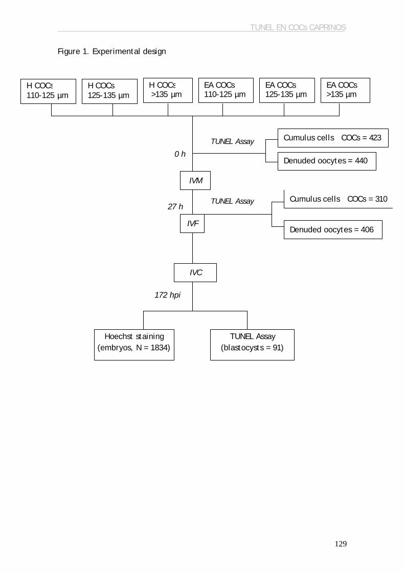
TUNEL EN COCs CAPRINOS
129
27 h
Figure 1. Experimental design
H COCs 110-125 µm
H COCs 125-135 µm
H COCs >135 µm µm
EA COCs 110-125 µm
EA COCs 125-135 µm
EA COCs >135 µm
Cumulus cells COCs = 423
Denuded oocytes = 440
TUNEL Assay
Denuded oocytes = 406
Cumulus cells COCs = 310
IVF
IVC
172 hpi
Hoechst staining (embryos, N = 1834)
0 h
IVM
TUNEL Assay
TUNEL Assay (blastocysts = 91)
ANNEXIN-V EN COCs CAPRINOS
133
EFFECT OF PREVALENCE OF APOPTOSIS IN OOCYTES AND CUMULUS CELLS,
ASSESSED BY ANNEXIN-V STAINING, ON EMBRYO DEVELOPMENT IN
PREPUBERTAL GOATS
Anguita B1, Paramio MT1, Romaguera R1, Jiménez-Macedo AR1, Morató R2,
Mogas T2, Izquierdo D1. 1Department of Animal and Food Sciences, Universitat Autònoma de
Barcelona, Bellaterra, Spain 2Department of Medicine and Surgery, Universitat Autònoma de
Barcelona, Bellaterra, Spain

ANNEXIN-V EN COCs CAPRINOS
134
ABSTRACT
Oocyte quality is the main factor that determine blastocyst yield. Any factor
that could affect oocyte quality, such as apoptosis, could impair the
subsequent embryonic development. Our aim was to investigate the incidence
of apoptosis in prepubertal goat oocytes and cumulus cells, assessed by
annexin-V staining, to evaluate its effect on embryo development. COCs from
slaughtered females were collected and classified in groups depending on
oocyte diameter: A: 110-125 µm, B: 125-135 µm and C: >135 µm. Each
diameter group was classified in two groups depending on COC morphology:
Healthy (H: compact cumulus layers and homogeneous cytoplasm) or Early
Atretic (EA: granulated cytoplasm and/or dispersed cumulus layers). COCs
were IVM for 27 h, IVF with fresh semen and IVC for 8 days after insemination.
Apoptosis analyses in oocytes and cumulus cells were performed after and
before maturation in all groups by Annexin V staining. Fisher’s exact test
showed that viable oocytes increased with diameter in the EA class both after
and before maturation (Immature oocytes: A: 70.9 %; B: 75.2 %; C: 72.4 %;
IVM-oocytes: A: 82.5 %; B: 95.2 %; C: 100 %), but not in the H class. Apoptotic
cumulus cells in immature COCs were less predominant in the largest oocytes.
There were less apoptotic cumulus cells in the H than in EA COCs. Blastocyst
yield obtained from the largest oocytes was the highest. In conclusion,
apoptosis in the smallest oocytes seems to be reversible. The highest
blastocyst rate is obtained from the largest healthy oocytes.
Keywords: apoptosis, oocyte, goat, Annexin-V

ANNEXIN-V EN COCs CAPRINOS
135
1. INTRODUCTION
The study of apoptosis incidence in oocytes as indicator of oocyte
quality and its effect on embryo development is of great relevance. Storage of
transcripts and proteins take place in the oocyte during its growth (Crozet et
al., 1981) and will determine the oocyte ability to maintain embryonic
development until the activation of the embryonic genome (Bachvarova,
1992; Gandolfi and Gandolfi, 2001). Therefore, storage of any molecule that
could affect oocyte quality could also impair embryo development. In fact,
cell survival/death regulatory molecules can be found in the stored pool of
maternal mRNA in mice and human (Exley et al., 1999; Jurisicova et al., 1998;
Metcalfe et al., 2004) and as a consequence they are likely to contribute in
determining the fate of the embryo. Not only the oocyte itself, but any factor
that could influence its quality, as cumulus cells, could affect blastocyst
yield. Oocyte and cumulus cells are connected through gap junctions, which
allow the interchange of molecules between these two cellular types (Tanghe
et al., 2002). Consequently, apoptosis incidence in cumulus cells could be
reflected in a lower oocyte developmental competence. Contradictory results
in this topic have been obtained: while many authors consider that early stage
of atresia in cumulus cells, assessed by morphological evaluation of COCs, is
reflected in a higher blastocyst rate (Blondin and Sirard, 1995; Boni et al.,
2002; de Wit et al., 2000; Nicholas et al., 2005), other studies observed that
apoptosis in cumulus cells, detected with TUNEL assay, affect embryo
development (Corn et al., 2005; Lee et al., 2001; Yuan et al., 2005; Zeuner et
al., 2003). These results suggest that COC morphology could not be related to
a higher incidence of apoptosis in cumulus cells, which should be further
studied.
Many approaches performed in order to find a marker of oocyte quality
are based in invasive methods (Anguita et al., 2006; Bilodeau-Goeseels and
Panich, 2002; De Sousa et al., 1998; Dode et al., 2006; Gutierrez-Adan et al.,
2004; Santos et al., 2006; Tamassia et al., 2004; Watson et al., 2000) and as a
consequence oocytes cannot be longer used for embryo production. It is

ANNEXIN-V EN COCs CAPRINOS
136
necessary to establish a relationship between the invasive methods studied
and morphological characteristics that allow an evaluation of oocyte without
impairing its developmental competence. In most laboratories, COC
morphology is a usual parameter to select the COCs that will be used for in
vitro embryo production. However, the low blastocyst rate obtained in our
laboratory following this parameter selection made necessary to use
additional selection parameters. In our study, we also select oocytes by
diameter, since it has been previously demonstrated in many species a
positive correlation between oocyte diameter and developmental competence
(Anguita et al., 2006; Gandolfi and Gandolfi, 2001; Pujol et al., 2004).
Moreover, oocyte diameter has been related to incidence of apoptosis in COC
(de Wit and Kruip, 2001) showing that a higher oocyte diameter was
accompanied by an increasing level of atresia in COC.
In normal conditions, there is an asymmetry among the outer and the
inner leaflet of the cell membrane (Bretscher, 1972): phosphatidyl-coline and
sphingomielin are located in the outer leaflet, and the phosphatidyl-serine
and phosphatidylethanolamine are located only in the inner leaflet. However,
translocation of phospatidyl-serine to the outer leaflet takes place in the
early stage of apoptosis, before DNA fragmentation (revised by (van Engeland
et al., 1998). Therefore, detection of phospatidyl-serine in the outer leaflet
can be used as a marker of early apoptosis. Annexin V is a molecule that binds
specifically to phospatidyl-serine (Andree et al., 1990; Tait et al., 1989).
Annexin V cannot cross the cell membrane of viable cells, but it can enter
dead cells that have lost membrane integrity and bind to the
posphatydilserine present in the inner leaflet. Consequently, it is necessary to
combine Annexin V staining with a vital stain, such as propidium iodide, in
order to distinguish between viable and dead cells (revised by (van Engeland
et al., 1998).
The objective of the present study was: 1) to evaluate the relationship
of oocyte diameter and COC morphology with oocyte and cumulus cells
apoptosis; 2) to establish whether COC morphology was related to a

ANNEXIN-V EN COCs CAPRINOS
137
differential incidence of apoptosis in oocytes or cumulus cells; 3) to study the
relationship between oocyte and cumulus cells apoptosis with embryo
development. In all experiments, apoptosis was assessed by Annexin-V
staining.
2. MATERIAL AND METHODS
Unless otherwise indicated, all chemicals were purchased from Sigma
Chemical Co. (St. Louis, MO).
2.1. Oocyte collection and IVM
Ovaries from prepubertal goats, approximately 2 months old, were
recovered from a local slaughterhouse and transported within 2 hours to the
laboratory at 38.5 ºC in PBS (Dulbecco’s phosphate-buffered saline, P-4417)
with 50 µg/mL gentamycin (G-3632). Ovaries were washed three times and
COCs (cumulus-oocyte complexes) were recovered by slicing in TCM199 (M-
2520) supplemented with 2.2 mg/mL NaHCO3, 2% (v/v) steer serum (Donor
Bovine Serum? , CanSera, Canada) and 50 ?g/mL gentamycin.
COCs were matured in TCM199 medium (M-7528) supplemented with
275 ? g/mL sodium pyruvate (P-3662), 146 µg/mL L-glutamine (G-5763), 10%
(v/v) steer serum, 10 µg/mL o-LH (L-5269), 10 µg/mL o-FSH (F-4520), 1 µg/mL
17-ß estradiol (E-2257), 400 µM cysteamine (M-9768) and 50 µg/mL
gentamycin (G-3632). Groups of 20-25 COCs were transferred to 100 µl
microdrops of maturation medium under mineral oil (M-3516) and incubated
for 27 hours at 38.5 ºC in a humidified air atmosphere with 5% CO2.
2.2. Sperm capacitation and IVF
Fresh semen was collected from two males of proven fertility and
transported to the laboratory at 38.5 ºC within 30 minutes. Motility of the
ejaculates was evaluated under a stereomicroscope. Motile sperm fraction
was separated by the swim-up method (Parrish et al., 1986), whereby 70 µL of
ANNEXIN-V EN COCs CAPRINOS
138
sperm were layered below 2 mL of mDM (Defined medium, (Brackett and
Oliphant, 1975); modified by (Younis et al., 1991), and incubated for 45 min
at 38.5 ºC in a humidified air atmosphere with 5% CO2. After incubation, the
supernatant was recovered and centrifuged at 170 x g for 3 minutes. The
pellet was resuspended in mDM (1:1; v/v) containing heparin (H-3149) and
ionomycin (I-0634) (Final concentration: 10 µg/mL heparin and 200 nM of
ionomycin) and incubated for 15 min in a humidified air atmosphere with
5%CO2 and 38.5 ºC (Wang et al., 2002). Sperm concentration was determined
using a Neubauer chamber cell.
After maturation, groups of 20 to 25 oocytes were transferred to 100 µL
microdrops of modified Tyrode’s medium (TALP) as described by Parrish et al.
(1986), supplemented with 1 µg/mL hypotaurine (H-1384) and 0.3 mg/mL
glutathione (G-6013) under mineral oil. Oocytes were co-cultured with
capacitated spermatozoa in a final concentration in the microdrop of 4 x 10⁶
sperm cells/mL.
2.3. In vitro embryo culture
At 24 hours post-insemination, presumptive zygotes were washed to
remove both remnant cumulus cells and attached sperm cells. Groups of 18-25
embryos were placed in 50 µl droplets of SOF medium (Synthetic Oviductal
Fluid, (Holm et al., 1999) and covered with mineral oil. The embryos were
incubated at 38.5°C in 5% CO2, 5% O2 and 90 % N2 for 7 days. At 48 hpi 0.1 µL
fetal bovine serum (F-7524) per embryo was added to the medium. At the end
of the culture period (8 days post-insemination), total cell number of embryos
was evaluated by Hoechst 33342 staining (B-2261) under a fluorescence
microscope.

ANNEXIN-V EN COCs CAPRINOS
139
2.4. Annexin-V staining
Detection of apoptosis was performed with Annexin-V-FLUOS Staining
kit (Roche, 1858777). During the whole process the reagents were kept at
38.5°C.
Staining was performed following the manufacturer’s instructions with
minor modifications. Briefly, samples were placed in 35 µl droplets containing
Annexin V buffer, Annexin V/FITC, PI and 1 mg/ml Hoechst 33342, and
incubated for 15 minutes at 38°C in the dark. Cells were counterstained with
propidium iodide (PI), a membrane impermeable stain, to distinguish between
live cells and dead cells. After incubation cells were mounted on siliconized
slides with vaseline bridges and observed under a fluorescence microscope.
Denuded oocytes were classified in three groups, representing (1) non-
apoptotic oocytes, with intact Hoechst positive nuclei and no annexin V
staining; (2) early apoptotic oocytes, with intact Hoechst positive nuclei and a
homogeneous annexin positive signal in the membrane; and (3) necrotic
oocytes which showed PI positive red nuclei, which is indicative for membrane
damage. In this last group, there were occasionally signs of annexin V staining
in the membrane. Oocytes showing a discontinuous green signal that came
from the remaining membrane of cumulus cells projections (Van Blerkom and
Davis, 1998) were also considered to be non-apoptotic oocytes.
COCs were evaluated as a whole mount preparation, and we evaluated
the proportion of the stained cumulus cells in the COC. The classification was
the following: (1) Healthy COCs, with no cells stained; (2) Apoptotic COCs,
with cells stained in with annexin V and, ocassionally, with propidium iodide.
It was also recorded if the percentage of stained cumulus cells represented
less than 25%, from 25 to 50%, from 50 to 75% or more than 75% of the total
cumulus cells.

ANNEXIN-V EN COCs CAPRINOS
140
2.5. Experimental design
All the COCs and oocytes necessary for one replicate of each
experiment were obtained the same day. At least three replicates of each
experiment were performed.
Recovered COCs from prepubertal goat ovaries were classified in two
groups depending on their morphology: Healthy (H): oocytes with
homogeneous cytoplasm and compact cumulus cells, and; Early atretic (EA):
oocytes with heterogeneous cytoplasm and/or cumulus cells with initial
expansion. Subsequently, oocyte diameter of COCs corresponding on each
morphological category was measured, and COCs were classified in three
additional groups: group A: 110-125 µm; group B: 125-135 µm; and group C:
>135 µm (Anguita et al., 2006).
Samples used for apoptosis analysis were randomly collected from each
one of the 6 experimental groups and evaluated at two points of time: before
IVM (0 h) and after maturation (27 h). Apoptosis evaluation was performed
both in cumulus cells and denuded oocytes by Annexin-V staining (see figure
1). COCs that were not used for apoptosis detection were fertilized in vitro
after 27 h of in vitro maturation, and at 24 hour post-insemination (hpi)
presumptive zygotes were cultured in vitro for additional 7 days. Each droplet
contained only embryos from the same morphology and diameter group. After
the in vitro culture, total number cell of embryos was evaluated by Hoechst
staining.
2.6. Statistical analysis
Data was analyzed using the statistical program Graph-Pad Instat 3.01
for Windows 95 (Graph-Pad software, San Diego, California, USA). Fisher’s
exact test was performed to compare the proportion of Annexin-V positive
cells in the different groups of oocytes and COC, and to assess the
developmental competence among diameter groups. Data of cell mean per

ANNEXIN-V EN COCs CAPRINOS
141
blastocyst were evaluated using analysis of variance with Tukey’s post-test.
Differences were considered significant when P<0.05.
3. RESULTS
3.1. Relationship between oocyte diameter and COC morphology and
prevalence of apoptosis in immature and mature oocytes
Annexin-V staining in immature oocytes (table 1) showed that the
percentage of apoptotic oocytes decreased with oocyte size in the EA class (A:
42.6%; B: 30.3%; 21%), but not in the H class (A: 29.1%; B: 24.8%; 27.5%). No
differences in the percentage of apoptotic oocytes were detected in oocytes
of the same diameter but different morphology group.
After maturation (table 1) a clear decrease of apoptotic oocytes (A:
17.5%; B: 4.8%; C: 0%) was observed with increasing oocyte diameter in the EA
class. However, in the H class, the percentage of apoptotic oocytes was lower
in the 125-135 µm oocytes (A: 5.5%; B: 0%; C: 3.7%). A reduction of the
incidence of apoptosis was also observed during maturation whatever the
diameter or the morphology group.
We observed that most apoptotic oocytes were at the latest stages, and
that necrotic ocytes were only detected in the EA class (data not shown).
3.2. Relationship between oocyte diameter and COC morphology and
prevalence of apoptosis in immature and mature cumulus-oocyte-complexes
Results are shown in table 2. At collection time, in both morphology
groups, the percentage of apoptotic cumulus cells decreased in the largest
COCs, and the percentage of COCs with less incidence of apoptosis was higher
in class H when compared to the same diameter group from class EA (80.5,
82.5 and 94.6% vs. 40.7, 48 and 60%, respectively). After maturation, the

ANNEXIN-V EN COCs CAPRINOS
142
incidence of apoptosis was lower in the largest Healthy oocytes. Moreover,
the percentage of COCs with more than 50% of apoptotic cumulus cells was
higher in >135 µm COCs from class EA when compared to the same diameter
group from class H.
The percentage of apoptotic cumulus cells varied during maturation
only in Healthy oocytes, and we detected a decrease in the percentage of
COCs with less than 25% of apoptotic cumulus cells, and an increase in the
percentage of COCs with 25-50% of apototic cumulus cells after maturation.
We observed that most of apoptotic cumulus cells in immature COCs
classified as Healthy was at the earliest stages of apoptosis, while in COCs
classified as Early atretic, most of apoptotic cumulus cells was at late stage of
apoptosis or necrosis. In addition, after IVM, apoptotic cumulus cells detected
in the largest oocytes were at an earliest stage of apoptosis in both
morphology classes, when compared to the other diameter groups. We
detected no necrotic COC in the largest group from the H class; however, the
proportion of necrotic COCs in the same diameter group from the EA class was
30% (data not shown).
3.3. Relationship between oocyte diameter and COC morphology and
prevalence of apoptosis in blastocysts.
Results of embryo development are shown in table 3. We observed a
clear relationship between oocyte diameter and embryo development in
terms of total embryos obtained and blastocyst rate in both morphology
classes, although in class EA no differences among 125-135 µm and >135 µm
oocytes were observed. In class H the blastocyst rate obtained in the largest
group was statistically higher than the blastocyst rate obtained by the same
diameter group in class EA (14.9% vs. 5.1%, respectively).

ANNEXIN-V EN COCs CAPRINOS
143
Blastocyst quality was evaluated in terms of percentage of hatched
blastocysts, and number of cells per blastocyst (table 3). No differences in
percentage of hatched blastocysts were found among diameter groups or
morphology classes. Cell number per blastocysts did not differ among
diameter nor do morphology groups, but the number of cells in the blastocysts
derived from the largest oocytes tended to be higher in the healthy group
(P<0.10)
4. DISCUSSION
In this study we have investigated the apoptosis incidence in oocytes
and cumulus cells related to morphological characteristics, in order to know
whether apoptosis incidence could affect blastocyst yield and quality. For
that purpose we have used Annexin-V staining, which detects translocation of
phosphatidylserine, a membrane phospholipid that flips from the inner to the
outer leaflet of the plasma membrane during early stage of apoptosis (Martin
et al., 1995; van Engeland et al., 1998). In addition, Annexin-V staining, in
combination to propidium iodide staining, lets us to distinguish between
viable and dead cells.
Apoptosis in prepubertal goat oocytes assessed by Annexin-V was
detected in both immature and IVM-oocytes. Our results showed a reduction
of incidence of apoptosis during maturation, which is contradictory to several
studies in mouse, that reported a negative effect of culture in vitro in terms
of oocyte apoptosis detected by TUNEL assay (Fujino et al., 1996; Takase et
al., 1995). However, (Van Blerkom and Davis, 1998) detected an increase and
a decrease of apoptosis, assessed by Annexin-V staining, during culture in
vitro in murine and human oocytes, respectively, while (Anguita et al., 2006)
reported a decrease of bovine oocyte apoptosis during in vitro maturation.
Discrepancies among authors may be caused by a differential incidence of
apoptosis between species or differences in the media composition, as was
suggested by Warzych et al. (2007). However, other hypotheses that could
ANNEXIN-V EN COCs CAPRINOS
144
explain reduction of apoptosis during in vitro maturation could be related to
the removal of oocyte from a follicular environment that may be promoting
apoptosis, the manipulation-induced apoptosis started in immature oocytes
and reverted during in vitro culture in specific conditions, or even a higher
sensitivity of immature oocytes to suffer apoptosis in comparison to mature
oocytes. In fact, in studies of oocyte cryopreservation, a reduced resistance
to cryopreservation in GV-oocytes in comparison to MII-oocytes has been
observed (Otoi et al., 1995; Parks and Ruffing, 1992), which may be related to
the higher incidence of apoptosis in immature oocytes, although further
studies should be performed in this matter.
Apoptosis is a regulatory mechanism that allows the maintenance of
homeostasis and the elimination of cells that are no longer required, and has
been also suggested as the mechanism to remove oocytes that have resumed
meiosis inside the follicle. However, in this study, oocyte nuclear stage did
not influence prevalence of apoptosis in oocytes after or before maturation,
which would indicate that apoptosis detected in prepubertal goat oocytes is
not a consequence of meiotic resumption inside the follicle. Only Early Atretic
oocytes larger than 135 µm showed a higher incidence of apoptosis in oocytes
at GV stage. These results are not in accordance with previous studies, which
detected higher apoptotic rate in “immature” oocytes that had resumed
meiosis (Anguita et al., 2006; Warzych et al., 2007). However, these studies
evaluated apoptotic rate in morphologically healthy oocytes, while we only
observe and effect of nuclear stage in morphologically Early Atretic oocytes,
which could explain the differences among studies.
Incidence of apoptosis in cumulus cells can be a good indicator of
oocyte developmental competence (Corn et al., 2005; Lee et al., 2001; Yuan
et al., 2005), due to the bidirectional communication established between
cumulus cells and oocytes through gap junctions (de Loos et al., 1991).
Cumulus cells play an important role in the regulation of nuclear and
cytoplasmic maturation in the oocyte (Tanghe et al., 2002) and in protecting
oocytes against the oxidative stress-induced apoptosis (Tatemoto et al.,

ANNEXIN-V EN COCs CAPRINOS
145
2000). In addition, presence of cumulus cells during in vitro maturation is
essential to improve blastocyst rate (Hashimoto et al., 1998). Several studies
have related mild signs of atresia in COCs with higher oocyte developmental
competence (Blondin and Sirard, 1995; Boni et al., 2002; de Wit et al., 2000;
Nicholas et al., 2005), while other studies reported lower blastocyst rate from
early atretic COCs (Corn et al., 2005; Lee et al., 2001; Yuan et al., 2005;
Zeuner et al., 2003). The use of different parameters to evaluate apoptosis in
a cell could explain the contradictory results from those studies. For that
reason, we investigated the relationship between morphological
characteristics and apoptosis incidence in COCs from prepubertal goats. We
observed that immature COCs classified morphologically as Early Atretic really
showed a higher percentage of apoptotic cells than those classified as
Healthy. Moreover, Early Atretic COCs yielded lower blastocyst rate than the
Healthy ones (3.2% vs. 6.2%, respectively). Our results enhance the
importance of the use of COC morphology as a selection parameter in
prepubertal goats. In addition to morphological selection, classification of
COCs by oocyte diameter was also useful as an additional selection
parameter, since the largest COCs presented the lowest incidence of
apoptosis, and their apoptotic cumulus cells were at the earliest stage of
apoptosis.
Our results showed a higher blastocyst rate achieved from the largest
oocytes, which is in agreement to previous studies which observed a
relationship between oocyte diameter and developmental competence
(Anguita et al., 2006; Gandolfi and Gandolfi, 2001). In prepubertal goats, the
use of early atretic COCs impaired embryo development from oocytes highest
than 135 µm. Taken together our results of embryonic development and
apoptosis in oocytes and cumulus cells, it seems that apoptosis in cumulus
cells is an important factor in determining differences in blastocyst yield
observed among oocytes of different diameter and oocytes of different
morphology. Thus, a high embryonic development is related to a reduced
incidence of apoptosis in both mature and immature COCs and a higher
prevalence of early stage of atresia in the apoptotic cumulus cells. These
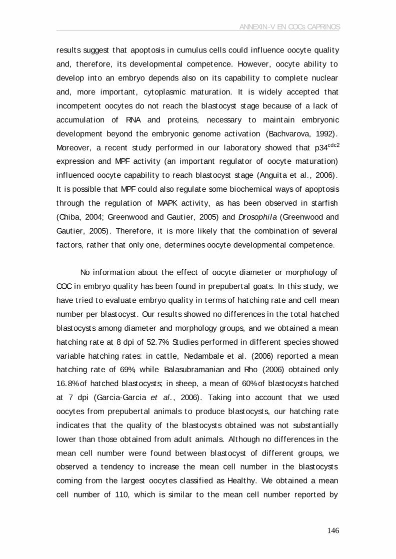
ANNEXIN-V EN COCs CAPRINOS
146
results suggest that apoptosis in cumulus cells could influence oocyte quality
and, therefore, its developmental competence. However, oocyte ability to
develop into an embryo depends also on its capability to complete nuclear
and, more important, cytoplasmic maturation. It is widely accepted that
incompetent oocytes do not reach the blastocyst stage because of a lack of
accumulation of RNA and proteins, necessary to maintain embryonic
development beyond the embryonic genome activation (Bachvarova, 1992).
Moreover, a recent study performed in our laboratory showed that p34cdc2
expression and MPF activity (an important regulator of oocyte maturation)
influenced oocyte capability to reach blastocyst stage (Anguita et al., 2006).
It is possible that MPF could also regulate some biochemical ways of apoptosis
through the regulation of MAPK activity, as has been observed in starfish
(Chiba, 2004; Greenwood and Gautier, 2005) and Drosophila (Greenwood and
Gautier, 2005). Therefore, it is more likely that the combination of several
factors, rather that only one, determines oocyte developmental competence.
No information about the effect of oocyte diameter or morphology of
COC in embryo quality has been found in prepubertal goats. In this study, we
have tried to evaluate embryo quality in terms of hatching rate and cell mean
number per blastocyst. Our results showed no differences in the total hatched
blastocysts among diameter and morphology groups, and we obtained a mean
hatching rate at 8 dpi of 52.7%. Studies performed in different species showed
variable hatching rates: in cattle, Nedambale et al. (2006) reported a mean
hatching rate of 69%, while Balasubramanian and Rho (2006) obtained only
16.8% of hatched blastocysts; in sheep, a mean of 60% of blastocysts hatched
at 7 dpi (Garcia-Garcia et al., 2006). Taking into account that we used
oocytes from prepubertal animals to produce blastocysts, our hatching rate
indicates that the quality of the blastocysts obtained was not substantially
lower than those obtained from adult animals. Although no differences in the
mean cell number were found between blastocyst of different groups, we
observed a tendency to increase the mean cell number in the blastocysts
coming from the largest oocytes classified as Healthy. We obtained a mean
cell number of 110, which is similar to the mean cell number reported by

ANNEXIN-V EN COCs CAPRINOS
147
Koeman et al. (2003) (120 ± 32) in blastocysts obtained from prepubertal goat
oocytes. The fact that Koeman et al. (2003) did not detect differences
between blastocysts obtained from prepubertal and adult goats reflects that
blastocyst quality in goats does not depend on animal age, and validates the
use of prepubertal goats as oocyte donors for embryo production.
In conclusion, in this study and in our experimental conditions, oocyte
quality, assessed by incidence of apoptosis in prepubertal goat oocytes and
cumulus cells, influenced blastocyst yield but not blastocyst quality. We
confirmed that COC morphology in prepubertal goats was related to a
differential incidence of apoptosis in cumulus cells and oocyte developmental
competence. We observed an effect of oocyte diameter on prevalence of
apoptosis in oocytes and cumulus cells, as well as in oocyte developmental
competence. Therefore, selection of prepubertal goat oocytes according to
oocyte diameter and COC morphology is a useful tool to select the COCs with
a lower incidence of apoptosis and a higher competence to reach blastocyst
stage.
5. ACKNOWLEDGEMENTS
This study was supported by a grant from the Ministry of Education and
Science of Spain (Project number: AGL2004-04737-C03-01/GAN) and a grant
from the Universitat Autònoma de Barcelona (Grant number: EME2004-25) and
from the Generalitat de Catalunya (2006FIC 00187).

ANNEXIN-V EN COCs CAPRINOS
148
6. REFERENCES
Andree H.A., Reutelingsperger C.P., Hauptmann R., Hemker H.C., Hermens
W.T., Willems G.M., 1990. Binding of vascular anticoagulant alpha (VAC alpha) to
planar phospholipid bilayers. J Biol Chem.265, 4923-4928.
Anguita B., Jimenez A.R., Izquierdo D., Mogas T., Paramio M.T., 2006. Effect
of oocyte diameter on meiotic competence, embryo development, P34 (cdc2) expression
and MPF activity in prepubertal goat oocytes. Theriogenology,
doi:10.1016/j.theriogenology.2006.09.003.
Anguita B., Vandaele L., Mateusen B., Maes D., Van Soom A., 2006.
Developmental competence of bovine oocytes is not related to apoptosis incidence in
oocytes, cumulus cells and blastocysts. Theriogenology.
doi:10.1016/j.theriogenology.2006.09.004.
Bachvarova R.F., 1992. A maternal tail of poly(A): the long and the short of it.
Cell.69, 895-897.
Balasubramanian S., Rho G.J., 2006. Effect of cysteamine supplementation of in
vitro matured bovine oocytes on chilling sensitivity and development of embryos. Anim
Reprod Sci. doi:10.1016/j.animreprosci.2006.03.011.
Bilodeau-Goeseels S., Panich P., 2002. Effects of oocyte quality on development
and transcriptional activity in early bovine embryos. Anim Reprod Sci.71, 143-155.
Blondin P., Sirard M.A., 1995. Oocyte and follicular morphology as determining
characteristics for developmental competence in bovine oocytes. Mol Reprod Dev.41,
54-62.
Boni R., Cuomo A., Tosti E., 2002. Developmental potential in bovine oocytes
is related to cumulus-oocyte complex grade, calcium current activity, and calcium
stores. Biol Reprod.66, 836-842.
Brackett B.G., Oliphant G., 1975. Capacitation of rabbit spermatozoa in vitro.
Biol Reprod.12, 260-274.
Bretscher M.S., 1972. Asymmetrical lipid bilayer structure for biological
membranes. Nat New Biol.236, 11-12.
Chiba K., 2004. MI arrest and apoptosis in starfish oocytes. Zoolog Sci.21, 1193.
Corn C.M., Hauser-Kronberger C., Moser M., Tews G., Ebner T., 2005.
Predictive value of cumulus cell apoptosis with regard to blastocyst development of
corresponding gametes. Fertil Steril.84, 627-633.

ANNEXIN-V EN COCs CAPRINOS
149
Crozet N., Motlik J., Szöllösi D., 1981. Nucleolar fine structure and RNA
synthesis in porcine oocytes during early stages of antrum formation. Biol Cell.41, 35-
42.
de Loos F., Kastrop P., Van Maurik P., Van Beneden T.H., Kruip T.A., 1991.
Heterologous cell contacts and metabolic coupling in bovine cumulus oocyte
complexes. Mol Reprod Dev.28, 255-259.
De Sousa P.A., Westhusin M.E., Watson A.J., 1998. Analysis of variation in
relative mRNA abundance for specific gene transcripts in single bovine oocytes and
early embryos. Mol Reprod Dev.49, 119-130.
de Wit A.A., Kruip T.A., 2001. Bovine cumulus-oocyte-complex quality is
reflected in sensivity for alpha-amanitin, oocyte-diameter and developmental capacity.
Anim Reprod Sci.65, 51-65.
de Wit A.A., Wurth Y.A., Kruip T.A., 2000. Effect of ovarian phase and follicle
quality on morphology and developmental capacity of the bovine cumulus-oocyte
complex. J Anim Sci.78, 1277-1283.
Dode M.A., Dufort I., Massicotte L., Sirard M.A., 2006. Quantitative expression
of candidate genes for developmental competence in bovine two-cell embryos. Mol
Reprod Dev.73, 288-297.
Exley G.E., Tang C., McElhinny A.S., Warner C.M., 1999. Expression of
caspase and BCL-2 apoptotic family members in mouse preimplantation embryos. Biol
Reprod.61, 231-239.
Fujino Y., Ozaki K., Yamamasu S., Ito F., Matsuoka I., Hayashi E., Nakamura
H., Ogita S., Sato E., Inoue M., 1996. DNA fragmentation of oocytes in aged mice.
Hum Reprod.11, 1480-1483.
Gandolfi T.A., Gandolfi F., 2001. The maternal legacy to the embryo:
cytoplasmic components and their effects on early development. Theriogenology.55,
1255-1276.
Garcia-Garcia R.M., Ward F., Fair S., O'Meara C M., Wade M., Duffy P.,
Lonergan P., 2006. Development and quality of sheep embryos cultured in commercial
G1.3/G2.3 sequential media. Anim Reprod Sci.
doi:10.1016/j.animreprosci.2006.03.007.
Greenwood J., Gautier J., 2005. From oogenesis through gastrulation:
developmental regulation of apoptosis. Semin Cell Dev Biol.16, 215-224.

ANNEXIN-V EN COCs CAPRINOS
150
Gutierrez-Adan A., Rizos D., Fair T., Moreira P.N., Pintado B., de la Fuente J.,
Boland M.P., Lonergan P., 2004. Effect of speed of development on mRNA expression
pattern in early bovine embryos cultured in vivo or in vitro. Mol Reprod Dev.68, 441-
448.
Hashimoto S., Saeki K., Nagao Y., Minami N., Yamada M., Utsumi K., 1998.
Effects of cumulus cell density during in vitro maturation of the deve lopmental
competence of bovine oocytes. Theriogenology.49, 1451-1463.
Holm P., Booth P.J., Schmidt M.H., Greve T., Callesen H., 1999. High bovine
blastocyst development in a static in vitro production system using SOFaa medium
supplemented with sodium citrate and myo-inositol with or without serum-proteins.
Theriogenology.52, 683-700.
Jurisicova A., Latham K.E., Casper R.F., Varmuza S.L., 1998. Expression and
regulation of genes associated with cell death during murine preimplantation embryo
development. Mol Reprod Dev.51, 243-253.
Koeman J., Keefer C.L., Baldassarre H., Downey B.R., 2003. Developmental
competence of prepubertal and adult goat oocytes cultured in semi-defined media
following laparoscopic recovery. Theriogenology.60, 879-889.
Lee K.S., Joo B.S., Na Y.J., Yoon M.S., Choi O.H., Kim W.W., 2001. Cumulus
cells apoptosis as an indicator to predict the quality of oocytes and the outcome of IVF-
ET. J Assist Reprod Genet.18, 490-498.
Martin S.J., Reutelingsperger C.P., McGahon A.J., Rader J.A., van Schie R.C.,
LaFace D.M., Green D.R., 1995. Early redistribution of plasma membrane
phosphatidylserine is a general feature of apoptosis regardless of the initiating stimulus:
inhibition by overexpression of Bcl-2 and Abl. J Exp Med.182, 1545-1556.
Metcalfe A.D., Hunter H.R., Bloor D.J., Lieberman B.A., Picton H.M., Leese
H.J., Kimber S.J., Brison D.R., 2004. Expression of 11 members of the BCL-2 family of
apoptosis regulatory molecules during human preimplantation embryo development and
fragmentation. Mol Reprod Dev.68, 35-50.
Nedambale T.L., Du F., Xu J., Chaubal S.A., Dinnyes A., Groen W., Faber D.,
Dobrinsky J.R., Yang X., Tian X.C., 2006. Prolonging bovine sperm-oocyte incubation
in modified medium 199 improves embryo development rate and the viability of
vitrified blastocysts. Theriogenology.1951-1960.

ANNEXIN-V EN COCs CAPRINOS
151
Nicholas B., Alberio R., Fouladi-Nashta A.A., Webb R., 2005. Relationship
between low-molecular-weight insulin- like growth factor-binding proteins, caspase-3
activity, and oocyte quality. Biol Reprod.72, 796-804.
Otoi T., Yamamoto K., Koyama N., Suzuki T., 1995. In vitro fertilization and
development of immature and mature bovine oocytes cryopreserved by ethylene glycol
with sucrose. Cryobiology.32, 455-460.
Parks J.E., Ruffing N.A., 1992. Factors affecting low temperature survival of
mammalian oocytes. Theriogenology.37, 59-73.
Parrish J., Susko-Parrish J., Leibfried-Rutledge M., Crister E., Eyeston W., First
N., 1986. Bovine in vitro fertilization with frozen thawed semen. Theriogenology.25,
591-600.
Pujol M., Lopez-Bejar M., Paramio M.T., 2004. Developmental competence of
heifer oocytes selected using the brilliant cresyl blue (BCB) test. Theriogenology.61,
735-744.
Santos T.A., El Shourbagy S., St John J.C., 2006. Mitochondrial content reflects
oocyte variability and fertilization outcome. Fertil Steril.85, 584-591.
Tait J.F., Gibson D., Fujikawa K., 1989. Phospholipid binding properties of
human placental anticoagulant protein-I, a member of the lipocortin family. J Biol
Chem.264, 7944-7949.
Takase K., Ishikawa M., Hoshiai H., 1995. Apoptosis in the degeneration
process of unfertilized mouse ova. Tohoku J Exp Med.175, 69-76.
Tamassia M., Nuttinck F., May-Panloup P., Reynier P., Heyman Y., Charpigny
G., Stojkovic M., Hiendleder S., Renard J.P., Chastant-Maillard S., 2004. In vitro
embryo production efficiency in cattle and its association with oocyte adenosine
triphosphate content, quantity of mitochondrial DNA, and mitochondrial DNA
haplogroup. Biol Reprod.71, 697-704.
Tanghe S., Van Soom A., Nauwynck H., Coryn M., de Kruif A., 2002.
Minireview: Functions of the cumulus oophorus during oocyte maturation, ovulation,
and fertilization. Mol Reprod Dev.61, 414-424.
Tatemoto H., Sakurai N., Muto N., 2000. Protection of porcine oocytes against
apoptotic cell death caused by oxidative stress during In vitro maturation: role of
cumulus cells. Biol Reprod.63, 805-810.

ANNEXIN-V EN COCs CAPRINOS
152
Van Blerkom J., Davis P.W., 1998. DNA strand breaks and phosphatidylserine
redistribution in newly ovulated and cultured mouse and human oocytes: occurrence
and relationship to apoptosis. Hum Reprod.13, 1317-1324.
van Engeland M., Nieland L.J., Ramaekers F.C., Schutte B., Reutelingsperger
C.P., 1998. Annexin V-affinity assay: a review on an apoptosis detection system based
on phosphatidylserine exposure. Cytometry.31, 1-9.
Wang B., Baldassarre H., Tao T., Gauthier M., Neveu N., Zhou J.F., Leduc M.,
Duguay F., Bilodeau A.S., Lazaris A., Keefer C., Karatzas C.N., 2002. Transgenic goats
produced by DNA pronuclear microinjection of in vitro derived zygotes. Mol Reprod
Dev.63, 437-443.
Warzych E., Peippo J., Szydlowski M., Lechniak D., 2007. Supplements to in
vitro maturation media affect the production of bovine blastocysts and their apoptotic
index but not the proportions of matured and apoptotic oocytes. Anim Reprod Sci.97,
334-343.
Watson A.J., De Sousa P., Caveney A., Barcroft L.C., Natale D., Urquhart J.,
Westhusin M.E., 2000. Impact of bovine oocyte maturation media on oocyte transcript
levels, blastocyst development, cell number, and apoptosis. Biol Reprod.62, 355-364.
Younis A.I., Zuelke K.A., Harper K.M., Oliveira M.A., Brackett B.G., 1991. In
vitro fertilization of goat oocytes. Biol Reprod.44, 1177-1182.
Yuan Y.Q., Van Soom A., Leroy J.L., Dewulf J., Van Zeveren A., de Kruif A.,
Peelman L.J., 2005. Apoptosis in cumulus cells, but not in oocytes, may influence
bovine embryonic developmental competence. Theriogenology.63, 2147-2163.
Zeuner A., Muller K., Reguszynski K., Jewgenow K., 2003. Apoptosis within
bovine follicular cells and its effect on oocyte development during in vitro maturation.
Theriogenology.59, 1421-1433.
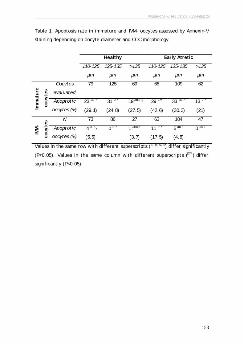
ANNEXIN-V EN COCs CAPRINOS
153
Table 1. Apoptosis rate in immature and IVM- oocytes assessed by Annexin-V
staining depending on oocyte diameter and COC morphology.
Healthy Early Atretic
110-125
µm
125-135
µm
>135
µm
110-125
µm
125-135
µm
>135
µm
Oocytes
evaluated
79 125 69 68 109 62
Imm
atur
e
oocy
tes
Apoptotic
oocytes (%)
23 ab ?
(29.1)
31 b ?
(24.8)
19 ab?? ?
(27.5)
29 a??
(42.6)
33 ab ?
(30.3)
13 b ?
(21)
N 73 86 27 63 104 47
IVM
-
oocy
tes
Apoptotic
oocytes (%)
4 a ? ?
(5.5)
0 c ?
1 abc??
(3.7)
11 b ?
(17.5)
5 ac ?
(4.8)
0 ac ?
Values in the same row with different superscripts (a, b, c, d) differ significantly
(P<0.05). Values in the same column with different superscripts (???? ) differ
significantly (P<0.05).

ANNEXIN-V EN COCs CAPRINOS
154
Table 2. Apoptotic rate in cumulus cells assessed by Annexin-V staining and
their distribution in immature and IVM- COCs depending on COC morphology
and oocyte diameter
Healthy Early Atretic
110-125
µm
125-135
µm
>135
µm
110-125
µm
125-135
µm
>135
µm
COCs
evaluated
87 103 56 54 77 70
Apoptotic
COCs (%)
86 ab
(98.8)
97 a
(94.2)
55 a
(98.2)
54 ab
(100)
77 b
(100)
70 ab
(100)
25 % 70 a ? ?
(81.4)
85 a ? ?
(87.6)
53 b?? ?
(96.4)
22 c
(40.7)
37 c
(48.05)
42 d
(60)
25-50 % 4 a ?
(4.6)
6 a ?
(6.2)
1 a ? ?
(1.8)
16 b
(29.6)
16 b
(20.8)
12 b
(17.1)
50-75 % 7 ab
(8.1)
3 b
(3.1)
0 c 9 a
(16.7)
9 a
(11.7)
6 ab
(8.6)
Imm
atur
e CO
Cs
>75 % 5 ab
(5.8)
3 b
(3.1)
1 b
(1.8)
7 ac
(13)
15 c
(19.5)
10 c
(14.3)
COCs
evaluated
59 75 50 45 65 60
Apoptotic
COCs (%)
59
(100)
75
(100)
50
(100)
45
(100)
65
(100)
60
(100)
25 % 29 a ?
(49.1)
45 ab ?
(60)
37 b ?
(74)
21 a
(46.7)
37 ab
(57.9)
32 ac
(53.3)
25-50 % 17 ?
(28.8)
17 ?
(22.7)
13 ? ?
(26)
9
(20)
10
(15.4)
9
(15)
50-75 % 7 a
(11.9)
8 a
(10.7)
0 b 9 a
(20)
9 a
(13.8)
9 a
(15)
IVM
- C
OC
s
>75 % 6 a
(10.2)
5 ab
(6.7)
0 b 6 a
(13.3)
9 a
(13.8)
10 a
(16.7)

ANNEXIN-V EN COCs CAPRINOS
155
Values in the same row with different superscripts (a, b, c, d) differ significantly
(P<0.05). Values in the same column with different superscripts (???? ) differ
significantly (P<0.05).

ANNEXIN-V EN COCs CAPRINOS
156
Table 3. Embryo development at 8 dpi depending on COC morphology and
oocyte diameter
Values in the same column with different superscripts (a, b) differ significantly
(P<0.05).
* These results are expressed as the hatched blastocysts related to the total
number of blastocysts
N: oocytes inseminated; Mor: Morulae; Blast: Blastocysts; Deg: Degenerated
embryos.
Ø
(µm)
N Total
Div
(%)
8-16
Cells
(%)
Mor
(%)
Blast.
(%)
Deg
(%)
Hatched
Blast.*
Cell
Mean
Blast.
110-
125 µm
222 71 a
(32)
6 a
(2.7)
4 a
(1.8)
0 a 18 ac
(8.1)
0
125-
135 µm
510 303 b
(59.4)
41 b
(8)
20 ab
(3.9)
27 b
(5.3)
31 c
(6.1)
14/27
(51.85)
92.52 ±
51.87
Hea
lthy
>135
µm
230 165 c
(71.7)
28 bc
(12.2)
16 bc
(7)
33 c
(14.3)
17 ac
(7.4)
16/33
(48.48)
128.90
± 81.40
110-
125 µm
284 95 a
(33.4)
12 a
(4.2)
5 a
(1.8)
1 a
(0.3)
34 ab
(12)
1/1
(100)
146
125-
135 µm
463 292 b
(63.1)
59 c
(12.7)
13 a
(2.8)
19 b
(4.1)
42 ac
(9.1)
9/19
(47.37)
106.7 ±
77.12
Earl
y at
reti
c
>135
µm
215 145 c
(68.8)
38 c
(17. 7)
17 c
(7.9)
11 b
(5.1)
31 b
(14.4)
8/11
(72.72)
107.38
± 67.73
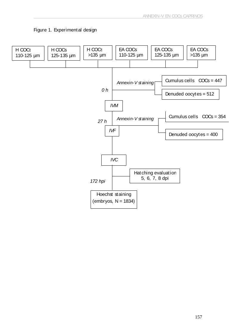
ANNEXIN-V EN COCs CAPRINOS
157
27 h
Figure 1. Experimental design
H COCs 110-125 µm
H COCs 125-135 µm
H COCs >135 µm µm
EA COCs 110-125 µm
EA COCs 125-135 µm
EA COCs >135 µm
Cumulus cells COCs = 447
Denuded oocytes = 512
Annexin-V staining
Denuded oocytes = 400
Cumulus cells COCs = 354
IVF
IVC
172 hpi
Hoechst staining (embryos, N = 1834)
0 h
IVM
Annexin-V staining
Hatching evaluation 5, 6, 7, 8 dpi

DISCUSIÓN GENERAL
8. DISCUSIÓN GENERAL La producción in vitro de blastocistos a partir de ovocitos de cabras
prepúberes (6%; Koeman et al., 2003) es bastante ineficiente, en comparación
con los resultados obtenidos en cabras adultas (33%, Keskintepe et al., 1996),
o en otras especies como el bovino (40%, revisado por Lonergan et al., 2003).
Se ha comprobado en numerosas especies que el uso de hembras prepúberes
como donantes de ovocitos perjudica el desarrollo embrionario posterior
(revisado por Armstrong, 2001), debido principalmente a deficiencias en su
maduración citoplasmática (Damiani et al., 1996; Salamone et al., 2001). Sin
embargo, se ha logrado obtener blastocistos, gestaciones y nacimientos a
partir de ovocitos de hembras prepúberes (Armstrong et al., 1992). En
numerosas especies, incluido el caprino, se ha observado que la competencia
del ovocito se incrementa con el tamaño folicular (vaca: Lonergan et al.,
1994; Blondin y Sirard, 1995; Lequarre et al., 2005; Kauffold et al., 2005;
cabra: Crozet et al., 1995; búfalo: Raghu et al., 2002; oveja: Ledda et al.,
1999), y éste, a su vez, se correlaciona con el diámetro ovocitario (vaca:
Arlotto et al., 1996; cabra: De Smedt et al., 1994; Crozet et al., 2000; búfalo:
Raghu et al., 2002; yegua: Goudet et al., 1997). Por lo tanto, el crecimiento
del ovocito es indispensable para que éste adquiera competencia para
mantener el desarrollo embrionario. En cabras adultas, los folículos mayores a
5 mm de diámetro son los que contienen los ovocitos que dan lugar a un
mayor porcentaje de blastocistos (Crozet et al., 1995). Sin embargo, la
mayoría de folículos presentes en los ovarios de cabras prepúberes no superan
los 3 mm de diámetro (Martino et al., 1994), lo que podría indicar que el
menor porcentaje de desarrollo embrionario obtenido a partir de ovocitos de
cabras prepúberes podría ser consecuencia de la mayor proporción de ovocitos
que todavía no han completado su crecimiento, pero el potencial para el
desarrollo de los ovocitos de cabras prepúberes podría ser el mismo que el de
los ovocitos de cabras adultas en el mismo estadío de crecimiento. Por otro
lado, en diversas especies se ha observado que el diámetro de los ovocitos
obtenidos a partir de folículos del mismo diámetro es menor en hembras
161

DISCUSIÓN GENERAL
prepúberes que en adultas (bovino: Gandolfi et al., 1998; ovino: Ledda et al.,
1999). El objetivo de esta tesis fue estudiar la competencia de los ovocitos de
cabras prepúberes a nivel meiótico, de fecundación y de desarrollo
embrionario, dependiendo de su diámetro y, así, poder determinar la utilidad
del diámetro ovocitario como un parámetro de selección de la calidad de los
ovocitos. Por otro lado, el diámetro ovocitario también ha sido utilizado para
investigar qué papel podrían desempeñar el MPF y la apoptosis en la
adquisición de la competencia ovocitaria, en un intento por entender un poco
más qué factores pueden estar influyendo en la maduración citoplasmática
del ovocito.
Se sabe que el MPF juega un papel muy importante en la progresión de
la maduración meiótica del ovocito, ya que es el responsable de la
desestructuración de la lámina nuclear (Peter et al., 1990a) y de los nucleolos
(Peter et al., 1990b), de la condensación cromosómica (Moreno y Nurse, 1990)
y de la reorganización de los microfilamentos (Morgan et al., 1989) y de la red
de filamentos intermedios (Chou et al., 1990). Nuestros resultados muestran
que, en cabras prepúberes, la competencia meiótica de los ovocitos, así como
la competencia para el desarrollo embrionario, se adquiere a medida que
aumenta la acumulación de Ciclina B1, p34cdc2 y la actividad del MPF en el
ovocito. Nuestros resultados concuerdan con otros estudios en los que se
observó que los dos componentes del MPF, la p34cdc2 y la Ciclina B1, se van
acumulando durante el crecimiento del ovocito y alcanzan niveles máximos a
medida que el ovocito adquiere competencia meiótica (ratón: Mitra y Schultz,
1996; de Vántery et al., 1996; cabras: Dedieu et al., 1998). Sin embargo, la
relación observada en cabras prepúberes entre la concentración y actividad
del MPF y la competencia para el desarrollo de los ovocitos indica que,
además del importante papel que juega en la progresión meiótica, el MPF
también jugaría un papel importante en la regulación de la maduración
citoplasmática del ovocito. Borsuk et al. (1991) y Naito et al. (1992) fueron los
primeros en sugerir la participación del MPF en la maduración citoplasmática
al observar que los niveles de MPF tras la MIV estaban relacionados con la
capacidad del ovocito para descondensar la cabeza del espermatozoide y
162
DISCUSIÓN GENERAL
formar el pronúcleo masculino. Por otro lado, la menor competencia de los
ovocitos de hembras prepúberes también ha sido relacionada con una menor
actividad del MPF en comparación con ovocitos de hembras adultas (bovino:
Salamone et al., 2001; ovino: Ledda et al., 2001).
En cabras prepúberes, la acumulación de mRNA de p34cdc2 y Ciclina B1
no parece ser la responsable de la competencia ovocitaria, ya que los niveles
fueron similares en todos los diámetros de ovocitos estudiados. En cambio, sí
se observaron diferencias en cuanto a los niveles de RNA de p34cdc2 y Ciclina
B1 almacenado en el ovocito antes de la maduración, pero no después,
mientras que las diferencias en la cantidad de proteína p34cdc2 se detectaron
después de la maduración, pero no antes. Estos resultados parecen indicar
que la acumulación de transcritos de las dos subunidades del MPF para su
utilización posterior determinará la actividad del MPF y, por lo tanto, la
adquisición de competencia meiótica y, posiblemente, de competencia para
el desarrollo. Sin embargo, la competencia del ovocito no sólo está
determinada por su capacidad para almacenar todos los factores necesarios
para el mantenimiento de la actividad celular hasta la activación del genoma
embrionario, sino también en su capacidad de movilizar los transcritos
almacenados y traducirlos a proteínas, como se demuestra en la menor
cantidad de RNA total detectado en los ovocitos más competentes después de
la maduración.
En general, nuestros resultados muestran que a lo largo de la
maduración in vitro se produjo acumulación de RNA de las subunidades del
MPF, sobretodo en los ovocitos más incompetentes; sin embargo, esta
acumulación no condujo a una mayor progresión meiótica en estos ovocitos.
De Vantéry et al. (1997) observaron que la inyección de mRNA p34cdc2 en
ovocitos meióticamente incompetentes daba lugar a la acumulación de
proteína p34cdc2 en niveles similares a los ovocitos competentes, pero no se
reiniciaba la meiosis. Estos resultados sugieren que otros factores que
probablemente intervienen en la regulación del MPF pueden estar en
deficiencia en los ovocitos que todavía no han completado su crecimiento.
163

DISCUSIÓN GENERAL
Diversas hipótesis pueden explicar los resultados: 1) aunque se produzca
acumulación de p34cdc2, es posible que el ovocito incompetente no sea capaz
de producir las modificaciones post-traduccionales que permitirán la
asociación de la p34cdc2 y la Ciclina B1 para formar el pre-MPF (Kanatsu-
Shinohara et al., 2000), posiblemente debido a la incapacidad para que el
p34cdc2 sea fosforilado, paso indispensable para que la subunidad catalítica
pueda asociarse con el pool endógeno de Ciclina B1 (de Vantéry et al., 1997);
2) la acumulación de p34cdc2 es suficiente para formar el heterodímero con
Ciclina B1, pero su activación puede estar comprometida si el ovocito no ha
sido capaz de acumular factores necesarios para su regulación, como la
fosfatasa Cdc25 o las kinasas Wee (Goudet et al., 1998; revisado por Jones,
2004); 3) es posible que la translocación del MPF del citoplasma al núcleo,
donde se encuentran algunos de sus sustratos, como la histona H1, esté
comprometida en ovocitos incompetentes (Kanatsu-Shinohara et al., 2000).
Está translocación está regulada por la fosforilación de la Ciclina B1, que
impide que el nuclear export sequence sea reconocido por el nuclear export
factor CRM1 (Yang et al., 1998), produciendo así su acumulación en el núcleo;
y 4) la incompetencia meiótica de los ovocitos puede estar ligado a la
inhabilidad para activar la MAPK, que es necesaria para dar lugar al reinicio
de la meiosis en muchas especies. No obstante, en cabras la activación de la
MAPK se produce después de la activación del MPF y la ruptura de vesícula
germinal, al igual que en cerdos (Inoue et al., 1995) y ratón (Verlhac et al.,
1994), con lo que esta hipótesis queda descartada. En cambio, se ha
comprobado que los ovocitos caprinos meióticamente incompetentes
contienen menos cantidad de Cdc25 fosfatasa que los ovocitos competentes
(Gall et al., 2002); sin embargo, es necesario realizar más estudios para
comprobar si algunos de los factores de regulación del MPF mencionados en
las otras hipótesis intervienen también en la adquisición de competencia
meiótica y para el desarrollo.
En cabras prepúberes la capacidad del ovocito para progresar
meióticamente hasta el estadío de metafase II, ser fecundado normalmente,
dividirse y alcanzar el estadío embrionario de blastocisto aumenta con el
164

DISCUSIÓN GENERAL
diámetro ovocitario, hecho que ha sido previamente observado en numerosas
especies (vaca: Arlotto et al., 1996; cabra: De Smedt et al., 1994; Martino et
al., 1994; Crozet et al., 2000; búfalo: Raghu et al., 2002; yegua: Goudet et
al., 1997). Sin embargo, nuestros resultados también muestran que el estadío
meiótico de los ovocitos inmediatamente después de su liberación de los
folículos varía con el diámetro; así, los ovocitos de mayor tamaño presentaban
una gran proporción que ya habían reanudado la meiosis dentro del folículo.
Los ovocitos que se encuentran dentro de los folículos están en parada
meiótica debido a sustancias inhibidoras de la meiosis que se encuentran en el
líquido folicular y que llegan al ovocito a través de las células del cumulus
(Dekel, 1988). Únicamente se produce la reanudación de la meiosis en el
ovocito que contiene el folículo dominante después del pico de LH, o en
folículos atrésicos debido a la expansión prematura de las células del
cumulus. En consecuencia, nuestros resultados parecían indicar que la
mayoría de ovocitos recuperados de cabras prepúberes eran atrésicos, y que
la atresia aumentaba con el diámetro ovocitario. Esta hipótesis parecía
concordar con estudios previos, en el que se observó que los ovocitos recién
recuperados de los folículos que ya habían reanudado la meiosis presentaban
mayor incidencia de apoptosis que aquellos que todavía permanecían en
parada meiótica (Warzych et al., 2006). Sin embargo, en el estudio realizado
sobre apoptosis en cabras prepúberes observamos que los ovocitos que habían
reanudado la meiosis en el momento de ser obtenidos del folículo presentaban
menor incidencia de apoptosis que los que se encontraban todavía en parada
meiótica, mientras que en ovocitos de vacas observamos lo contrario. Es
posible que la dinámica de reanudación meiótica tras la liberación del folículo
en ovocitos de cabras prepúberes sea mucho más rápida que en cabras adultas
(Le Gal et al., 1992), que presentan el 100% de los ovocitos en estadío de
vesícula germinal a las 3 h de la liberación del folículo. En cambio, en bovino,
los ovocitos de menor tamaño que han reanudado la meiosis en el momento
de ser liberados de los folículos son los que mayoritariamente provienen de
folículos atrésicos.
165

DISCUSIÓN GENERAL
Diversos estudios han descrito que los COCs que muestran signos
tempranos de atresia dan lugar a un mayor porcentaje de blastocistos
(Blondin y Sirard, 1995; Boni et al., 2002; de Wit et al., 2000; Nicholas et al.,
2005), debido a que estos COCs sufren un proceso similar a la maduración
dentro del folículo (Assey et al., 1994) que les proporciona mayor
competencia para el desarrollo. Sin embargo, en cabras prepúberes el efecto
de la atresia del COC en su capacidad para el desarrollo no había sido
evaluado hasta el momento. Para ello, la incidencia de apoptosis en los
ovocitos y las células del cumulus fue evaluada mediante dos técnicas
complementarias: la tinción mediante Annexin-V, que permite distinguir las
células en estadíos tempranos de apoptosis de las células en estadíos más
tardíos; y la técnica de TUNEL, que permite observar las células que se
encuentran en las últimas fases de la apoptosis. Estas técnicas fueron puestas
a punto mediante la realización de un estudio previo en COCs bovinos, y
posteriormente se aplicaron en COCs de cabras prepúberes. En general,
pudimos observar que los COCs de mayor diámetro, que son más competentes
para dar lugar a un embrión viable, son también los que sufren menor
incidencia de apoptosis, tanto en vacas como en cabras prepúberes. La
incidencia de apoptosis en ovocitos fue similar en las dos especies, según el
tamaño y la técnica de detección utilizada. En ambos casos se pudo observar
una reducción de la apoptosis ovocitaria durante la maduración in vitro, como
había sido observada previamente en ovocitos humanos por Van Blerkom y
Davis (1998). Sin embargo, estudios realizados en otras especies muestran un
incremento de los niveles de apoptosis en ovocitos en cultivo (Fujino et al.,
1996; Takase et al., 1995; Van Blerkom y Davis, 1998), que puede ser
indicativo del posible efecto que los diferentes medios de cultivo pueden
tener sobre estos ovocitos. En cabras prepúberes, al contrario de lo observado
en otras especies, los COCs que no presentan ningún signo morfológico de
atresia son los que dan lugar a un mayor porcentaje de blastocistos. La
morfología del COC y el diámetro ovocitario en cabras prepúberes no parece
afectar a la calidad de los blastocistos obtenidos, evaluada según el
porcentaje de blastómeros apoptóticos (ACR), capacidad de eclosión y número
de células del blastocisto. Curiosamente, el porcentaje de blastómeros
166

DISCUSIÓN GENERAL
apoptóticos detectado en blastocistos de cabras prepúberes fue menor que el
obtenido en blastocistos bovinos, aunque estos últimos estuvieron menos
tiempo en cultivo. No obstante, los índices de apoptosis en los blastocistos de
ambas especies se encuentran dentro de los índices que ya habían sido
observados previamente en otros estudios (2.89 %, Warzych et al., 2006; 3-7 %
Boelhauve et al., 2005; 4-9 %, Fabian et al., 2005; 10.3 %, Pomar et al., 2005;
12 %, Yuan et al., 2005; 9.8-14.2 %, Vandaele et al., 2006)
Teniendo en consideración todos los estudios realizados en esta tesis,
podemos concluir que la capacidad de los ovocitos para llegar al estadío de
blastocisto es mayor en ovocitos de mayor diámetro, con mayor actividad de
MPF y menor incidencia de apoptosis, tanto a nivel ovocitario como en las
células del cumulus. Parece ser que existen indicios de la existencia de una
conexión entre la regulación de la maduración ovocitaria y la de la muerte
celular, según diversos estudios realizados en invertebrados (Drosophila:
Ivanovska et al., 2004; starfish: Sasaki y Chiba, 2004; Greenwood y Gautier,
2005; Xenopus: Smith et al., 2000; revisado por Greenwood y Gautier, 2005;
Caenorhabditis elegans: Kritikou et al., 2006) y en mamíferos (ratón: Tatone
et al., 2006; cerdo: Ma et al., 2005). Sin embargo, sería necesario realizar
estudios complementarios para averiguar de qué modo se relacionan las dos
rutas de regulación, y cual sería el efecto en la competencia del ovocito.
167

CONCLUSIONES
171
9. CONCLUSIONES
Las conclusiones de esta tesis fueron las siguientes:
? El diámetro del ovocito y la morfología del COC son buenos indicadores
no invasivos de la competencia ovocitaria para producir embriones en
cabras prepúberes. Así, para obtener el mayor porcentaje de
blastocistos deben seleccionarse los COCs con un diámetro ovocitario
superior a 135 µm y sin signos visibles de atresia.
? Los ovocitos de cabras prepúberes con una mayor competencia para el
desarrollo también presentaron un mayor nivel de expresión de Ciclina
B1-ARN en ovocitos inmaduros, una mayor acumulación de proteína
p34cdc2 y mayor actividad kinasa del Maturation Promoting Factor
después de la maduración in vitro. Por lo tanto, estos parámetros
pueden utilizarse como indicadores invasivos de calidad ovocitaria.
? En vacas y cabras prepúberes, el porcentaje de apoptosis en ovocitos y
células del cumulus varía con el diámetro ovocitario. Además, una
mayor incidencia de apoptosis en ovocitos y células del cumulus no
afectó a la calidad de los blastocistos resultantes, evaluada según la
incidencia de apoptosis en estos blastocistos.
? Las diferencias de morfología entre COCs sin signos y con signos visibles
de atresia en cabras prepúberes se relaciona con un diferente grado de
apoptosis en ovocitos y células del cumulus. La selección de COCs
mediante su morfología antes de la MIV incrementa el porcentaje de
blastocistos sólo en los ovocitos que han completado su crecimiento.

BIBLIOGRAFÍA
10. BIBLIOGRAFÍA
Algriany O, Bevers M, Schoevers E, Colenbrander B, Dieleman S. 2004. Follicle
size-dependent effects of sow follicular fluid on in vitro cumulus
expansion, nuclear maturation and blastocyst formation of sow cumulus
oocytes complexes. Theriogenology 62(8):1483-1497.
Alikani M, Calderon G, Tomkin G, Garrisi J, Kokot M, Cohen J. 2000. Cleavage
anomalies in early human embryos and survival after prolonged culture
in-vitro. Hum Reprod 15(12):2634-2643.
Alikani M, Cohen J, Tomkin G, Garrisi GJ, Mack C, Scott RT. 1999. Human
embryo fragmentation in vitro and its implications for pregnancy and
implantation. Fertil Steril 71(5):836-842.
Andree HA, Reutelingsperger CP, Hauptmann R, Hemker HC, Hermens WT,
Willems GM. 1990. Binding of vascular anticoagulant alpha (VAC alpha)
to planar phospholipid bilayers. J Biol Chem 265(9):4923-4928.
Anifandis G, Koutselini E, Stefanidis I, Liakopoulos V, Leivaditis C, Mantzavinos
T, Vamvakopoulos N. 2005. Serum and follicular fluid leptin levels are
correlated with human embryo quality. Reproduction 130(6):917-921.
Ankarcrona M, Dypbukt JM, Bonfoco E, Zhivotovsky B, Orrenius S, Lipton SA,
Nicotera P. 1995. Glutamate-induced neuronal death: a succession of
necrosis or apoptosis depending on mitochondrial function. Neuron
15(4):961-973.
Arlotto T, Schwartz JL, First NL, Leibfried-Rutledge ML. 1996. Aspects of
follicle and oocyte stage that affect in vitro maturation and
development of bovine oocytes. Theriogenology 45:943-956.
Armstrong DT. 2001. Effects of maternal age on oocyte developmental
competence. Theriogenology 55(6):1303-1322.
Armstrong DT, Holm P, Irvine B, Petersen BA, Stubbings RB, McLean D, Stevens
G, Seamark RF. 1992. Pregnancies and live birth from in vitro
fertilization of calf oocytes collected by laparoscopic follicular
aspiration. Theriogenology 38(4):667-678.
175

BIBLIOGRAFÍA
Armstrong DT, Irvine BJ, Earl CR, McLean D, Seamark RF. 1994. Gonadotropin
stimulation regimens for follicular aspiration and in vitro embryo
production from calf oocytes. Theriogenology 42(7):1227-1236.
Ashkenazi A, Dixit VM. 1998. Death receptors: signaling and modulation.
Science 281(5381):1305-1308.
Asselin E, Xiao CW, Wang YF, Tsang BK. 2000. Mammalian follicular
development and atresia: role of apoptosis. Biol Signals Recept 9(2):87-
95.
Assey RJ, Hyttel P, Greve T, Purwantara B. 1994. Oocyte morphology in
dominant and subordinate follicles. Mol Reprod Dev 37(3):335-344.
Atef A, Francois P, Christian V, Marc-Andre S. 2005. The potential role of gap
junction communication between cumulus cells and bovine oocytes
during in vitro maturation. Mol Reprod Dev 71(3):358-367.
Bachvarova RF. 1992. A maternal tail of poly(A): the long and the short of it.
Cell 69(6):895-897.
Badouel C, Korner R, Frank-Vaillant M, Couturier A, Nigg EA, Tassan JP. 2006.
M-phase MELK activity is regulated by MPF and MAPK. Cell Cycle
5(8):883-889.
Baker TG. 1963. A Quantitative and Cytological Study of Germ Cells in Human
Ovaries. Proc R Soc Lond B Biol Sci 158:417-433.
Baldassare H, Karatzas CN. 2004. Advanced assisted reproduction technologies
(ART) in goats. Anim Reprod Sci 82-83: 255-266.
Bar-Ami S, Tsafriri A. 1981. Acquisition of meiotic competence in the rat: role
of gonadotropin and estrogen. Gamete Res 4:463-472.
Bilby TR, Block J, do Amaral BC, Sa Filho O, Silvestre FT, Hansen PJ, Staples
CR, Thatcher WW. 2006. Effects of dietary unsaturated fatty acids on
oocyte quality and follicular development in lactating dairy cows in
summer. J Dairy Sci 89(10):3891-3903.
Billig H, Furuta I, Hsueh AJ. 1994. Gonadotropin-releasing hormone directly
induces apoptotic cell death in the rat ovary: biochemical and in situ
detection of deoxyribonucleic acid fragmentation in granulosa cells.
Endocrinology 134(1):245-252.
176
BIBLIOGRAFÍA
Bilodeau-Goeseels S, Panich P. 2002. Effects of oocyte quality on
development and transcriptional activity in early bovine embryos. Anim
Reprod Sci 71(3-4):143-155.
Blondin P, Sirard MA. 1995. Oocyte and follicular morphology as determining
characteristics for developmental competence in bovine oocytes. Mol
Reprod Dev 41(1):54-62.
Boelhauve M, Sinowatz F, Wolf E, Paula-Lopes FF. 2005. Maturation of bovine
oocytes in the presence of leptin improves development and reduces
apoptosis of in vitro-produced blastocysts. Biol Reprod 73(4):737-744.
Boni R, Cuomo A, Tosti E. 2002. Developmental potential in bovine oocytes is
related to cumulus-oocyte complex grade, calcium current activity, and
calcium stores. Biol Reprod 66(3):836-842.
Boone DL, Tsang BK. 1997. Identification and localization of
deoxyribonuclease I in the rat ovary. Biol Reprod 57(4):813-821.
Borsuk E. 1991. Anucleate fragments of parthenogenetic eggs and of maturing
oocytes contain complementary factors required for development of a
male pronucleus. Mol Reprod Dev 29(2):150-156.
Bosco L, Ruvolo G, Morici G, Manno M, Cittadini E, Roccheri MC. 2005.
Apoptosis in human unfertilized oocytes after intracytoplasmic sperm
injection. Fertil Steril 84(5):1417-1423.
Brackett BG, Zuelke KA. 1993. Analysis of factors involved in the in vitro
production of bovine embryos. Theriogenology 39:43-64.
Bretscher MS. 1972. Asymmetrical lipid bilayer structure for biological
membranes. Nat New Biol 236(61):11-12.
Brevini-Gandolfi TA, Favetta LA, Mauri L, Luciano AM, Cillo F, Gandolfi F.
1999. Changes in poly(A) tail length of maternal transcripts during in
vitro maturation of bovine oocytes and their relation with
developmental competence. Mol Reprod Dev 52(4):427-433.
Brison DR, Schultz RM. 1997. Apoptosis during mouse blastocyst formation:
evidence for a role for survival factors including transforming growth
factor alpha. Biol Reprod 56(5):1088-1096.
177

BIBLIOGRAFÍA
Brison DR, Schultz RM. 1998. Increased incidence of apoptosis in transforming
growth factor alpha-deficient mouse blastocysts. Biol Reprod 59(1):136-
144.
Brower PT, Schultz RM. 1982. Intercellular communication between granulosa
cells and mouse oocytes: existence and possible nutritional role during
oocyte growth. Dev Biol 90(1):144-153.
Calvin HI, Grosshans K, Blake EJ. 1986. Estimation and manipulation of
glutathione levels in prepubertal mouse ovaries and ova: relevance to
sperm nucleus transformation in the fertilized egg. Gamete Res 14:265-
275.
Caralco PG. 1995. Polarization of mitocondria in the unfertilized mouse
oocyte. Dev Genet 16:503-512.
Chang CL, Wang TH, Horng SG, Wu HM, Wang HS, Soong YK. 2002. The
concentration of inhibin B in follicular fluid: relation to oocyte
maturation and embryo development. Hum Reprod 17(7):1724-1728.
Chesnel F, Eppig JJ. 1995. Synthesis and accumulation of p34cdc2 and cyclin B
in mouse oocytes during acquisition of competence to resume meiosis.
Mol Reprod Dev 40(4):503-508.
Chian RC, Niwa K, Sirard MA. 1994. Effects of cumulus cells on male
pronuclear formation and subsequent early development of bovine
oocytes in vitro. Theriogenology 41(7):1499-1508.
Chiu TT, Rogers MS, Law EL, Briton-Jones CM, Cheung LP, Haines CJ. 2002.
Follicular fluid and serum concentrations of myo-inositol in patients
undergoing IVF: relationship with oocyte quality. Hum Reprod
17(6):1591-1596.
Choi T, Aoki F, Mori M, Yamashita M, Nagahama Y, Kohmoto K. 1991.
Activation of p34cdc2 protein kinase activity in meiotic and mitotic cell
cycles in mouse oocytes and embryos. Development 113:789-795.
Chou YH, Bischoff JR, Beach D, Goldman RD. 1990. Intermediate filament
reorganization during mitosis is mediated by p34cdc2 phosphorylation
of vimentin. Cell 62(6):1063-1071.
Christmann L, Jung T, Moor RM. 1994. MPF components and meiotic
competence in growing pig oocytes. Mol Reprod Dev 38(1):85-90.
178
BIBLIOGRAFÍA
Chun SY, Billig H, Tilly JL, Furuta I, Tsafriri A, Hsueh AJ. 1994. Gonadotropin
suppression of apoptosis in cultured preovulatory follicles: mediatory
role of endogenous insulin-like growth factor I. Endocrinology
135(5):1845-1853.
Cohen GM, Sun XM, Snowden RT, Dinsdale D, Skilleter DN. 1992. Key
morphological features of apoptosis may occur in the absence of
internucleosomal DNA fragmentation. Biochem J 286 ( Pt 2):331-334.
Collas P, Sullivan EJ, Barnes FL. 1993. Histone H1 kinase activity in bovine
oocytes following calcium stimulation. Mol Reprod Dev 34(2):224-231.
Colledge WH, Carlton MB, Udy GB, Evans MJ. 1994. Disruption of c-mos causes
parthenogenetic development of unfertilized mouse eggs. Nature
370(6484):65-68.
Collins JA, Schandi CA, Young KK, Vesely J, Willingham MC. 1997. Major DNA
fragmentation is a late event in apoptosis. J Histochem Cytochem
45(7):923-934.
Collins RJ, Harmon BV, Gobe GC, Kerr JF. 1992. Internucleosomal DNA
cleavage should not be the sole criterion for identifying apoptosis. Int J
Radiat Biol 61(4):451-453.
Columbano A. 1995. Cell death: current difficulties in discriminating apoptosis
from necrosis in the context of pathological processes in vivo. J Cell
Biochem 58(2):181-190.
Corn CM, Hauser-Kronberger C, Moser M, Tews G, Ebner T. 2005. Predictive
value of cumulus cell apoptosis with regard to blastocyst development
of corresponding gametes. Fertil Steril 84(3):627-633.
Cran DG. 1985. Qualitative and quantitative structural changes during pig
oocyte maturation. J Reprod Fertil 74(1):237-245.
Crozet N, Ahmed-Ali M, Dubos MP. 1995. Developmental competence of goat
oocytes from follicles of different size categories following maturation,
fertilization and culture in vitro. J Reprod Fertil 103(2):293-298.
Crozet N, Dahirel M, Gall L. 2000. Meiotic competence of in vitro grown goat
oocytes. J Reprod Fertil 118(2):367-373.
Dalin AM. 1987. Ovarian follicular activity during the luteal phase in gilts.
Zentralbl Veterinarmed A 34(8):592-601.
179

BIBLIOGRAFÍA
Damiani P, Fissore RA, Cibelli JB, Long CR, Balise JJ, Robl JM, Duby RT. 1996.
Evaluation of developmental competence, nuclear and ooplasmic
maturation of calf oocytes. Mol Reprod Dev 45(4):521-534.
Danforth DR. 1995. Endocrine and paracrine control of oocyte development.
Am J Obstet Gynecol 172(2 Pt 2):747-752.
de Loos F, Kastrop P, Van Maurik P, Van Beneden TH, Kruip TA. 1991.
Heterologous cell contacts and metabolic coupling in bovine cumulus
oocyte complexes. Mol Reprod Dev 28(3):255-259.
de Smedt V, Crozet N, Gall L. 1994. Morphological and functional changes
accompanying the acquisition of meiotic competence in ovarian goat
oocyte. J Exp Zool 269(2):128-139.
de Sousa PA, Westhusin ME, Watson AJ. 1998. Analysis of variation in relative
mRNA abundance for specific gene transcripts in single bovine oocytes
and early embryos. Mol Reprod Dev 49(2):119-130.
de Vantéry C, Gavin A, Vassali J, Schorderet-Slatkine S. 1996. An
accumulation of p34cdc2 at the end of mouse oocyte growth correlates
with the acquisition of meiotic competence. Dev Biol 174:335-344.
de Vantéry C, Stutz A, Vassalli JD, Schorderet-Slatkine S. 1997. Acquisition of
meiotic competence in growing mouse oocytes is controlled at both
translational and posttranslational levels. Dev Biol 187(1):43-54.
de Wit AA, Kruip TA. 2001. Bovine cumulus-oocyte-complex quality is
reflected in sensivity for alpha-amanitin, oocyte-diameter and
developmental capacity. Anim Reprod Sci 65:51-65.
de Wit AA, Wurth YA, Kruip TA. 2000. Effect of ovarian phase and follicle
quality on morphology and developmental capacity of the bovine
cumulus-oocyte complex. J Anim Sci 78(5):1277-1283.
Dedieu T, Gall L, Crozet N, Sevellec C, Ruffini S. 1996. Mitogen-activated
protein kinase activity during goat oocyte maturation and the
acquisition of meiotic competence. Mol Reprod Dev 45(3):351-358.
Dedieu T, Gall L, Hue I, Ledan E, Crozet N, Ruffini S, Sevellec C. 1998.
p34cdc2 expression and meiotic competence in growing goat oocytes.
Mol Reprod Dev 50(3):251-262.
180

BIBLIOGRAFÍA
Dekel N. 1988. Regulation of oocyte maturation. The role of cAMP. Ann N Y
Acad Sci 541:211-216.
Dekel N. 2005. Cellular, biochemical and molecular mechanisms regulating
oocyte maturation. Mol Cell Endocrinol 234(1-2):19-25.
Diaz C, Schroit AJ. 1996. Role of translocases in the generation of
phosphatidylserine asymmetry. J Membr Biol 151(1):1-9.
Ding HF, Fisher DE. 1998. Mechanisms of p53-mediated apoptosis. Crit Rev
Oncog 9(1):83-98.
Dode MA, Dufort I, Massicotte L, Sirard MA. 2006. Quantitative expression of
candidate genes for developmental competence in bovine two-cell
embryos. Mol Reprod Dev 73(3):288-297.
Dode MA, Graves C. 2002. Involvement of steroid hormones on in vitro
maturation of pig oocytes. Theriogenology 57(2):811-821.
Driancourt MA, Fair T, Reynaud K. 1998. Oocyte apoptosis: when, how, why?.
Contracept Fertil Sex 26(7-8):522-527.
Driancourt MA, Thatcher WW, Terqui M, Andrieu D. 1991. Dynamics of ovarian
follicular development in cattle during the estrous cycle, early
pregnancy and in response to PMSG. Domest Anim Endocrinol 8(2):209-
221.
Ducibella T, Duffy P, Buetow J. 1994. Quantification and localization of
cortical granules during oogenesis in the mouse. Biol Reprod 50(3):467-
473.
Eppig JJ. 1982. The relationship between cumulus cell-oocyte coupling,
oocyte meiotic maturation, and cumulus expansion. Dev Biol 89(1):268-
272.
Eppig JJ, Ward-Bailey PF, Coleman DL. 1985. Hypoxanthine and adenosine in
murine ovarian follicular fluid: concentrations and activity in
maintaining oocyte meiotic arrest. Biol Reprod 33(5):1041-1049.
Evan G, Littlewood T. 1998. A matter of life and cell death. Science
281(5381):1317-1322.
Exley GE, Tang C, McElhinny AS, Warner CM. 1999. Expression of caspase and
BCL-2 apoptotic family members in mouse preimplantation embryos.
Biol Reprod 61(1):231-239.
181

BIBLIOGRAFÍA
Fabian D, Koppel J, Maddox-Hyttel P. 2005. Apoptotic processes during
mammalian preimplantation development. Theriogenology 64(2):221-
231.
Fadok VA, Laszlo DJ, Noble PW, Weinstein L, Riches DW, Henson PM. 1993.
Particle digestibility is required for induction of the phosphatidylserine
recognition mechanism used by murine macrophages to phagocytose
apoptotic cells. J Immunol 151(8):4274-4285.
Fadok VA, Voelker DR, Campbell PA, Cohen JJ, Bratton DL, Henson PM. 1992.
Exposure of phosphatidylserine on the surface of apoptotic lymphocytes
triggers specific recognition and removal by macrophages. J Immunol
148(7):2207-2216.
Fair T, Hyttel P, Greve T. 1995. Bovine oocyte diameter in relation to
maturational competence and transcriptional activity. Mol Reprod Dev
42(4):437-442.
Fatehi AN, Bevers MM, Schoevers E, Roelen BA, Colenbrander B, Gadella BM.
2006. DNA damage in bovine sperm does not block fertilization and
early embryonic development but induces apoptosis after the first
cleavages. J Androl 27(2):176-188.
Frank-Vaillant M, Haccard O, Ozon R, Jessus C. 2001. Interplay between Cdc2
kinase and the c-Mos/MAPK pathway between metaphase I and
metaphase II in Xenopus oocytes. Dev Biol 231(1):279-288.
Fujino Y, Ozaki K, Yamamasu S, Ito F, Matsuoka I, Hayashi E, Nakamura H,
Ogita S, Sato E, Inoue M. 1996. DNA fragmentation of oocytes in aged
mice. Hum Reprod 11(7):1480-1483.
Fulka J, Jr., First NL, Moor RM. 1998. Nuclear and cytoplasmic determinants
involved in the regulation of mammalian oocyte maturation. Mol Hum
Reprod 4(1):41-49.
Fulka J, Jr., Jung T, Moor RM. 1992. The fall of biological maturation
promoting factor (MPF) and histone H1 kinase activity during anaphase
and telophase in mouse oocytes. Mol Reprod Dev 32(4):378-382.
Furger C, Cronier L, Poirot C, Pouchelet M. 1996. Human granulosa cells in
culture exhibit functional cyclic AMP-regulated gap junctions. Mol Hum
Reprod 2(8):541-548.
182

BIBLIOGRAFÍA
Furher F, Matr B, Schellander K, Kalat M, Schleger W. 1989. Maturation
competence and chromatin behaviour in growing and fully grown cattle
oocytes. J Vet Med 36:285-291.
Galeati G, Modina S, Lauria A, Mattioli M. 1991. Follicle somatic cells
influence pig oocyte penetrability and cortical granule distribution. Mol
Reprod Dev 29(1):40-46.
Gall L, Ruffini S, Le Bourhis D, Boulesteix C. 2002. Cdc25C expression in
meiotically competent and incompetent goat oocytes. Mol Reprod Dev
62(1):4-12.
Galli C, Crotti G, Notari C, Turini P, Duchi R, Lazzari G. 2001. Embryo
production by ovum pick up from live donors. Theriogenology
55(6):1341-1357.
Gandolfi F, Luciano AM, Modina S, Ponzini A, Pocar P, Armstrong DT, Lauria A.
1997. The in vitro developmental competence of bovine oocytes can be
related to the morphology of the ovary. Theriogenology 48(7):1153-
1160.
Gandolfi F, Milanesi E, Pocar P, Luciano AM, Brevini TA, Acocella F, Lauria A,
Armstrong DT. 1998. Comparative analysis of calf and cow oocytes
during in vitro maturation. Mol Reprod Dev 49(2):168-175.
Gandolfi TA, Gandolfi F. 2001. The maternal legacy to the embryo:
cytoplasmic components and their effects on early development.
Theriogenology 55(6):1255-1276.
Gautier J, Minshull J, Lohka M, Glotzer M, Hunt T, Maller JL. 1990. Cyclin is a
component of maturation-promoting factor from Xenopus. Cell
60(3):487-494.
Gavrieli Y, Sherman Y, Ben-Sasson SA. 1992. Identification of programmed cell
death in situ via specific labeling of nuclear DNA fragmentation. J Cell
Biol 119(3):493-501.
Giampietro F, Sancilio S, Tiboni GM, Rana RA, Di Pietro R. 2006. Levels of
apoptosis in human granulosa cells seem to be comparable after
therapy with a gonadotropin-releasing hormone agonist or antagonist.
Fertil Steril 85(2):412-419.
183
BIBLIOGRAFÍA
Ginther OJ, Beg MA, Bergfelt DR, Donadeu FX, Kot K. 2001. Follicle selection
in monovular species. Biol Reprod 65(3):638-647.
Gjorret JO, Knijn HM, Dieleman SJ, Avery B, Larsson LI, Maddox-Hyttel P.
2003. Chronology of apoptosis in bovine embryos produced in vivo and
in vitro. Biol Reprod 69(4):1193-1200.
Goudet G, Belin F, Bezard J, Gerard N. 1998. Maturation-promoting factor
(MPF) and mitogen activated protein kinase (MAPK) expression in
relation to oocyte competence for in-vitro maturation in the mare. Mol
Hum Reprod 4(6):563-570.
Goudet G, Bezard J, Duchamp G, Gerard N, Palmer E. 1997. Equine oocyte
competence for nuclear and cytoplasmic in vitro maturation: effect of
follicle size and hormonal environment. Biol Reprod 57(2):232-245.
Gould KL, Nurse P. 1989. Tyrosine phosphorylation of the fission yeast cdc2/
protein kinase regulates entry into mitosis. Nature 342(39-45).
Green DR, Reed JC. 1998. Mitochondria and apoptosis. Science
281(5381):1309-1312.
Greenwood J, Gautier J. 2005. From oogenesis through gastrulation:
developmental regulation of apoptosis. Semin Cell Dev Biol 16(2):215-
224.
Grutter MG. 2000. Caspases: key players in programmed cell death. Curr Opin
Struct Biol 10(6):649-655.
Guraya SS. 1982. Recent progress in the structure, origin, composition, and
function of cortical granules in animal egg. Int Rev Cytol 78:257-360.
Guthrie HD, Garrett WM, Cooper BS. 2000. Inhibition of apoptosis in cultured
porcine granulosa cells by inhibitors of caspase and serine protease
activity. Theriogenology 54(5):731-740.
Gutiérrez-Adán A, Rizos D, Fair T, Moreira PN, Pintado B, de la Fuente J,
Boland MP, Lonergan P. 2004. Effect of speed of development on mRNA
expression pattern in early bovine embryos cultured in vivo or in vitro.
Mol Reprod Dev 68(4):441-448.
Han Z, Chung YG, Gao S, Latham KE. 2005. Maternal factors controlling
blastomere fragmentation in early mouse embryos. Biol Reprod
72(3):612-618.
184

BIBLIOGRAFÍA
Handyside AH, Hunter S. 1986. Cell division and death in the mouse blastocyst
before implantation. Dev Biol 195:519-526.
Hao Y, Lai L, Mao J, Im GS, Bonk A, Prather RS. 2004. Apoptosis in
parthenogenetic preimplantation porcine embryos. Biol Reprod
70(6):1644-1649.
Hardy K. 1999. Apoptosis in the human embryo. Rev Reprod 4(3):125-134.
Hardy K, Handyside AH, Winston RM. 1989. The human blastocyst: cell
number, death and allocation during late preimplantation development
in vitro. Development 107(3):597-604.
Hardy K, Spanos S. 2002. Growth factor expression and function in the human
and mouse preimplantation embryo. J Endocrinol 172(2):221-236.
Hardy K, Spanos S, Becker D, Iannelli P, Winston RM, Stark J. 2001. From cell
death to embryo arrest: mathematical models of human
preimplantation embryo development. Proc Natl Acad Sci U S A
98(4):1655-1660.
Hardy K, Stark J, Winston RM. 2003. Maintenance of the inner cell mass in
human blastocysts from fragmented embryos. Biol Reprod 68(4):1165-
1169.
Hashimoto N, Watanabe N, Furuta Y, Tamemoto H, Sagata N, Yokoyama M,
Okazaki K, Nagayoshi M, Takeda N, Ikawa Y, et al. 1994.
Parthenogenetic activation of oocytes in c-mos-deficient mice. Nature
370(6484):68-71.
Heikinheimo O, Gibbons WE. 1998. The molecular mechanisms of oocyte
maturation and early embryonic development are unveiling new
insights into reproductive medicine. Mol Hum Reprod 4(8):745-756.
Hendriksen PJ, Vos PL, Steenweg WN, Bevers MM, Dieleman SJ. 2000. Bovine
follicular development and its effect on the in vitro competence of
oocytes. Theriogenology 53(1):11-20.
Higgins CF. 1994. Flip-flop: the transmembrane translocation of lipids. Cell
79(3):393-395.
Hirshfield AN. 1991. Development of follicles in the mammalian ovary. Int Rev
Cytol 124:43-101.
185
BIBLIOGRAFÍA
Hoffmann I, Clarke PR, Marcote MJ, Karsenti E, Draetta G. 1993.
Phosphorylation and activation of human cdc25-C by cdc2--cyclin B and
its involvement in the self-amplification of MPF at mitosis. Embo J
12(1):53-63.
Host E, Mikkelsen AL, Lindenberg S, Smidt-Jensen S. 2000. Apoptosis in human
cumulus cells in relation to maturation stage and cleavage of the
corresponding oocyte. Acta Obstet Gynecol Scand 79(11):936-940.
Huarte J, Stutz A, O'Connell ML, Gubler P, Belin D, Darrow AL, Strickland S,
Vassalli JD. 1992. Transient translational silencing by reversible mRNA
deadenylation. Cell 69(6):1021-1030.
Hue I, Dedieu T, Huneau D, Ruffini S, Gall L, Crozet N. 1997. Cyclin B1
expression in meiotically competent and incompetent goat oocytes. Mol
Reprod Dev 47(2):222-228.
Hughes FM, Jr., Gorospe WC. 1991. Biochemical identification of apoptosis
(programmed cell death) in granulosa cells: evidence for a potential
mechanism underlying follicular atresia. Endocrinology 129(5):2415-
2422.
Hussein MR. 2005. Apoptosis in the ovary: molecular mechanisms. Hum Reprod
Update 11(2):162-177.
Hussein MR, Haemel AK, Wood GS. 2003. Apoptosis and melanoma: molecular
mechanisms. J Pathol 199(3):275-288.
Hyttel P, Fair T, Callesen H, Greve T. 1997. Oocyte growth, capacitation and
final maturation in cattle. Theriogenology 47:23-32.
Hyttel P, Greve T, Callesen H. 1989. Ultrastructural aspects of oocyte
maturation and fertilization in cattle. J Reprod Fertil Suppl 38:35-47.
Hyttel P, Xu KP, Smith S, Greve T. 1986. Ultrastructure of in-vitro oocyte
maturation in cattle. J Reprod Fertil 78(2):615-625.
Inoue M, Naito K, Aoki F, Toyoda Y, Sato E. 1995. Activation of mitogen-
activated protein kinase during meiotic maturation in porcine oocytes.
Zygote 3(3):265-271.
Irving-Rodgers HF, van Wezel IL, Mussard ML, Kinder JE, Rodgers RJ. 2001.
Atresia revisited: two basic patterns of atresia of bovine antral
follicles. Reproduction 122(5):761-775. Ivanovska I, Lee E, Kwan KM,
186

BIBLIOGRAFÍA
Fenger DD, Orr-Weaver TL. 2004. The Drosophila MOS ortholog is not
essential for meiosis. Curr Biol 14(1):75-80.
Izquierdo D, Villamediana P, Lopez-Bejar M, Paramio MT. 2002. Effect of in
vitro and in vivo culture on embryo development from prepubertal goat
IVM-IVF oocytes. Theriogenology 57(5):1431-1441.
Izquierdo D, Villamediana P, Paramio MT. 1999. Effect of culture media on
embryo development from prepubertal goat IVM-IVF oocytes.
Theriogenology 52(5):847-861.
Jelinkova L, Kubelka M, Motlik J, Guerrier P. 1994. Chromatin condensation
and histone H1 kinase activity during growth and maturation of rabbit
oocytes. Mol Reprod Dev 37(2):210-215.
Jiménez-Macedo AR, Izquierdo D, Urdaneta A, Anguita B, Paramio MT. 2006.
Effect of roscovitine on nuclear maturation, MPF and MAP kinase
activity and embryo development of prepubertal goat oocytes.
Theriogenology 65(9):1769-1782.
Jones KT. 2004. Turning it on and off: M-phase promoting factor during
meiotic maturation and fertilization. Mol Hum Reprod 10(1):1-5.
Josefsberg LB, Galiani D, Dantes A, Amsterdam A, Dekel N. 2000. The
proteasome is involved in the first metaphase-to-anaphase transition of
meiosis in rat oocytes. Biol Reprod 62(5):1270-1277.
Jurisicova A, Latham KE, Casper RF, Varmuza SL. 1998. Expression and
regulation of genes associated with cell death during murine
preimplantation embryo development. Mol Reprod Dev 51(3):243-253.
Kaipia A, Hsueh AJ. 1997. Regulation of ovarian follicle atresia. Annu Rev
Physiol 59:349-363.
Kajihara Y, Blakewood EG, Myers MW, Kometani N, Goto K, Godke RA. 1991.
In vitro
maturation and fertilization of follicular oocytes obtained from calves.
Theriogenology 35:220.
Kanatsu-Shinohara M, Schultz RM, Kopf GS. 2000. Acquisition of meiotic
competence in mouse oocytes: absolute amounts of p34(cdc2), cyclin
B1, cdc25C, and wee1 in meiotically incompetent and competent
oocytes. Biol Reprod 63(6):1610-1616.
187

BIBLIOGRAFÍA
Kauffold J, Amer HA, Bergfeld U, Weber W, Sobiraj A. 2005. The in vitro
developmental competence of oocytes from juvenile calves is related
to follicular diameter. J Reprod Dev 51(3):325-332.
Kelekar A, Thompson CB. 1998. Bcl-2-family proteins: the role of the BH3
domain in apoptosis. Trends Cell Biol 8(8):324-330.
Kerr JFR, Winterford CM, Harmon BV. 1994. Morphological criteria
for identifying apoptosis. In Cell Biology: A Laboratory JC Celis (ed) San
Diego, CA, Academic Press:319-329.
Kerr JF, Wyllie AH, Currie AR. 1972. Apoptosis: a basic biological phenomenon
with wide-ranging implications in tissue kinetics. Br J Cancer 26(4):239-
257.
Keskintepe L, Luvoni GC, Bassiony MM, Brackett BG. 1996. Procedural
improvements for in vitro production of viable uterine stage caprine
embryos. Small Ruminant Research 20:247-254.
Kidson A, Rubio-Pomar FJ, Van Knegsel A, Van Tol HT, Hazeleger W, Ducro-
Steverink DW, Colenbrander B, Dieleman SJ, Bevers MM. 2004. Quality
of porcine blastocysts produced in vitro in the presence or absence of
GH. Reproduction 127(2):165-177.
Koeman J, Keefer CL, Baldassarre H, Downey BR. 2003. Developmental
competence of prepubertal and adult oocytes cultures in semi-defines
media following laparoscopic recovery. Theriogenology 60: 879-889.
Kritikou EA, Milstein S, Vidalain PO, Lettre G, Bogan E, Doukoumetzidis K,
Gray P, Chappell TG, Vidal M, Hengartner MO. 2006. C. elegans GLA-3 is
a novel component of the MAP kinase MPK-1 signaling pathway required
for germ cell survival. Genes Dev 20(16):2279-2292.
Ko LJ, Prives C. 1996. p53: puzzle and paradigm. Genes Dev 10(9):1054-1072.
Kochhar HP, Wu B, Morris LH, Buckrell BC, Pollard JW, Basrur PK, King WA.
2002. Maturation status, protein synthesis and developmental
competence of oocytes derived from lambs and ewes. Reprod Domest
Anim 37(1):19-25.
Koeman J, Keefer CL, Baldassarre H, Downey BR. 2003. Developmental
competence of prepubertal and adult goat oocytes cultured in semi-
188
BIBLIOGRAFÍA
defined media following laparoscopic recovery. Theriogenology
60(5):879-889.
Kruip TA, Dieleman SJ. 1982. Macroscopic classification of bovine follicles and
its validation by micromorphological and steroid biochemical
procedures. Reprod Nutr Dev 22(3):465-473.
Kruip TA, Dieleman SJ. 1985. Steroid hormone concentrations in the fluid of
bovine follicles relative to size, quality and stage of the oestrus cycle.
Theriogenology 24(4):395-408.
Kruip TA, Dieleman SJ. 1989. Intrinsic and extrinsic factors influencing steroid
production in vitro by bovine follicles. Theriogenology 31(3):531-544.
Kumar NM, Gilula NB. 1996. The gap junction communication channel. Cell
84(3):381-388.
Lazar S, Galiani D, Dekel N. 2002. cAMP-Dependent PKA negatively regulates
polyadenylation of c-mos mRNA in rat oocytes. Mol Endocrinol
16(2):331-341.
Lazar S, Gershon E, Dekel N. 2004. Selective degradation of cyclin B1 mRNA in
rat oocytes by RNA interference (RNAi). J Mol Endocrinol 33(1):73-85.
Le Gal F, Gall L, De Smedt V. 1992. Changes in protein synthesis pattern
during in vitro maturation of goat oocytes. Mol Reprod Dev 32(1):1-8.
Ledda S, Bogliolo L, Calvia P, Leoni G, Naitana S. 1997. Meiotic progression
and developmental competence of oocytes collected from juvenile and
adult ewes. J Reprod Fertil 109(1):73-78.
Ledda S, Bogliolo L, Leoni G, Naitana S. 1999. Production and lambing rate of
blastocysts derived from in vitro matured oocytes after gonadotropin
treatment of prepubertal ewes. J Anim Sci 77(8):2234-2239.
Lee KS, Joo BS, Na YJ, Yoon MS, Choi OH, Kim WW. 2001. Cumulus cells
apoptosis as an indicator to predict the quality of oocytes and the
outcome of IVF-ET. J Assist Reprod Genet 18(9):490-498.
Leibfried L, First NL. 1979. Characterization of bovine follicular oocytes and
their ability to mature in vitro. J Anim Sci 48(1):76-86.
Leist M, Single B, Castoldi AF, Kuhnle S, Nicotera P. 1997. Intracellular
adenosine triphosphate (ATP) concentration: a switch in the decision
between apoptosis and necrosis. J Exp Med 185(8):1481-1486.
189
BIBLIOGRAFÍA
Lemasters JJ, Nieminen AL, Qian T, Trost LC, Elmore SP, Nishimura Y, Crowe
RA, Cascio WE, Bradham CA, Brenner DA, Herman B. 1998. The
mitochondrial permeability transition in cell death: a common
mechanism in necrosis, apoptosis and autophagy. Biochim Biophys Acta
1366(1-2):177-196.
Leoni G, Ledda S, Bogliolo L, Succu S, Rosati I, Bebbere D, Pintus PP, Naitana
S. 2004. Ovine
prepubertal oocyte shows alterate gene expression and low developmental
competence. Reprod Fert Dev 16:240.
Lequarre AS, Vigneron C, Ribaucour F, Holm P, Donnay I, Dalbies-Tran R,
Callesen H, Mermillod P. 2005. Influence of antral follicle size on
oocyte characteristics and embryo development in the bovine.
Theriogenology 63(3):841-859.
Levesque JT, Sirard MA. 1996. Resumption of meiosis is initiated by the
accumulation of cyclin B in bovine oocytes. Biol Reprod 55(6):1427-
1436.
Lévy R. 2005. [Apoptosis in oocyte]. Gynecol Obstet Fertil 33(9):645-652.
Liu HC, He ZY, Mele CA, Veeck LL, Davis O, Rosenwaks Z. 2000. Expression of
apoptosis-related genes in human oocytes and embryos. J Assist Reprod
Genet 17(9):521-533.
Lonergan P, Monaghan P, Rizos D, Boland MP, Gordon I. 1994. Effect of follicle
size on bovine oocyte quality and developmental competence following
maturation, fertilization, and culture in vitro. Mol Reprod Dev 37(1):48-
53.
Lonergan P, Rizos D, Gutierrez-Adan A, Fair T, Boland MP. 2003. Oocyte and
embryo quality: effect of origin, culture conditions and gene expression
patterns. Reprod Domest Anim 38(4):259-267.
Ma W, Zhang D, Hou Y, Li YH, Sun QY, Sun XF, Wang WH. 2005. Reduced
expression of MAD2, BCL2, and MAP kinase activity in pig oocytes after
in vitro aging are associated with defects in sister chromatid
segregation during meiosis II and embryo fragmentation after
activation. Biol Reprod 72(2):373-383.
190
BIBLIOGRAFÍA
Madison V, Avery B, Greve T. 1992. Selection of immature bovine oocytes for
developmental potential in vitro. Anim Reprod Sci 27:1-11.
Makarevich AV, Markkula M. 2002. Apoptosis and cell proliferation potential of
bovine embryos stimulated with insulin-like growth factor I during in
vitro maturation and culture. Biol Reprod 66(2):386-392.
Marchal R, Vigneron C, Perreau C, Bali-Papp A, Mermillod P. 2002. Effect of
follicular size on meiotic and developmental competence of porcine
oocytes. Theriogenology 57(5):1523-1532.
Marchal R, Feugang JM, Perreau C, Venturi E, Terqui M, Mermillod P. 2001.
Meiotic and developmental competence of prepubertal and adult swine
oocytes. Theriogenology 56(1):17-29.
Martin SJ, Reutelingsperger CP, McGahon AJ, Rader JA, van Schie RC, LaFace
DM, Green DR. 1995. Early redistribution of plasma membrane
phosphatidylserine is a general feature of apoptosis regardless of the
initiating stimulus: inhibition by overexpression of Bcl-2 and Abl. J Exp
Med 182(5):1545-1556.
Martino A, Mogas T, Palomo M, Paramio M. 1994a. Meiotic competence of
prepubertal goat oocytes. Theriogenology 41:969-980.
Martino A, Mogas T, Palomo M, Paramio M. 1995. In vitro maturation and
fertilization of prepubertal goat oocytes. Theriogenology 43:473-485.
Martino A, Palomo MJ, Mogas T, Paramio MT. 1994b. Influence of the
collection technique of prepubertal goat oocytes on in vitro maturation
and fertilization. Theriogenology 42(5):859-873.
Masui Y, Markert CL. 1971. Cytoplasmic control of nuclear behavior during
meiotic maturation of frog oocytes. J Exp Zool 177(2):129-145.
Mayor P, Lopez-Bejar M, Rodriguez-Gonzalez E, Paramio MT. 2001. Effects of
the addition of glutathione during maturation on in vitro fertilisation of
prepubertal goat oocytes. Zygote 9(4):323-330.
Mermillod P, Oussaid B, Cognie Y. 1999. Aspects of follicular and oocyte
maturation that affect the developmental potential of embryos. J
Reprod Fertil Suppl 54:449-460.
Metcalfe AD, Hunter HR, Bloor DJ, Lieberman BA, Picton HM, Leese HJ,
Kimber SJ, Brison DR. 2004. Expression of 11 members of the BCL-2
191

BIBLIOGRAFÍA
family of apoptosis regulatory molecules during human preimplantation
embryo development and fragmentation. Mol Reprod Dev 68(1):35-50.
Mitra J, Schultz RM. 1996. Regulation of the acquisition of meiotic
competence in the mouse: changes in the subcellular localization of
cdc2, cyclin B1, cdc25C and wee1, and in the concentration of these
proteins and their transcripts. J Cell Sci 109 ( Pt 9):2407-2415.
Mogas T, Palomo MJ, Izquierdo MD, Paramio MT. 1997a. Developmental
capacity of in vitro matured and fertilized oocytes from prepubertal
and adult goats. Theriogenology 47(6):1189-1203.
Mogas T, Palomo MJ, Izquierdo MD, Paramio MT. 1997b. Morphological events
during in vitro fertilization of prepubertal goat oocytes matured in
vitro. Theriogenology 48(5):815-829.
Moor RM, Gandolfi F. 1987. Molecular and cellular changes associated with
maturation and early development of sheep eggs. J Reprod Fertil Suppl
34:55-69.
Moor RM, Smith MW, Dawson RM. 1980. Measurement of intercellular coupling
between oocytes and cumulus cells using intracellular markers. Exp Cell
Res 126(1):15-29.
Moreno S, Nurse P. 1990. Substrates for p34cdc2: in vivo veritas? Cell 61:549-
551.
Morgan DO, Kaplan JM, Bishop JM, Varmus HE. 1989. Mitosis specific
phosphorylation of p60c-src by p34cdc2-associated protein kinase. Cell
57:775-786.
Morgan K, Wiemer K, Steuerwald N, Hoffman D, Maxson W, Godke R. 1995.
Use of videocinematography to assess morphological qualities of
conventionally cultured and cocultured embryos. Hum Reprod
10(9):2371-2376.
Motlik J, Crozet N, Fulka J. 1984. Meiotic competence in vitro of pig oocytes
isolated from early antral follicles. J Reprod Fertil 72(2):323-328.
Moussa M, Tremoleda JL, Duchamp G, Bruyas JF, Colenbrander B, Bevers MM,
Daels PF. 2004. Evaluation of viability and apoptosis in horse embryos
stored under different conditions at 5 degrees C. Theriogenology
61(5):921-932.
192

BIBLIOGRAFÍA
Munne S, Lee A, Rosenwaks Z, Grifo J, Cohen J. 1993. Diagnosis of major
chromosome aneuploidies in human preimplantation embryos. Hum
Reprod 8(12):2185-2191.
Nagata S. 1997. Apoptosis by death factor. Cell 88(3):355-365.
Naito K, Daen FP, Toyoda Y. 1992. Comparison of histone H1 kinase activity
during meiotic maturation between two types of porcine oocytes
matured in different media in vitro. Biol Reprod 47(1):43-47.
Naito K, Toyoda Y. 1991. Fluctuation of histone H1 kinase activity during
meiotic maturation in porcine oocytes. J Reprod Fertil 93(2):467-473.
Nicholas B, Alberio R, Fouladi-Nashta AA, Webb R. 2005. Relationship between
low-molecular-weight insulin-like growth factor-binding proteins,
caspase-3 activity, and oocyte quality. Biol Reprod 72(4):796-804.
Nowshari MA. 2005. The effect of harvesting technique on efficiency of oocyte
collection and different maturation media on the nuclear maturation of
oocytes in camels (Camelus dromedarius). Theriogenology 63(9):2471-
2481.
Oltvai ZN, Milliman CL, Korsmeyer SJ. 1993. Bcl-2 heterodimerizes in vivo with
a conserved homolog, Bax, that accelerates programmed cell death.
Cell 74(4):609-619.
Ortiz R, Echeverria OM, Salgado R, Escobar ML, Vazquez-Nin GH. 2006. Fine
structural and cytochemical analysis of the processes of cell death of
oocytes in atretic follicles in new born and prepubertal rats. Apoptosis
11(1):25-37.
Palma GA, Tortonese DJ, Sinowatz F. 2001. Developmental capacity in vitro of
prepubertal oocytes. Anat Histol Embryol 30(5):295-300.
Pampfer S, Cordi S, Vanderheyden I, Van Der Smissen P, Courtoy PJ, Van
Cauwenberge A, Alexandre H, Donnay I, De Hertogh R. 2001. Expression
and role of Bcl-2 in rat blastocysts exposed to high D-glucose. Diabetes
50(1):143-149.
Paula-Lopes FF, Hansen PJ. 2002. Heat shock-induced apoptosis in
preimplantation bovine embryos is a developmentally regulated
phenomenon. Biol Reprod 66(4):1169-1177.
193

BIBLIOGRAFÍA
Perreault SD, Barbee RR, Slott VL. 1988. Importance of glutathione in the
acquisition and maintenance of sperm nuclear decondensing activity in
maturing hamster oocytes. Dev Biol 125(1):181-186.
Peter M, Nakagawa J, Doree M, Labbe JC, Nigg EA. 1990a. Identification of
major nucleolar proteins as candidate mitotic substrates of cdc2
kinase. Cell 60(5):791-801.
Peter M, Nakagawa J, Doree M, Labbe JC, Nigg EA. 1990b. In vitro disassembly
of the nuclear lamina and M phase-specific phosphorylation of lamins
by cdc2 kinase. Cell 61(4):591-602.
Pincus G, Enzmann EV. 1935. The comparative behaviour of mammalian eggs
in vivo and in vitro. J Exp Med 62:665-675.
Pomar FJ, Teerds KJ, Kidson A, Colenbrander B, Tharasanit T, Aguilar B,
Roelen BA. 2005. Differences in the incidence of apoptosis between in
vivo and in vitro produced blastocysts of farm animal species: a
comparative study. Theriogenology 63(8):2254-2268.
Ponderato N, Lagutina I, Crotti G, Turini P, Galli C, Lazzari G. 2001. Bovine
oocytes treated prior to in vitro maturation with a combination of
butyrolactone I and roscovitine at low doses maintain a normal
developmental capacity. Mol Reprod Dev 60(4):579-585.
Prather RS, Day BN. 1998. Practical considerations for the in vitro production
of pig embryos. Theriogenology 49(1):23-32.
Raghu HM, Nandi S, Reddy SM. 2002. Follicle size and oocyte diameter in
relation to developmental competence of buffalo oocytes in vitro.
Reprod Fertil Dev 14(1-2):55-61.
Reynaud K, Driancourt MA. 2000. Oocyte attrition. Mol Cell Endocrinol 163(1-
2):101-108.
Rizos D, Fair T, Papadopoulos S, Boland MP, Lonergan P. 2002.
Developmental, qualitative, and ultrastructural differences between
ovine and bovine embryos produced in vivo or in vitro. Mol Reprod Dev
62(3):320-327.
Robert C, Hue I, McGraw S, Gagne D, Sirard MA. 2002. Quantification of cyclin
B1 and p34(cdc2) in bovine cumulus-oocyte complexes and expression
194
BIBLIOGRAFÍA
mapping of genes involved in the cell cycle by complementary DNA
macroarrays. Biol Reprod 67(5):1456-1464.
Robles R, Tao XJ, Trbovich AM, Maravel DV, Nahum R, Perez GI, Tilly KI, Tilly
JL. 1999. Localization, regulation and possible consequences of
apoptotic protease-activating factor-1 (Apaf-1) expression in granulosa
cells of the mouse ovary. Endocrinology 140(6):2641-2644.
Rodríguez-González E, López-Béjar M, Izquierdo D, Paramio MT. 2003a.
Developmental competence of prepubertal goat oocytes selected with
brilliant cresyl blue and matured with cysteamine supplementation.
Reprod Nutr Dev 43(2):179-187.
Rodríguez-González E, López-Béjar M, Mertens MJ, Paramio MT. 2003b.
Effects on in vitro embryo development and intracellular glutathione
content of the presence of thiol compounds during maturation of
prepubertal goat oocytes. Mol Reprod Dev 65(4):446-453.
Rodríguez-González E, López-Béjar M, Velilla E, Paramio M. 2002. Selection of
prepubertal goat oocytes using the brilliant cresyl blue test.
Theriogenology 57(5):1397-1409.
Rosales-Torres AM, Avalos-Rodriguez A, Vergara-Onofre M, Hernandez-Perez
O, Ballesteros LM, Garcia-Macedo R, Ortiz-Navarrete V, Rosado A. 2000.
Multiparametric study of atresia in ewe antral follicles: histology, flow
cytometry, internucleosomal DNA fragmentation, and lysosomal
enzyme activities in granulosa cells and follicular fluid. Mol Reprod Dev
55(3):270-281.
Roy LM, Haccard O, Izumi T, Lattes BG, Lewellyn AL, Maller JL. 1996. Mos
proto-oncogene function during oocyte maturation in Xenopus.
Oncogene 12(10):2203-2211.
Salamone DF, Adams GP, Mapletoft RJ. 1999. Changes in the cumulus-oocyte
complex of subordinate follicles relative to follicular wave status in
cattle. Theriogenology 52(4):549-561.
Salamone DF, Damiani P, Fissore RA, Robl JM, Duby RT. 2001. Biochemical and
developmental evidence that ooplasmic maturation of prepubertal
bovine oocytes is compromised. Biol Reprod 64(6):1761-1768.
195
BIBLIOGRAFÍA
Santos TA, El Shourbagy S, St John JC. 2006. Mitochondrial content reflects
oocyte variability and fertilization outcome. Fertil Steril 85(3):584-591.
Sasaki K, Chiba K. 2004. Induction of apoptosis in starfish eggs requires
spontaneous inactivation of MAPK (extracellular signal-regulated
kinase) followed by activation of p38MAPK. Mol Biol Cell 15(3):1387-
1396.
Schramm RD, Tennier MT, Boatman DE, Bavister BD. 1993. Chromatin
configurations and meiotic competence of oocytes are related to
follicular diameter in nonstimulated rhesus monkeys. Biol Reprod
48(2):349-356.
Sirard MA, Picard L, Dery M, Coenen K, Blondin P. 1999. The time interval
between FSH administration and ovarian aspiration influences the
development of cattle oocytes. Theriogenology 51(4):699-708.
Smith JJ, Evans EK, Murakami M, Moyer MB, Moseley MA, Woude GV, Kornbluth
S. 2000. Wee1-regulated apoptosis mediated by the crk adaptor protein
in Xenopus egg extracts. J Cell Biol 151(7):1391-1400.
Sorensen RA, Wassarman PM. 1976. Relationship between growth and meiotic
maturation of the mouse oocyte. Dev Biol 50(2):531-536.
Staigmiller RB, Moor RM. 1984. Effect of follicle cells on
the maturation and developmental competence of ovine oocytes
matured outside the follicle. Gamete Res 9:221-229.
Steller H. 1995. Mechanisms and genes of cellular suicide. Science
267(5203):1445-1449.
Stojkovic M, Machado SA, Stojkovic P, Zakhartchenko V, Hutzler P, Goncalves
PB, Wolf E. 2001. Mitochondrial distribution and adenosine triphosphate
content of bovine oocytes before and after in vitro maturation:
correlation with morphological criteria and developmental capacity
after in vitro fertilization and culture. Biol Reprod 64(3):904-909.
Sun Q, Lai L, Bonk A, Prather R, Schatten H. 2001. Cytoplasmic changes in
relation to nuclear maturation and early embryo developmental
potential of porcine oocytes: Effects of gonadotropins, cumulus cells,
follicular size, and protein synthesis inhibition. Mol Reprod Dev 59:192-
198.
196
BIBLIOGRAFÍA
Tait JF, Gibson D, Fujikawa K. 1989. Phospholipid binding properties of human
placental anticoagulant protein-I, a member of the lipocortin family. J
Biol Chem 264(14):7944-7949.
Tajima K, Orisaka M, Hosokawa K, Amsterdam A, Kotsuji F. 2002. Effects of
ovarian theca cells on apoptosis and proliferation of granulosa cells:
changes during bovine follicular maturation. Biol Reprod 66(6):1635-
1639.
Takahashi M, Nagai T, Hamano S, al. e. 1993. Effect of thiol compounds on in
vitro development and intracellular glutathione content of bovine
embryos. Biol Reprod 49:228-232.
Takase K, Ishikawa M, Hoshiai H. 1995. Apoptosis in the degeneration process
of unfertilized mouse ova. Tohoku J Exp Med 175(1):69-76.
Tatemoto H, Sakurai N, Muto N. 2000. Protection of porcine oocytes against
apoptotic cell death caused by oxidative stress during In vitro
maturation: role of cumulus cells. Biol Reprod 63(3):805-810.
Tatone C, Carbone MC, Gallo R, Delle Monache S, Di Cola M, Alesse E,
Amicarelli F. 2006. Age-associated changes in mouse oocytes during
postovulatory in vitro culture: possible role for meiotic kinases and
survival factor BCL2. Biol Reprod 74(2):395-402.
Telford NA, Watson AJ, Schultz GA. 1990. Transition from maternal to
embryonic control in early mammalian development: a comparison of
several species. Mol Reprod Dev 26(1):90-100.
Temeles GL, Schultz RM. 1997. Transient polyadenylation of a maternal mRNA
following fertilization of mouse eggs. J Reprod Fertil 109(2):223-228.
Tilly JL. 1996. Apoptosis and ovarian function. Rev Reprod 1(3):162-172.
Tilly JL, Kowalski KI, Johnson AL, Hsueh AJ. 1991. Involvement of apoptosis in
ovarian follicular atresia and postovulatory regression. Endocrinology
129(5):2799-2801.
Tilly JL, Tilly KI. 1995. Inhibitors of oxidative stress mimic the ability of
follicle-stimulating hormone to suppress apoptosis in cultured rat
ovarian follicles. Endocrinology 136(1):242-252.
Tsafriri A, Braw RH. 1984. Experimental approaches to atresia in mammals.
Oxf Rev Reprod Biol 6:226-265.
197
BIBLIOGRAFÍA
Tseng JK, Tang PC, Ju JC. 2006. In vitro thermal stress induces apoptosis and
reduces development of porcine parthenotes. Theriogenology
66(5):1073-1082.
Urdaneta A, Jiménez AR, Izquierdo D, Paramio MT. 2003a. Effect of the
addition of glutathione and glucose to the culture medium on embryo
development of IVM-IVF prepubertal goat oocytes. Zygote 11(2):131-
138.
Urdaneta A, Jiménez AR, Paramio MT, Izquierdo D. 2004. Cysteamine,
glutathione and ionomycin treatments improve in vitro fertilization of
prepubertal goat oocytes. Zygote 12(4):277-284.
Urdaneta A, Jiménez-Macedo AR, Izquierdo D, Paramio MT. 2003b.
Supplementation with cysteamine during maturation and embryo
culture on embryo development of prepubertal goat oocytes selected
by the brilliant cresyl blue test. Zygote 11(4):347-354.
Van Blerkom J, Davis P, Alexander S. 2001. A microscopic and biochemical
study of fragmentation phenotypes in stage-appropriate human
embryos. Hum Reprod 16(4):719-729.
Van Blerkom J, Davis PW. 1998. DNA strand breaks and phosphatidylserine
redistribution in newly ovulated and cultured mouse and human
oocytes: occurrence and relationship to apoptosis. Hum Reprod
13(5):1317-1324.
van Engeland M, Nieland LJ, Ramaekers FC, Schutte B, Reutelingsperger CP.
1998. Annexin V-affinity assay: a review on an apoptosis detection
system based on phosphatidylserine exposure. Cytometry 31(1):1-9.
Vandaele L, Mateusen B, Maes D, de Kruif A, Van Soom A. 2006. Is apoptosis in
bovine in vitro produced embryos related to early developmental
kinetics and in vivo bull fertility? Theriogenology 65(9):1691-1703.
Vanderhyden BC, Armstrong DT. 1989. Role of cumulus cells and serum on the
in vitro maturation, fertilization, and subsequent development of rat
oocytes. Biol Reprod 40(4):720-728.
Vassena R, Mapletoft RJ, Allodi S, Singh J, Adams GP. 2003. Morphology and
developmental competence of bovine oocytes relative to follicular
status. Theriogenology 60(5):923-932.
198
BIBLIOGRAFÍA
Vayssiere JL, Petit PX, Risler Y, Mignotte B. 1994. Commitment to apoptosis is
associated with changes in mitochondrial biogenesis and activity in cell
lines conditionally immortalized with simian virus 40. Proc Natl Acad
Sci U S A 91(24):11752-11756.
Verlhac MH, Kubiak JZ, Clarke HJ, Maro B. 1994. Microtubule and chromatin
behavior follow MAP kinase activity but not MPF activity during meiosis
in mouse oocytes. Development 120(4):1017-1025.
Vigneron C, Perreau C, Dupont J, Uzbekova S, Prigent C, Mermillod P. 2004.
Several signaling pathways are involved in the control of cattle oocyte
maturation. Mol Reprod Dev 69(4):466-474.
Villamediana P, Vidal F, Paramio MT. 2001. Cytogenetic analysis of caprine 2-
to 4-cell embryos produced in vitro. Zygote 9(3):193-199.
Wang X. 2001. The expanding role of mitochondria in apoptosis. Genes and
Development 15:2922-2933.
Warzych E, Peippo J, Szydlowski M, Lechniak D. 2006. Supplements to in vitro
maturation media affect the production of bovine blastocysts and their
apoptotic index but not the proportions of matured and apoptotic
oocytes. Anim Reprod Sci.
Wassarman PM, Liu C, Litscher ES. 1996. Constructing the mammalian egg
zona pellucida: some new pieces of an old puzzle. J Cell Sci 109 ( Pt
8):2001-2004.
Watson AJ, De Sousa P, Caveney A, Barcroft LC, Natale D, Urquhart J,
Westhusin ME. 2000. Impact of bovine oocyte maturation media on
oocyte transcript levels, blastocyst development, cell number, and
apoptosis. Biol Reprod 62(2):355-364.
Wert SE, Larsen WJ. 1989. Meiotic resumption and gap junction modulation in
the cultured rat cumulus-oocyte complex. Gamete Res 22(2):143-162.
Williams JR, Little JB, Shipley WU. 1974. Association of mammalian cell death
with a specific endonucleolytic degradation of DNA. Nature
252(5485):754-755.
Wongsrikeao P, Kaneshige Y, Ooki R, Taniguchi M, Agung B, Nii M, Otoi T.
2005. Effect of the removal of cumulus cells on the nuclear maturation,
199

BIBLIOGRAFÍA
fertilization and development of porcine oocytes. Reprod Domest Anim
40(2):166-170.
Wu J, Zhang L, Wang X. 2000. Maturation and apoptosis of human oocytes in
vitro are age-related. Fertil Steril 74(6):1137-1141.
Wunder DM, Kretschmer R, Bersinger NA. 2005. Concentrations of leptin and
C-reactive protein in serum and follicular fluid during assisted
reproductive cycles. Hum Reprod 20(5):1266-1271.
Wyllie AH, Kerr JF, Currie AR. 1980. Cell death: the significance of apoptosis.
Int Rev Cytol 68:251-306.
Yamauchi N, Nagai T. 1999. Male pronuclear formation in denuded porcine
oocytes after in vitro maturation in the presence of cysteamine. Biol
Reprod 61(3):828-833.
Yang J, Bardes ESG, Moore JD, Brennan J, Powers MA, Kornbluth S. 1998.
Control of cyclin B1 localization through rergulated binding of the
nuclear export factor CRM1. Genes Dev 12:2131-2143.
Yang HW, Hwang KJ, Kwon HC, Kim HS, Choi KW, Oh KS. 1998. Detection of
reactive oxygen species (ROS) and apoptosis in human fragmented
embryos. Hum Reprod 13(4):998-1002. Yang J, Liu X, Bhalla K, Kim CN,
Ibrado AM, Cai J, Peng TI, Jones DP, Wang X. 1997. Prevention of
apoptosis by Bcl-2: release of cytochrome c from mitochondria blocked.
Science 275(5303):1129-1132.
Yang MY, Rajamahendran R. 2000. Involvement of apoptosis in the atresia of
nonovulatory dominant follicle during the bovine estrous cycle. Biol
Reprod 63(5):1313-1321.
Yang MY, Rajamahendran R. 2002. Expression of Bcl-2 and Bax proteins in
relation to quality of bovine oocytes and embryos produced in vitro.
Anim Reprod Sci 70(159-169).
Yoshida M, Ishigaki K, Nagai T, Chikyu M, Pursel VG. 1993. Glutathione
concentration during maturation and after fertilization in pig oocytes:
relevance to the ability of oocytes to form male pronucleus. Biol
Reprod 49(1):89-94.
Yuan YQ, Van Soom A, Leroy JL, Dewulf J, Van Zeveren A, de Kruif A, Peelman
LJ. 2005. Apoptosis in cumulus cells, but not in oocytes, may influence
200

BIBLIOGRAFÍA
bovine embryonic developmental competence. Theriogenology
63(8):2147-2163.
Zamai L, Falcieri E, Marhefka G, Vitale M. 1996. Supravital exposure to
propidium iodide identifies apoptotic cells in the absence of
nucleosomal DNA fragmentation. Cytometry 23(4):303-311.
Zamzami N, Hirsch T, Dallaporta B, Petit PX, Kroemer G. 1997. Mitochondrial
implication in accidental and programmed cell death: apoptosis and
necrosis. J Bioenerg Biomembr 29(2):185-193.
Zamzami N, Marchetti P, Castedo M, Zanin C, Vayssiere JL, Petit PX, Kroemer
G. 1995. Reduction in mitochondrial potential constitutes an early
irreversible step of programmed lymphocyte death in vivo. J Exp Med
181(5):1661-1672.
Zeuner A, Muller K, Reguszynski K, Jewgenow K. 2003. Apoptosis within bovine
follicular cells and its effect on oocyte development during in vitro
maturation. Theriogenology 59(5-6):1421-1433.
Zou H, Henzel WJ, Liu X, Lutschg A, Wang X. 1997. Apaf-1, a human protein
homologous to C. elegans CED-4, participates in cytochrome c-
dependent activation of caspase-3. Cell 90(3):405-413.
201


![[curso de bolsa] 01 chartismo by mpf](https://img.pdfslide.es/doc/110x75/568caa271a28ab186da06e5d/curso-de-bolsa-01-chartismo-by-mpf.jpg)