Embed Size (px)
Citation preview
On-line version: http://www.revistadeosteoporosisymetabolismomineral.com
Submit originals: [email protected]
EDITORIALSPaget's disease of boneTorrijos Eslava A
Osteoporosis, a look into the future from PrimaryCareOlmo Quintana V, Martín Torres M
ORIGINAL ARTICLESStudy of the deletions in the GSTM1 and GSTT1 genesand of the Ile105Val polymorphism of the GSTP1gene in patients with Paget’s disease of boneUsategui‐Martín R, Corral E, Alonso M, Calero‐Paniagua I, Carranco‐Medina TE, Quesada‐Moreno A,Sánchez‐González MD, Hidalgo‐Calleja C, Pérez‐Garrido L, Montilla Morales C, Mirón‐Canelo JA,González‐Sarmiento R, del Pino‐Montes J
Knowledge of osteoporosis, and the pharmaceuticalexpenditure it entails, in the primary health caresystem of the Canary IslandsHigueras Linares T, Sosa Cabrera N, Blanco Blanco J,Fernández Palacio LM, Sosa Henríquez M
Effect of spinal cord injury recently in bone turnoverand in bone mass evolution of complete motor.Preliminary findingsGifre L, Vidal J, Ruiz‐Gaspà S, Portell E, Monegal A,Muxi A, Guañabens N, Peris P
REVIEWSAdvances in the study of the mechanisms involvedin the modulation of the expression of sclerostin inhuman cellsDelgado‐Calle J, Pérez‐Campo FM, Riancho JA
Osteoclasts: much more than bone remodellingcellsArboleya L, Castañeda S
SPECIAL ARTICLEDoctors handwritingRobaina Bordón JM, Morales Castellano E, LópezRodríguez JF, Sosa Henríquez M
LETTERS TO THE EDITORClinical case debate: therapeutic holidays, yes or no?Nogués Solán X, Casado Burgos E, Díaz Curiel M, JódarGimeno E, Torrijos Eslava A
77
79
83
89
97
103
122
127
109
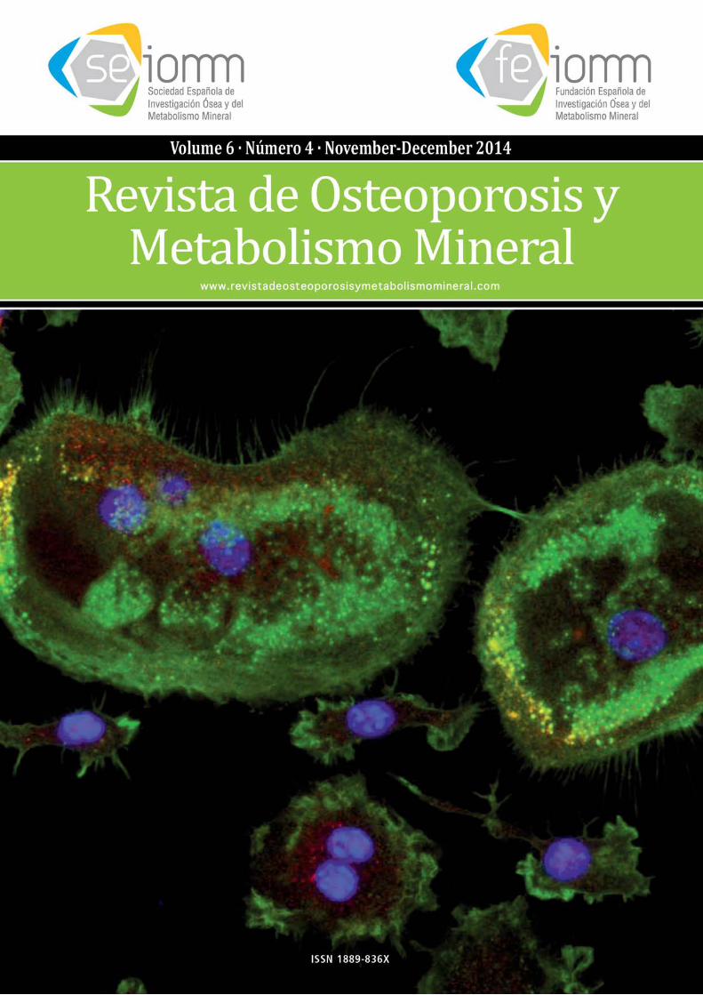
SUMMARY Vol. 6 - Nº 4 - November-December 2014Our coverOsteoclasts
Author:Marta Martín Millán
Sociedad Española de Investigación Ósea y del Metabolismo Mineral (SEIOMM)
PresidentFrancesc Xavier Nogués Solán
Vice-presidentJosé Manuel Olmos Martínez
SecretariatCarmen Gómez Vaquero
TreasureArancha Rodríguez de Cortazar
Vocal 1Cristina Carbonell Abella
Vocal 2Antonio Cano Sánchez
Paseo de la Castellana, 135 (7ª planta)28046 Madrid (Spain)
Telf: +34-917906834Fax: +34-917906869
e-mail: [email protected]
http://www.seiomm.org
Editing
Avda. Reina Victoria, 47 (6º D)28003 Madrid (Spain)
Telf. +34-915 538 297 e-mail: [email protected]://www.ibanezyplaza.com
Graphic designConcha García García
English translationAndrew Stephens
ImpresionGráficas 82, S.L.
Valid Support32/09-R-CM
Legal DepositM-3643-2013
ISSN 1889-836X
DirectorManuel Sosa Henríquez
Editor HeadMª Jesús Gómez de Tejada Romero
Reviewers Volume 6 (2014)
The Board and the Directorate SEIOMM Magazine thanks you for yourinvaluable assistance.
Luis Arboleya Rodríguez Teresita BellidoJosé Ramón Caeiro ReyJavier Calvo CataláCristina Carbonell AbellaSantos Castañeda Sanz Adolfo Díez PérezJuan José García Borrás Mª Jesús Gómez de Tejada RomeroDaniel Grinberg Vaisman Roberto Güerri Fernández Diego Hernández Hernández Gabriel Herrero-Beaumont CuencaFernando Marín Díez
Manuel Mesa RamosAna Monegal Broncós Mª Jesús Moro ÁlvarezXavier Nogués SolánJosé Luis Olmos Martínez Ramón Pérez Cano Pilar Peris BernalJavier del Pino MontesLuis del Río Barquero Arancha Rodríguez de Gortázar
Alonso-VillalobosRafael Sánchez Borrego Oscar Torregrosa Suau Carmen Valdés y Llorca
75COMMITTEES / Rev Osteoporos Metab Miner 2014 6;4:75
Pilar Aguado AcínMaría José Amérigo GarcíaAbdón Arbelo RodríguezMiguel Arias PacienciaEmilia Aznar VillacampaChesús Beltrán AuderaPere Benito RuizSantiago Benito UrbinaMiguel Bernard PinedaJosep Blanch i RubióJosé Antonio Blázquez CabreraJosé Ramón Caeiro ReyJavier Calvo CataláMª Jesús Cancelo HidalgoJorge Cannata AndíaAntonio Cano SánchezCristina Carbonell AbellaJordi Carbonell AbellóPedro Carpintero BenítezEnrique Casado BurgosSantos Castañeda SanzFidencio Cons MolinaSonia Dapia RobledaJesús Delgado CalleBernardino Díaz LópezCasimira Domínguez CabreraFernando Escobar JiménezJosé Filgueira RubioJordi Fiter AresteJuan José García BorrásJuan Alberto García Vadillo
Eduardo Girona QuesadaCarlos Gómez AlonsoMilagros González BéjarJesús González MacíasEmilio González ReimersJenaro Graña GilSilvana di GregorioDaniel Grinberg VaismanNuria Guañabens GayRoberto Güerri FernándezFederico Hawkins CarranzaDiego Hernández HernándezJosé Luis Hernández HernándezGabriel Herrero-Beaumont CuencaEsteban Jódar GimenoPau Lluch MezquidaJosé Andrés López-Herce CidMª Luisa Mariñoso BarbaGuillermo Martínez Díaz-GuerraMaría Elena Martínez RodríguezLeonardo Mellivobsky SaldierManuel Mesa RamosPedro Mezquita RayaAna Monegal BrancosJosefa Montoya GarcíaMaría Jesús Moro ÁlvarezManuel Muñoz TorresLaura Navarro CasadoManuel Naves GarcíaJosé Luis Neyro BilbaoXavier Nogués Solán
Joan Miquel Nolla SoléJosé Antonio Olmos MartínezNorberto Ortego CentenoSantiago Palacios Gil-AntuñanoEsteban Pérez AlonsoRamón Pérez CanoJosé Luis Pérez CastrillónPilar Peris BernalConcepción de la Piedra GordoJosé Manuel Quesada GómezEnrique Raya ÁlvarezRebeca Reyes GarcíaJosé Antonio Riancho MoralLuis de Río BarqueroLuis Rodríguez ArboleyaArancha Rodríguez de Gortázar
Alonso-Villalobos Minerva Rodríguez GarcíaAntonia Rodríguez HernándezManuel Rodríguez PérezInmaculada Ros VillamajóRafael Sánchez BorregoOscar Torregrosa SuauArmando Torres RamírezAntonio Torrijos EslavaCarmen Valdés y LlorcaCarmen Valero Díaz de LamadridAna Weruaga ReyMETHODOLOGY AND DESIGN OF DATA
Pedro Saavedra SantanaJosé María Limiñana Cañal
Committee of experts
Editorial Committee
Teresita Bellido. PhDDepartment of Medicine, Division of Endocrinology. IndianaUniversity School of Medicine. Indianapolis, Indiana. EstadosUnidos
Ernesto Canalis. MD, PhDDirector, Center for Skeletal Research. Professor of OrthopedicSurgery and Medicine New England Musculoskeletal InstituteUniversity of Connecticut Health Center. Farmington, CT.Estados Unidos
Oswaldo Daniel MessinaFacultad de Medicina. Universidad de Buenos Aires. HospitalCosme Argerich. Buenos Aires. Argentina
Patricia Clark Peralta. MD, PhDFacultad de Medicina, UNAM. Unidad Clínica Epidemiológica.Hospital Infantil Federico Gómez. México DF. México
Lilian I Plotkin. PhDAnatomy and Cell Biology. Indiana University School ofMedicine. Indianapolis, Indiana. Estados Unidos
Manuel Díaz CurielUniversidad Autónoma de Madrid. Unidad de MetabolismoÓseo. Hospital Fundación Jiménez Díaz. Instituto deInvestigación FJD. Fundación Hispana de Osteoporosis yMetabolismo Mineral (FHOEMO). Madrid. España
Adolfo Díez PérezUniversidad de Barcelona. Servicio de Medicina Interna.Instituto Municipal de Investigación Médica. (IMIM). Hospitaldel Mar. Barcelona. España
Francesc Xavier Nogués SolánUniversidad Autónoma de Barcelona. Unidad de Investigaciónen Fisiopatología Ósea y Articular (URFOA). Departamento deMedicina Interna, Parc de Salut Mar – RETICEF. Barcelona.España
Manuel Sosa Henríquez (Director)Universidad de Las Palmas de Gran Canaria. Grupo deInvestigación en Osteoporosis y Metabolismo Mineral. HospitalUniversitario Insular. Servicio de Medicina Interna. UnidadMetabólica Ósea. Las Palmas de Gran Canaria. España
María Jesús Gómez de Tejada Romero (Redactora Jefe)Universidad de Sevilla. Departamento de Medicina. Sevilla.España
EDITORIALS / Rev Osteoporos Metab Miner 2014 6;4:77-7877
Torrijos Eslava AResponsable de la Unidad Metabólica Ósea - Servicio de Reumatología - Hospital Universitario La Paz - Madrid
Paget’s disease of bone
n 14th November 1877, the British doc-tor James Paget presented to theMedical and Surgical Society ofLondon five cases of a condition whichwas called “Osteitis Deformans”, aslowly developing bone disease cha-
racterised by the lengthening, softening and defor-mation of the bones, above all affecting the cranialbones and the long bones of the lower limbs. Hepublished the first report in Medical-SurgicalTransactions in 1877, in which he described in detaila man he had treated over a period of 20 years1. Hesubsequently published, more cases in 1882 as wellas saying that he had not known that Czerney hadused the term “Osteitis Deformans” in 1873.Since this date many cases have been publishedand a large amount of information has beengathered relating to its etiology, prevalence, epi-demiology, diagnosis and treatment, and “OsteitisDeformans” is now known as Paget’s disease.Today, Paget’s disease of bone (PDB) is defined asa non-diffuse bone disease characterised by anincrease in bone remodelling whose principleagent is the osteoclast. It is an entity of unknownetiology, sited segmentally in different areas of theskeleton. PDB may affect any bone and may bemonostotic or polyostotic. The bones most affec-ted are the pelvis (up to 70%), femur (30-55%),lumbar spine (25-50%) cranium (20-40%) and tibia(15-30%). The disease progresses along the affec-ted bone and the appearance of a new locationsome years after the first diagnosis is very rare.This affectation leads to deformation of the bonewith an increase in its size and deformity whichmay produce bone pain, arthralgia and nervecompression syndromes in the cranial nerve pairs,spinal stenosis or compression of the spinal cord.It also results in a greater risk of fracture in theaffected long bones. It should also not be forgot-ten that pagetic tissue may suffer a neoplastictransformation with a higher incidence of sarco-mas, especially in the polyostotic type whichdevelop in 0.3-1% of cases2,3.PDB is asymptomatic in 50-75% of cases, and thedoctor is alerted when the typical deformitiesappear (increased growth in the skull or bowing
of the tibia), or when an increased level of alkali-ne phosphatase is detected in a routine analysis,or findings in an X-ray examination for anotherreason. In many cases the diagnosis of PDB ismade after the complications have occurred, andif the Paget’s is active, the markers for bone turno-ver are elevated. Among the markers for bone tur-nover the most useful appear to be amino-termi-nal telopeptide of collagen type 1, bone-specificalkaline phosphatase and amino-terminal propep-tide of procollagen 1. However, taking intoaccount its ease of use and low cost, the determi-nation of concentrations of alkaline phosphataseis still a valid alternative.The diagnosis of PDB is carried out primarilyusing X-rays with its characteristic images. Bonegammagraphy is not a specific method, but for usis useful to see the locations and spread of thedisease. CAT and MRI scans are useful in evalua-ting neurological symptoms in the context of PDBand may also be of use to determine the extentand nature of neoplastic degeneration of thePagetic tissue.PDB has an interesting geographic distribution.The highest incidence is found in the UnitedKingdom (4.5% in those over 55 years of age) andwithin this country the highest incidence is in thenortheast, with the best known concentrationbeing Lancashire in which 7% of the populationover 55 years of age is affected. It is quite com-mon in the northeast of France, Spain and Italy. InSpain the prevalence of PPDB is at least 1% inpeople over 55 years of age, with notable varia-tions according to geography and age. The bestknown predominant concentrations of PDB in ourcountry are those of the province of Salamancaand the Sierra Norte de Madrid (Northern Sierra ofMadrid) among others. It also occurs in the majo-rity of other European countries, with the excep-tion of the Scandinavian countries. In the rest ofthe world it is also common in countries whichhave seen high levels of immigration from Britainand other European countries during the 19th and20th centuries such as: Australia, New Zealand, theUnited States and some regions of Canada. PDB israre in the Indian sub-continent, Malaysia,
Oe-mail: [email protected]
Indonesia, China and Japan. The disease affectsboth sexes, with a slight predominance in men inmost series (the male/female ratio is approxima-tely 1.4:1 in the United Kingdom), is rare beforethe age of 50, and its prevalence increases withage and affects up to 5-8% in the eighth decadeof life in some countries4-7. Although there is nodoubt that PDB has a genetic basis, the incidenceand seriousness of this disease has diminishedover recent decades8-10. Those patients with PDB often have a family his-tory of the disease and it is estimated that the riskof PDB affectation in a first degree relative isincreased seven-fold. In many families the diseaseis inherited in an autosomal dominant fashionwith high incomplete penetrance, increasing withage. Great advances have been made in the last 15years in the understanding of the genetics of PDB.Linkage analysis has identified some loci whichare potential candidates in the chromosomes2p36, 5q31, 5q35, 10p13 y 18q2111.The genes and loci which predispose for PDBhave been identified through a correlation analy-sis of families. Among those genes and loci whichhave been associated with PDB or related syndro-mes are: CSF1 (located in 1p13), SQSTM1 (locatedin 5q35), in the 7q33 chromosome (the genesNUP205, SLC13A,4, and CNOT4), TM7SF4 (alsoknown as DCSTAMP, located in 8q22) TNFRSF11B(located in 8q24), VCP (located in 9p13), OPTN(located in 10p13), TNFRSF11A (located in 18q21)and RIN3 (located in 14q33) and in the chromo-some 15q24 (genes GOLGA6 and PML). In someof these the causal variant remains to be discove-red. More studies are still required to determinethe association of the different genes, as well asthe importance of environmental factors whichinfluence the development of PDB with thesegenetic alterations11-16.Some mutations of SQSTM1 may act as predispo-sing factors but are not sufficient to induce PDB,with additional factors (genetic or environmental)possibly being necessary10-12,17,18. Mutations of thisgene are the most common cause of familial PDB.Transverse studies indicate that 80% of the carriersof SQSMT1 mutations develop PDB in the eighthdecade of their lives. There are data which showthat the age of onset of the disease in families withPDB in the current generation, in those withSQSMT1 mutation, is delayed in comparison withtheir parents’ generation10-12,15. This emphasises theimportance of environmental factors in triggeringthe disease.To date, the precise molecular mechanisms whichlead to the development of pagetic lesions incarriers of the SQSMT1 mutation have not yetbeen defined. On the other hand, recent experi-mental data suggest that one or a number of envi-ronmental factors may be required to induce thecomplete pagetic phenotype in the presence ofthe SQSMT1 mutation. Since environmental fac-tors, some of them toxic, play a significant role inthe development of PDB, and as the response tothese factors is genetically conditioned, presented
in this number is a work by Dr Usategui-Martín etal.19, designed to determine whether the variabilityof some of the genes involved in the metabolismof exogenous toxins are related to the risk ofdeveloping PDB.
Bibliography
1. Paget J. On a form of chronic inflammation of bones(osteitis deformans). Med Chir Trans 1877;60:37-63.
2. Mankin HJ, Hornicek FJ. Paget’s sarcoma: A historical andoutcome review. Clin Orthop Relat Res 2005;438:97-102.
3. Hansen MF, Seton M, Merchant A. Osteosarcoma inPaget’s disease of bone. J Bone Miner Res 2006;21(Suppl 2):P58-63.
4. Ralston SH. Clinical practice. Paget's disease of bone.N Engl J Med 2013;368:644-50.
5. Del Pino J, Corral L, Miron JA, Morales A. Enfermedadde Paget: epidemiología y fisiopatología. En: TorrijosEslava A, coordinador. Enfermedad Ósea de Paget.Madrid: Medea;2001.p.11-42.
6. Del Pino J, Rodríguez M. Epidemiologia: consideracio-nes actuales. En: Guañabens Gay N, coordinadora.Enfermedad Ósea de Paget. Barcelona: SCM;2006.p.3-12.
7. Guañabens N, Garrido J, Gobbo M, Morales Piga A,Del Pino J, Torrijos A, et al. On behalf of the PAGETStudy Group. Prevalence of Paget's disease of bone inSpain. Bone 2008;43:1006-9.
8. Poor G, Donath J, Fornet B, Cooper C. Epidemiologyof Paget's disease in Europe: the prevalence is decrea-sing. J Bone Miner Res 2006;21:1545-9.
9. Cundy HR, Gamble G, Wattie D, Rutland M, Cundy T.Paget's disease of bone in New Zealand: continued decli-ne in disease severity. Calcif Tissue Int 2004;75:358-64.
10. Bolland MJ, Tong PC, Naot D, Callon KE, Wattie DJ,Gamble GD, et al. Delayed development of Paget'sDisease in offspring inheriting SQSTM1 mutations. JBone Miner Res 2007;22:411-5.
11. Ralston SH, Albagha OME, Genetics of Paget’s Diseaseof Bone. Curr Osteoporos Rep 2014;12:263-71.
12. Gennari L, Merlotti D, Rendina D, Gianfrancesco F,Esposito T, Ranuccio N. Paget’s disease of bone: epi-demiology, pathogenesis and pharmacotherapy.Expert Opin Orphan Drugs 2014;2:591-603.
13. Morissette J, Laurin N, Brown JP. Sequestosome 1:mutation frequencies, haplotypes, and phenotypes infamilial Paget's Disease of bone. J Bone Miner Res2006;21(Suppl 2):38-44.
14. Albagha OM, Visconti MR, Alonso N, Wani S,Goodman K, Fraser WD, et al. Common susceptibilityalleles and SQSTM1 mutations predict disease extentand severity in a multinational study of patients withPaget's disease. J Bone Miner Res 2013;28:2238-46.
15. Visconti MR, Langston AL, Alonso N, Goodman K, SelbyPL, Fraser WD, et al. Mutations of SQSTM1 are associa-ted with severity and clinical outcome in Paget's diseaseof bone. J Bone Miner Res 2010;25:2368-73.
16. Albagha OME, Wani S, Visconti MR, Alonso N,Goodman K, Cundy T, et al. Genome-wide associationidentifies three new susceptibility loci for Paget's dise-ase of bone. Nat Genet 2011;43:685-9.
17. Laurin N, Brown JP, Morissette J, Raymond V.Recurrent mutation of the gene encoding sequestoso-me 1 (SQSTM1/p62) in Paget Disease of bone. Am JHum Genet 2002;70:1582-8.
18. Hocking LJ, Lucas GJA, Daroszewska A, Mangion J,Olavesen M, Nicholson GC, et al. Domain specific muta-tions in Sequestosome 1 (SQSTM1) cause familial andsporadic Paget's disease. Hum Mol Genet 2002;11:2735-9.
19. Usategui-Martín R, Corral E, Alonso M, Calero-Paniagua I, Carranco-Medina TE, Quesada-Moreno A,et al. Estudio de las deleciones de los genes GSTM1 yGSTT1 y del polimorfismo Ile105Val del gen GSTP1 enpacientes con enfermedad ósea de Paget. RevOsteoporos Metab Miner 2014 6;4:83-8.
78EDITORIALS / Rev Osteoporos Metab Miner 2014 6;4:77-78
Olmo Quintana V1,2 y Martín Torres M1
1 Farmacéutico de Atención Primaria - Gerencia de Atención Primaria - Las Palmas de Gran Canaria2 Comité de Investigación y Ensayos Clínicos - Complejo Hospitalario Universitario Insular Materno Infantil - Las Palmas de Gran Canaria
Osteoporosis: a look into the future fromPrimary Care
rimary health care (PHC) is the firstpoint of contact for patients in thehealth care system and is key to thesuspicion of osteoporosis in postmeno-pausal women (PMO), as well as in theapproach to its diagnosis and treat-ment and the establishment of the risk
of fracture.Osteoporosis is the most common bone metabolicdisease in our environment, representing a seriouspublic health problem world-wide1, and specifi-cally in our country2. The prevalence of osteopo-rosis determined by bone densitometry in thelumbar spine is especially high after the menopau-se3,4. It is estimated that in Spain, one in threewomen over 50 years of age suffer from osteopo-rosis, increasing to one in two for those over 70years of age. Most of these patients are located inthe 55-80 years age range3,4, and it is estimated that4% of those patients over 50 years of age with ahip fracture will die during hospitalisation, and24% in the first year after fracture5. Vertebral frac-ture is the most common, and that of the hip themost serious and with a greater cost to the healthsystem, while there may also be other fragilityfractures such as the distal radius, humerus, riband tibia6. The different guides consider those patients inwhom two of the following factors exist, combi-ned with low bone mineral density (BMD)7, to beat high risk: being over 65 years of age; havingfamily history, especially maternal, of femoral frac-ture; prolonged consumption of corticoids: andlastly, having had falls.The aim of treatment is the prevention of fragilityfractures, in both the short- and long-term. Toachieve this, the correct identification of the originof the fracture – traumatic or due to fragility – willenable the correct diagnosis and the correct clini-cal decision regarding treatment. In our setting,we simply calculate the risk of the main osteopo-
rotic fractures, so-called major fractures (vertebral,hip, humerus and forearm), and of the hip at 10years, using the FRAX tool8. These do not indica-te the decision as to who to treat, but are the cli-nical criteria which govern that decision, whichmay follow the decision thresholds proposed bythe European Guide to Osteoporosis9, and aboveall, the guides of the different scientific societieswhich better fit our work environment. Thus, following the criteria of the CanadianScientific Advisory Council on Osteoporosis10, forwhom the risk is defined on the basis of the FRAXscores as: low risk (risk of fracture after 10 years<10%), intermediate risk (10-19%) and high risk(≥20%) for the main osteoporotic fractures, andlow risk (<3%) or high risk (≥3%) for hip fractu-res; or Qfracture®11,12. Both tools would both sup-port clinical decisions by identifying patients athigh/low risk of osteoporotic fracture, as well asdecision to treat, thus improving the predictiveparameters for Spanish women in a way which ismore cost-effective than the traditional modelbased on a T-score of ≤-2.5 in the DXA13. In respect of the increase in bone mass measuredthrough densitometry, frequently used the primarycare system in patients over the age of 65, this isnot a good variable with which to measure theefficacy of drugs14, this being one of the tests iden-tified as having little clinical value in a study in201215. In fact, a number of clinical trials have indi-cated that antiresorptive drugs continue to preventthe appearance of fractures, even though the BMDdiminishes. Therefore, DXA should not be carriedout in a generalised and indiscriminate way, notsimply on the basis of the age of the patient orwhen a postmenopausal women presents herself,but rather it should be requested on the basis ofthe presence of risk factors14. Regarding the efficacy of drugs used in the treat-ment of osteoporosis we must consider this to bevery limited in secondary prevention (previous
PCorrespondence: Vicente Olmo Quintanae-mail: [email protected]
EDITORIALS / Rev Osteoporos Metab Miner 2014 6;4:79-8279
fragility fractures), and, practically-speaking, notdemonstrated in primary prevention. There hasrecently been published a systematic review16,comparing the effectiveness of drugs used for thetreatment of osteoporosis, which updates thereview carried out in 201215, whose objective wasto review the evidence to determine the salientaspects of the efficacy and safety of drugs indica-ted for the prevention of fractures. A striking con-clusion of the study was the existence of goodquality evidence supporting the idea that somemedicines reduce the risk of fractures in peoplewith a BMD in the osteoporotic range and/or pre-vious vertebral or hip fractures, with a great varia-tion in in efficacy between bisphosphonates,denosumab and teriparatide, and with serious butvery uncommon adverse effects, such as atypicalsubtrochanteric fracture or osteonecrosis of thejaw. Also highlighted is the lack of direct compa-risons of the benefits, and the harm which resultsfrom indirect comparisons, which do not allowone to indicate which drug is more efficaciousthan another17.There is no evidence that early treatment in peo-ple below the age of 65 brings any benefits, nor isthere sufficient evidence to recommend treatmentfrom the age of 8016,18. So, in a study by Sanfélix-Genovés et al.19, they draw attention to the con-trast in the high levels of pharmacological treat-ment which exists in the region of Valencia andthe low prevalence of risk factors in adults (50-65years) which, coupled with an overuse of BMD,translates into a very significant impact on healthspending. We should not forget that the treatmentof first choice is still to consider changing harmfulhabits, taking physical exercise or avoiding falls,combined with a sufficient intake of calcium andvitamin D, which are as effective as the increase inBMD obtained through drug treatment, or evenmore so. Doctors in the Spanish health system areaware of the existence of the high comorbidity ofosteoporosis with cardiovascular risk factorswhich could indicate a closer physiopathologicalrelationship. For this reason, 9 out of 10 patientswho attend a clinic for osteoporosis receive infor-mation on healthy life styles, balanced diets andon how to achieve a sufficient intake of calcium19. However, in spite of the low efficacy of thesedrugs their consumption has shot up, multiplyingsix-fold in the last ten years. A report by the natio-nal health service in the UK which examined theuse of medicines for osteoporosis developedcountries put Spain as the top country in the pres-cription of these treatments, which is seriouslyincongruous if we take into account the fact thatthe incidence of osteoporosis in Spain is thelowest, not only in Europe, but in the world20. Thisis why drug treatment should be limited to highrisk patients who are going to be those who reallybenefit16,18,20. In all the guides to clinical practice consulted, thefirst choice pharmacological treatment, due totheir efficacy, safety and efficiency, are the bis-phosphonates, essentially alendronate and/or rise-
dronate2,9,10,17, which are those which have beenshown to be the most efficient in the differenttypes of fractures, with as a possible alternativethe oral administration of zoledronate in patientsat high risk, and/or with osteoporosis.Given the controversy over safety in recent years,in addition to the studies of new drugs and theircombinations, studies are also being conducted ofefficacy21, of efficacy compared with new molecu-les22,23, of treatment in men24 and of the optimumduration of treatments25. To date, the scientific evi-dence only justifies prolonged use over more thanfive years in highly selected patients.In those patients with intolerance or contraindica-tions for bisphosphonates, denosumab26, a SERMor strontium ranelate may be recommended. All ofthese have safety problems notified by PRAC (thePharmacovigilance Risk Assessment Committee ofthe European Medicines Agency), such as the tem-porary suspension of the sale in January 2014 ofstrontium ranelate due to an increase in seriouscardiac events, thromboembolisms and/or skinreactions, or the risk of atypical femoral fractureswith the use of denosumab, following the criticalreview which was carried out of the FREEDOMstudy26. In short, once a new medicine is authorised by theregulatory agencies and more in this type ofpathology, the information regarding its efficacyand safety comes from the baseline studies and,therefore, the data are very limited. This obliges usto limit its prescription or, at least be “conservati-ve” and, consequently, before prescribing a newdrug for this pathology a careful risk-benefitassessment must be carried out, as well as asses-sing its suitability for each patient as against alter-native more efficient therapies, with criticalreviews carried out by professionals which allowcomparison of the available information. This is anapproach which addresses the patients and notthe disease.One of the issues which most concerns medicalstaff is adherence to treatment by the patient. Inthe work of Martínez et al. two out of three parti-cipating doctors considered that there was a levelof non-adherence of 20%. Hence, one of the majorchallenges which doctors face is to successfullyincrease the adherence of patients to the recom-mendations and treatments they provide27, invol-ving all those concerned: patients, pharmacistsand doctors. Adherence is understood to be asdefined by the WHO in 2003, for whom it is: “thedegree to which the conduct of a patient, in rela-tion to the taking of medication, the following ofa diet or the modifications of lifestyle correspondwith the recommendations given by health profes-sionals”.It can be deduced from this definition that, apartfrom the professionals and their communicationwith their patients, adherence also depends on thepsychological connotations, experiences and kno-wledge of the patients themselves. Those peopleat risk of fragility fracture should take the oppor-tunity to take informed decisions about their care
EDITORIALS / Rev Osteoporos Metab Miner 2014 6;4:79-8280
and treatment, in collaboration with the healthprofessionals treating them. If the patient is inagreement, the families and carers should have theopportunity to participate in the decisions aboutthe patient’s treatment and care. The families andthe carers should also have the information andsupport they need.Good communication between health professio-nals and their patients is essential. This should besupported by written information based on theevidence, which suit the patients’ needs andwhich should be culturally appropriate. It shouldalso be accessible to those with additional needssuch as physical, sensory or learning disabilities.In short, in primary health care new paradigms arebeing opened up in relation to the management ofa chronic disease, as is osteoporosis, in which thepatients, in their environment, and the professio-nals involved should be jointly responsible for thedevelopment of their disease. It is known that awell-informed patient and a high degree ofempathy with their family doctor enables animprovement in the doctor-patient relationshipand, therefore, brings better health outcomes27.The health professionals face new challenges inimproving therapeutic efficiency at this first level ofcare. For this reason they must carry out an effecti-ve selection of patients according to their risk (weought not to forget that most patients with fracturesare older, polymedicated and with multiple comor-bidities and, up until now, not included in clinicaltrials), as well as the costs associated with their tre-atment and hospitalisation. In view of what liesahead, this requires the establishment of protocolsand guides to clinical practice agreed among thescientific societies in primary and specialised care(called “guides to guides”, given the multidiscipli-nary nature of treatment in this pathology) andbased on scientific evidence, which provide thoseaforementioned elements, creating integrated mul-tidisciplinary educational programmes for themanagement of osteoporosis which allow the con-tinuous updating and perfecting of the skills of doc-tors in primary care through active training pro-grammes based on28:1. Diagnostic and therapeutic decision algorithms.2. Criteria for referral/monitoring.3. Instructions for improving compliance4. Criteria for the quality of care, of treatments, of life.5. Cost criteria with evaluation of information onthe economic impact of osteoporosis and associa-ted fractures on their revenue.In the primary care sector we should be more acti-ve in the search for new diagnostics, essentiallypromoting the use of the FRAX® tool, in womenwho attend clinics with diseases in which there isdocumented a high comorbidity with osteoporosis.It would seem to be essential to improve the qua-lity of treatments of pluripathological and polyme-dicated patients, in whom new approaches, whichincorporate the use of new drugs with demonstra-ble action on the risk of fracture and with widely-spaced doses, may help to resolve problems ofadherence.
Throughout this process there will be key aspectsof the management of osteoporosis, recommenda-tions related to healthy lifestyles, diet, physicalexercise, prior to the initiation of drug treatment,together with the need to emphasise that patientsshould take the recommended daily doses of vita-min D and calcium.
Declaration of conflict of interest: The authorsdeclare that they have no conflicts of interest.
Bibliography
1. National Osteoporosis Foundation. Clinician, guide toprevention and treatment of osteoporosis. Washington,2010.
2. González J, Guañabens N, Gómez C, Del Rio L, Muñoz M,Delgado M, et al. Guías de práctica clínica en la osteoporo-sis posmenopáusica, glucocorticoidea y del varón.(SEIOMM) Rev Clin Esp 2008; 2008 (suppp1):1-28.
3. De Felipe R, Cáceres C, Cima M, Dávila G, FernándezS, Ruiz T. Características clínicas de los pacientes contratamiento para la osteoporosis en un centro deAtención Primaria. ¿A quien tratamos en nuestras con-sultas? Aten Prim 2010;42:559-63.
4. Díaz Curiel M. Actualización de osteoporosis Madrid,FHOEMO; 2001.
5. Osteoporosis en Atención Primaria. Anónimo.Protocolos 4/2011 FMC 2011:7-32 Disponible en:http://www.elsevierinstituciones.com/ficheros/pdf/45/45v18nProtocolo_4a90034624pdf001.pdf [Accedido06/12/2014].
6. Sosa M, Hernández D. Protocolo de actuación ante dossituaciones en osteoporosis frecuentes en AtenciónPrimaria: Cuándo tratar siempre y cuándo evitar trata-mientos innecesarios. Medicine 2014;11:3567-70.
7. Pérez Edo L, Alonso A, Roig D, García A, GuañabensN, Peris P, et al. Actualización 2011 del ConsensoSociedad Española de Reumatología de Osteoporosis.Reumatol Clin 2011;7:357-79.
8. Prieto-Alhambra D, Pagés Castellá A. Estimación delriesgo de fractura mediante la Escala FRAX®. FMC2010;17:473-4.
9. Kanis JA, Burlet N, Cooper C, Delmas PD, Reginster JY,Borgstrom F, et al. European guidance for the diagno-sis and management of osteoporosis in postmenopau-sal women. Osteoporos Int 2008;19:399-428.
10. Papaioannou A, Morin S, Cheung AM, Atkinson S,Brown JP, Feldman S, et al. 2010 clinical practice gui-deline for the diagnosis and management of osteopo-rosis in Canada: Summary. CMAJ 2010;182:1864-73.
11. Hippisley-Cox J, Coupland C. Derivation and valida-tion of updated QFracture algorithm to predict risk ofosteoporotic fracture in primary care in the UnitedKingdom: prospective open cohort study. BMJ2012;344:e3427.
12. Hippisley-Cox J, Coupland C. Predicting risk of osteo-porotic fracture in men and women in England andWales: prospective derivation and validation ofQfracture Scores. BMJ 2009;339:b4229.
13. Azagra R, Roca G, Martín-Sánchez JC, Casado E,Encabo G, Zwart M, et al. Umbrales de FRAX® queidentifica personas con alto o bajo riesgo de fracturaosteoporótica en población femenina española. MedClin (Barc) 2014;144:1-8.
14. Jamart L, Herrero S, Barrera C. ¿Está justificado el gastoen fármacos contra la osteoporosis? FMC 2011;18:317-20.
15. Qassem A, Alguire P, Dalla P, Feinberg LE, FitzgeraldFT, Horwitch C, et al. Appropriate Use of Screeningand Diagnostic Tests to Foster High-Value, Cost-Conscious Care. Ann Intern Med 2012;156:147-9.
16. Crandall CJ, Newbery SJ, Diamant A, Lim YW, Gellad WF,Booth MJ, et al. Comparative effectiveness of pharmaco-logic treatments to prevent fractures: An update
EDITORIALS / Rev Osteoporos Metab Miner 2014 6;4:79-8281
Systematic Review. Ann Intern Med 2014;161:711-23. 17. Montes E, Bruno S, Cantabrana A, Sosa M, Arnaíz A,
Plasencia M, et al. Osteoporosis en la postmenopáusi-ca. Boletín Canario de Uso Racional del Medicamento(Bolcan) 2012;41-8.
18. Effective health Care Program. Treatment To PreventFractures in Men and Women With Low Bone Densityor Osteoporosis: Update of a 2007 Report. Disponibleen: http://effectivehealthcare.ahrq.gov/ehc/products/160/1007/CER53_LowBoneDensity_FinalReport_20120823.pdf [accedido 06/12/2014].
19. Sanfélix-Genovés J, Sánfelix-Genovés G, Peíro S,Hurtado C, Fluixá C, Fuertes J, et al. Prevalence ofosteoporotic fracture risk factors and anti-osteoporotictreatment in the Valencia región, Spain. The Baselinecharacteristis of the ESOSVAL cohort. Osteoporos Int2013;24:1045-55.
20. Kanis JA, Johnell O, De Laet C, Jonsson B, Oden A,Ogelsby AK. International variations in hip fractureprobabilities: implications for risk assesment. J BoneMiner Res 2002;17:1237-44.
21. McClung MR, Grauer A, Boonen S, Boloñesa MA,Marrón JP, Diez-Pérez A, et al. Romosozumab in postme-nopausal women with low bone mineral density. NEngl J Med 2014;370:412-20.
22. Tsai JN, Uihlein AV, Lee H, Kumbhani R, Siwila-Sackman E, McKay EA, et al. Teriparatide andDenosumab, alone or combined, in women with pos-tmenopausal osteoporosis: the DATA study randomi-
sed Trial DATOS. Lancet 2013;382:50-6. 23. Vertebral Fracture Treatment Comparisons in
Osteoporotic Women (VERO) Bethesda, MD: InstitutoNacional de Salud; Octubre 16 2012 [actualizado 11 dejulio 2014] [accedido 07/12/2014 http://clinicaltrials.gov/ct2/show/study/NCT01709110].
24. Combination Risedronate-Parathyroid Hormone Trialin Male Osteoporosis (RPM) Bethesda, MD: InstitutoNacional de Salud; 29 de mayo 2012 [actualizado 15 deenero 2014] [Accedido 07/12/2014 http://clinicaltrials.gov/show/NCT01611571].
25. Comparison of the Effect of an Ongoing TreatmentWith Alendronate or a Drug Holiday on the FractureRisk in Osteoporotic Patients With BisphosphonateLong Term Therapy (BILANZ) Disponible en:http://clinicaltrials.gov/show/NCT01512446 [accedido06/12/2014].
26. Cummings S, San-Martín J, McClung MR, Siris ES,Eastell R, Reid AR, et al. Denosumab for prevention offractures in posmenopausal women with osteoporosis.N Engl J Med 2009;361:756-65.
27. Vargas Negrín F. Adherencia al tratamiento: un retodifícil pero posible. Rev Osteoporos Metab Miner2014;6:5-7.
28. Martínez D, Abad P, Orero A, Navarro A, González J.Olmo V. Estudio socio sanitario de la osteoporosis pos-tmenopáusica en Atención Primaria. Estudio ESTOP-MAP. IMC International Marketing Communication.Madrid 2013..
EDITORIALS / Rev Osteoporos Metab Miner 2014 6;4:79-8282
Usategui-Martín R1,2, Corral E1, Alonso M1, Calero-Paniagua I2,3, Carranco-Medina TE2,3, Quesada-Moreno A2,3, Sánchez-González MD2,3,Hidalgo-Calleja C2,3, Pérez-Garrido L2,3, Montilla Morales C2,3,4, Mirón-Canelo JA5, González-Sarmiento R1,2,4, del Pino-Montes J2,3,4
1 Unidad de Medicina Molecular - Departamento de Medicina - Universidad de Salamanca2 Instituto de Investigación Biomédica de Salamanca (IBSAL)3 Servicio de Reumatología - Hospital Universitario de Salamanca4 Red Temática de Investigación Cooperativa en Envejecimiento y Fragilidad (RETICEF)5 Departamento de Medicina Preventiva, Salud Pública y Microbiología Médica - Universidad de Salamanca
Study of the deletions in the GSTM1 and GSTT1genes and of the Ile105Val polymorphism of theGSTP1 gene in patients with Paget’s disease of bone
Correspondence: Javier del Pino Montes - Servicio de Reumatología - Hospital Universitario de Salamanca - Pº SanVicente, 182 - 37007 Salamanca (Spain)e-mail: [email protected]
Date of receipt: 29/08/2014Date of acceptance: 23/11/2014
ORIGINALS / Rev Osteoporos Metab Miner 2014 6;4:83-8883
SummaryBackground: Paget’s disease of bone (PDB) is a disorder focussed on the bone with an increase in thenumber, size and activity of the osteoclasts. Some epidemiological data support the theory of its relations-hip with toxic or infectious environmental agents, whose interaction with some predisposing genetic alte-rations may lead to PDB. The glutathione S-transferases (GST) are involved in the metabolism of toxins,by catalysing the nucleophilic attack of the physiological substrate, reduced glutathione or GSH (g-Glu-Cys-Gly) on the electrophilic centre of a great number of toxic structures. We studied whether the varia-bility of the GSTM1, GSTP1 and GSTT1 genes is related to the risk of developing PDB. Patients and methods: We analysed 148 patients diagnosed with PDB, and 207 control individuals mat-ched in sex and age with no history of bone alterations. Using genomic DNA obtained from peripheralblood the presence-absence of the GSTM1 and GSTT1 genes was studied by means of multiplex PCR.The study of the Ile105Val GSTP1 gene was carried out using PCR and subsequent digestion with the res-triction enzyme BsmAI. The distribution of genotypes was analysed by means of the Pearson chi-squaretest. When statistically significant differences were found we carried out a multivariate logistical regres-sion to determine the risk which the presence of a particular genotype could generate. We used the CSPSS21.0 program. Differences were considered to be statistically significant when the value of p<0.05.Results: We found differences in the distribution of the presence-absence of the deletion in the GSTM1gene; not being a carrier for the deletion or being a heterozygous carrier in the GSTM1 gene confers alower risk of developing PDB (OR=0.56, 95% CI: 0.36-0.87; p=0.011). In the study of the GSTT1 andGSTP1 genes there were no significant differences.Conclusion: The detoxifying activity diminishes when two copies of the GSTM1 gene with deletions areinherited by reducing in enzyme activity, which has been associated with a greater susceptibility to somecancers, alcoholic hepatopathy and other inflammatory problems. We are not aware of any descriptionof its association with PDB. PDB is observed more frequently in carriers of the homozygous deletion inthe GSTM1 gene. This fact could explain the epidemiological findings which link PDB to exposure to cer-tain environmental agents.
Key words: Paget’s disease of bone, glutathione S-transferase, genetics, polymorphism.
Introduction Paget’s disease of bone is the most common bonemetabolic disease after osteoporosis1. It is a bonedisorder characterised by an increase in bone tur-nover in a disorganised way: a large increase inbone resorption, followed by bone formation ofthe same proportions. The result is bone with astructure which is irregular and anarchic, whichalters its morphology and mechanical properties.Some patients are asymptomatic, while othershave pain, degenerative arthropathy, fractures,bone deformities, deafness or other syndromes ofnerve compression. The main change resides inthe osteoclasts which increase in number size andactivity2,3.
There are currently two etiopathogenic hypo-theses which attempt to explain the origin of PDB:the influence of environmental factors and theexistence of genetic determinants1.
There is evidence that genetic changes play asignificant role in the development of the disease.There is a strong tendency to family aggregation(15-40%), with a seven-fold increase in relativerisk of suffering the disease in families of patientswith PDB3-6. In most of these families there is anautosomal dominant pattern with high penetrancein the sixth decade3. Recently, alterations in thegenes SQSTM1, CSF1, OPTN, TNFRSF andTM7SF47,8, have been associated with a higher riskof developing PDB. Some epidemiological data,such as its heterogeneous distribution, or morerecent changes in its incidence and seriousness,support the idea of the influence of environmen-tal factors in the development of the disease. Itsassociation with diets poor in calcium and vitaminD during infancy9,10, exposure to environmentaltoxins11, contact with animals during infancy12-14,consumption of non-controlled meat15, consump-tion of untreated water16 and infectious agentssuch as viruses (paramixoviridae)14,17,18, have beenreported.
Neither environmental nor genetic factorsalone explain the etiopathogeny of this disease.The most accepted model considers PDB to be theresult of the synergetic action of both environmen-tal and genetic factors. The genetic determinationwould explain the individual susceptibility todeveloping the disease following the exposure tothe participating environmental factor2.
Involved in the metabolization of toxins are theglutathione S-transferases (GSTs). These are afamily of enzymes which participate in cell deto-xification. These enzymes catalyse the nucleophi-lic attack of the physiological substrate, reducedglutathione or GSH (g-Glu-Cys-Gly) on the elec-trophilic centres of a great number of toxic struc-tures, allowing their degradation. They are classi-fied in seven families (alpha, kappa, mu, pi,sigma, theta and zeta) which are differentiatedboth in their sequences, as well as in their immu-nological properties and physiological roles19,20.GSTM1, GSTP1 and GSTT are the most studiedGSTs, and those which have been most commonlyassociated with human pathologies21.
The role of environmental factors, some ofwhich are toxic, appears to be significant in thedevelopment of the disease. Given that the indivi-dual response to the toxic factors is geneticallydetermined, we have designed this study with theaim of trying to determine whether the variabilityof the GSTM1 GSTP1 and GSTT1 genes (involvedin the metabolization of exogenous toxins) is rela-ted to the risk of developing PDB.
Materials and methodsPatients and controlsWe studied 148 patients diagnosed with PDB. Inthe case of patients with family history of the dise-ase, we only selected one patient per family toavoid familial genotype bias. The patients werediagnosed by the rheumatology service of theUniversity Hospital of Salamanca. As a controlgroup, we analysed 207 individuals, matched insex and age with the group of patients, withouthistory of bone alterations, and coming from thesame geographic area. From each of the patientswere gathered clinical characteristics such as sex,age at diagnosis, family history, number of bonesaffected, presence of fractures, affectation of skulland affectation of cranial nerve pairs. All the sub-jects studied, both in the group of patients and thecontrol group, gave their informed consent to par-ticipate in the study, which was approved by theethics committee of the hospital.
DNA extraction and analysis of polymorphismsIn both the patient and control groups the extrac-tion of genomic DNA from peripheral blood wascarried out using the standard phenol-chloroformprocedure.
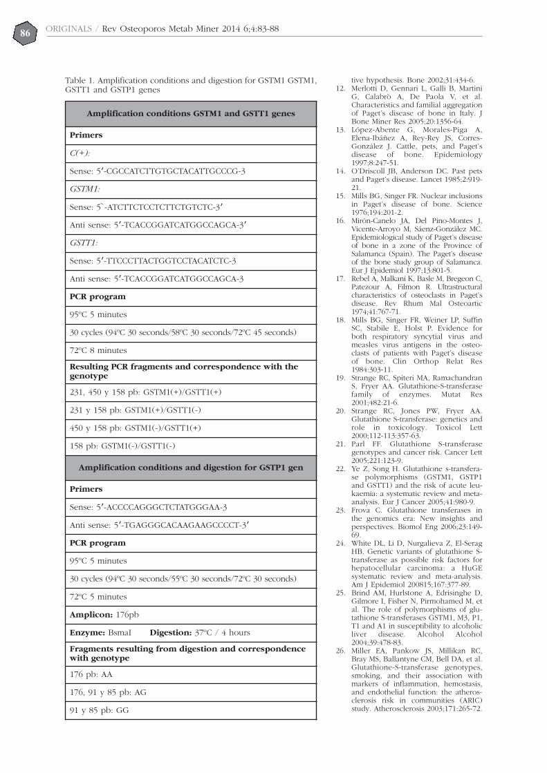
The study of the presence-absence of the dele-tions in the GSTM1 and GSST1 genes was carriedout using multiplex PCR under conditions descri-bed in Table 1. The study of the Ile105Val poly-morphism of the GSTP1 gene was conductedusing PCR and subsequent digestion with the res-triction enzyme BsmAl. The conditions used areset out in Table 1.
Statistical analysisThe distribution of genotypes among patients andcontrols was analysed using the Pearson chi-squa-re test. In those polymorphisms in which statisti-cally significant differences were found we carriedout a multivariate logistical regression to determi-ne the risk which the presence of a particulargenotype could generate. The statistical analysiswas carried out using the SPSS 21.0 program.Those differences whose p value was <0.05 wereconsidered as statistically significant.
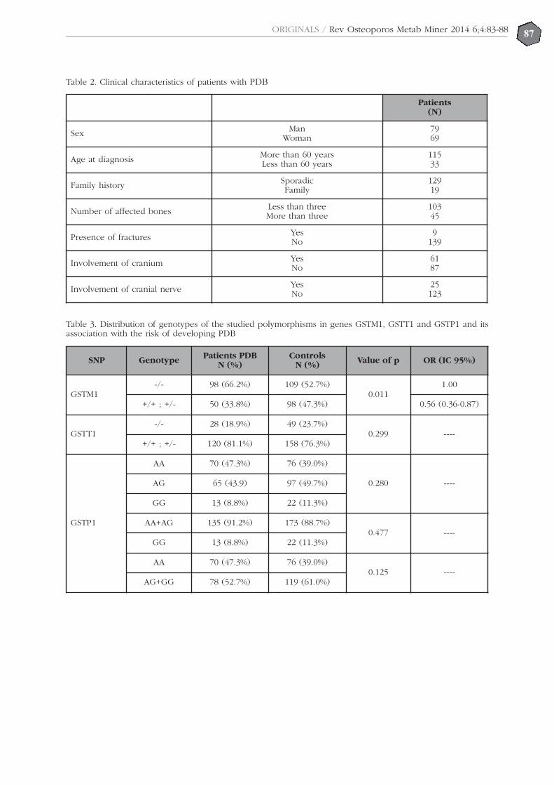
ResultsWe studied a total of 148 patients and 207 con-trols. The clinical characteristics of the patients areset out in Table 2. The distribution of the presen-ce-absence of deletions in the GSTM1 and GSST1genes and the distribution of the genotypes for theIle105Val polymorphism in the GSTP1 gene, and
ORIGINALS / Rev Osteoporos Metab Miner 2014 6;4:83-8884
their relationship to the risk of developing PDBare shown in Table 3.
We found statistically significant differences inthe distribution of the presence-absence of dele-tion in the GSTM1 gene: not being a carrier for thehomozygous deletion in the GSTM1 gene confersa lower risk of developing PDB (OR=0.56, CI 95%:0.36-0.87; p=0.011). In the study of the GSTTT1and GSTP1 genes we found no statistically signifi-cant differences (Table 3).
No statistically significant differences werefound in the analysis of the clinical characteristicsof the patients in relation to the variability of theGSTM1, GSTT1 and GSTP1 genes.
DiscussionPDB lesions occur as the result of an increase inbone resorption followed by an increase in its for-mation. The main change is located in the osteo-clasts which increase in number, size and activity.There is a range of evidence which indicates thatthe etiopathogeny of the disease is a synergy bet-ween a series of environmental factors and theexistence of certain genetic determinants2.Through a study of the variability of the GSTM1,GSTT1 and GSTP1 genes (involved in the metabo-lism of endogenous toxins) we intended to eva-luate the relationship between these variables andthe risk of developing PDB. As far as we know,this is the first work which examines the influen-ce of the changes in these genes on the develop-ment of this disease.
The GSTM gene is located in the 1p13 chromo-some, and to date, five allelic variants are known:GSTM1, GSTM2, GSTM3, GSTM4 and GSTM5. Areduction in detoxification activity occurs whenthe deletion in gene GSTM1 is inherited, meaningthat being a homozygous carrier of a deletion inthe GSTM1 gene causes a reduction in enzymeactivity. The theta class of GSTs comprises twogenes which code for the two proteins GSTT1 andGSTT2. As with the GSTM1 gene, if a homozygousdeletion in the GSTT1 gene is inherited, there is areduction in detoxification activity. In terms of thesub-family of GSTP, it comprises a single geneGSTP1 in which have been described two allelicvariants which differ in the base 313 of the cDNA,one adenine (A) being substitute by a guanine(G). This difference results in a change of a valine(Val) to an isoleucine (Ile) in the 105 codon of theamino-acid sequence, causing a defective bondbetween the enzyme and the substrate, and thus,a reduction detoxification activity19,20,22,23.
Being a homozygous carrier for deletion in theGSTM1 and/or GSTT1 genes has been associatedwith a greater susceptibility to developing diffe-rent types of cancer21,22,24, alcohol-related liver dise-ase25 and other inflammatory diseases25,26, becauseit causes poorer metabolization of toxic agents,with the synthesis of free radicals which damageDNA20. Our results show that not being a homozy-gous carrier for the deletion in the GSTM1 genebrings a lower risk of suffering PDB. In the studyof the GSTT1 and GSTP1 genes we found no sta-
tistically significant difference between the patientand control groups. We found no statistically sig-nificant differences between the clinical expres-sion, extent and activity of the disease in relationto the variability in the GSTM1, GSTT1 and GSTP1genes in the group of patients with PDB.
One of the causes postulated as the origin ofPDB is exposure to environmental toxins from theproduction of cotton, meat or drinking waterwithout adequate control of sanitation, which mayalter the maturation and activity of the osteoclasts,the increase in activity fostering the developmentof PDB11,15,16. Our hypothesis is that to have thehomozygous deletion in the GSTM1 gene assumespoor metabolization of environmental toxinswhich, by a mechanism yet unknown, may incre-ase the function of the osteoclasts and osteoclastprecursors which, combined with other geneticchanges not yet well described, could result in thedevelopment of PDB.
In conclusion, in those individuals who arecarriers of the GSTM1 gene with homozygousdeletions, PDB is more frequently observed. Thisfact could explain the epidemiological findingswhich associate PDB with exposure to certainenvironmental agents. Even so, functional studiesof these polymorphisms are required in order tovalidate our hypothesis.
Bibliography
1. Ralston SH, Layfield R. Pathogenesis of Paget Diseaseof Bone. Calcif Tissue Int 2012;91:97-113.
2. Singer FR, Mills BG, Gruber HE, Windle JJ, RoodmanGD. Ultrastructure of bone cells in Paget’s disease ofbone. J Bone Miner Res 2006;21(Suppl 2):P51-4.
3. Morales-Piga AA, Rey-Rey JS, Corres-González J,García-Sagredo JM, López-Abente G. Frequency andcharacteristics of familial aggregation of Paget’s disea-se of bone. J Bone Miner Res 1995;10:663-70.
4. Morissette J, Laurin N, Brown JP. Sequestosome 1:mutation frequencies, haplotypes, and phenotypes infamilial Paget’s disease of bone. J Bone Miner Res2006;21(Suppl 2):P38-44.
5. Siris ES, Ottman R, Flaster E, Kelsey JL. Familial aggre-gation of Paget’s disease of bone. J Bone Miner Res1991;6:495-500.
6. Hocking LJ, Herbert CA, Nicholls RK, Williams F, BennettST, Cundy T, et al. Genomewide search in familial Pagetdisease of bone shows evidence of genetic heteroge-neity with candidate loci on chromosomes 2q36, 10p13,and 5q35. Am J Hum Genet 2001;69:1055-61.
7. Albagha OM, Visconti MR, Alonso N, Langston AL,Cundy T, Dargie R, et al. Genome wide associationstudy identifies variants at CSF1, OPTN andTNFRSF11A as genetic risk factors for Paget’s diseaseof bone. Nat Genet 2010;42:520-4.
8. Albagha OME, Wani SE, Visconti MR, Alonso N,Goodman K, Brandi ML, et al. Genome-wide associa-tion identifies three new susceptibility loci for Paget’sdisease of bone. Nat Genet 2011;43:685-9.
9. Barker DJ, Gardner MJ. Distribution of Paget’s diseasein England, Wales and Scotland and a possible rela-tionship with vitamin D deficiency in childhood. Br JPrev Soc Med 1974;28:226-32.
10. Siris ES. Epidemiological aspects of Paget’s disease:family history and relationship to other medical condi-tions. Semin Arthritis Rheum 1994;23:222-5.
11. Lever JH. Paget’s disease of bone in Lancashire andarsenic pesticide in cotton mill wastewater: a specula-
ORIGINALS / Rev Osteoporos Metab Miner 2014 6;4:83-8885
ORIGINALS / Rev Osteoporos Metab Miner 2014 6;4:83-8886
tive hypothesis. Bone 2002;31:434-6. 12. Merlotti D, Gennari L, Galli B, Martini
G, Calabrò A, De Paola V, et al.Characteristics and familial aggregationof Paget’s disease of bone in Italy. JBone Miner Res 2005;20:1356-64.
13. López-Abente G, Morales-Piga A,Elena-Ibáñez A, Rey-Rey JS, Corres-González J. Cattle, pets, and Paget’sdisease of bone. Epidemiology1997;8:247-51.
14. O’Driscoll JB, Anderson DC. Past petsand Paget’s disease. Lancet 1985;2:919-21.
15. Mills BG, Singer FR. Nuclear inclusionsin Paget’s disease of bone. Science1976;194:201-2.
16. Mirón-Canelo JA, Del Pino-Montes J,Vicente-Arroyo M, Sáenz-González MC.Epidemiological study of Paget’s diseaseof bone in a zone of the Province ofSalamanca (Spain). The Paget’s diseaseof the bone study group of Salamanca.Eur J Epidemiol 1997;13:801-5.
17. Rebel A, Malkani K, Basle M, Bregeon C,Patezour A, Filmon R. Ultrastructuralcharacteristics of osteoclasts in Paget’sdisease. Rev Rhum Mal Osteoartic1974;41:767-71.
18. Mills BG, Singer FR, Weiner LP, SuffinSC, Stabile E, Holst P. Evidence forboth respiratory syncytial virus andmeasles virus antigens in the osteo-clasts of patients with Paget’s diseaseof bone. Clin Orthop Relat Res1984:303-11.
19. Strange RC, Spiteri MA, RamachandranS, Fryer AA. Glutathione-S-transferasefamily of enzymes. Mutat Res2001;482:21-6.
20. Strange RC, Jones PW, Fryer AA.Glutathione S-transferase: genetics androle in toxicology. Toxicol Lett2000;112-113:357-63.
21. Parl FF. Glutathione S-transferasegenotypes and cancer risk. Cancer Lett2005;221:123-9.
22. Ye Z, Song H. Glutathione s-transfera-se polymorphisms (GSTM1, GSTP1and GSTT1) and the risk of acute leu-kaemia: a systematic review and meta-analysis. Eur J Cancer 2005;41:980-9.
23. Frova C. Glutathione transferases inthe genomics era: New insights andperspectives. Biomol Eng 2006;23:149-69.
24. White DL, Li D, Nurgalieva Z, El-SeragHB. Genetic variants of glutathione S-transferase as possible risk factors forhepatocellular carcinoma: a HuGEsystematic review and meta-analysis.Am J Epidemiol 200815;167:377-89.
25. Brind AM, Hurlstone A, Edrisinghe D,Gilmore I, Fisher N, Pirmohamed M, etal. The role of polymorphisms of glu-tathione S-transferases GSTM1, M3, P1,T1 and A1 in susceptibility to alcoholicliver disease. Alcohol Alcohol2004;39:478-83.
26. Miller EA, Pankow JS, Millikan RC,Bray MS, Ballantyne CM, Bell DA, et al.Glutathione-S-transferase genotypes,smoking, and their association withmarkers of inflammation, hemostasis,and endothelial function: the atheros-clerosis risk in communities (ARIC)study. Atherosclerosis 2003;171:265-72.
Amplification conditions GSTM1 and GSTT1 genes
Primers
C(+):
Sense: 5′-CGCCATCTTGTGCTACATTGCCCG-3
GSTM1:
Sense: 5`-ATCTTCTCCTCTTCTGTCTC-3′
Anti sense: 5′-TCACCGGATCATGGCCAGCA-3′
GSTT1:
Sense: 5′-TTCCCTTACTGGTCCTACATCTC-3
Anti sense: 5′-TCACCGGATCATGGCCAGCA-3
PCR program
95ºC 5 minutes
30 cycles (94ºC 30 seconds/58ºC 30 seconds/72ºC 45 seconds)
72ºC 8 minutes
Resulting PCR fragments and correspondence with thegenotype
231, 450 y 158 pb: GSTM1(+)/GSTT1(+)
231 y 158 pb: GSTM1(+)/GSTT1(-)
450 y 158 pb: GSTM1(-)/GSTT1(+)
158 pb: GSTM1(-)/GSTT1(-)
Amplification conditions and digestion for GSTP1 gen
Primers
Sense: 5′-ACCCCAGGGCTCTATGGGAA-3
Anti sense: 5′-TGAGGGCACAAGAAGCCCCT-3′
PCR program
95ºC 5 minutes
30 cycles (94ºC 30 seconds/55ºC 30 seconds/72ºC 30 seconds)
72ºC 5 minutes
Amplicon: 176pb
Enzyme: BsmaI Digestion: 37ºC / 4 hours
Fragments resulting from digestion and correspondencewith genotype
176 pb: AA
176, 91 y 85 pb: AG
91 y 85 pb: GG
Table 1. Amplification conditions and digestion for GSTM1 GSTM1,GSTT1 and GSTP1 genes
87ORIGINALS / Rev Osteoporos Metab Miner 2014 6;4:83-88
Table 2. Clinical characteristics of patients with PDB
Table 3. Distribution of genotypes of the studied polymorphisms in genes GSTM1, GSTT1 and GSTP1 and itsassociation with the risk of developing PDB
Patients(N)
Sex ManWoman
7969
Age at diagnosis More than 60 yearsLess than 60 years
11533
Family history SporadicFamily
12919
Number of affected bones Less than threeMore than three
10345
Presence of fractures YesNo
9139
Involvement of cranium YesNo
6187
Involvement of cranial nerve YesNo
25123
SNP Genotype Patients PDBN (%)
ControlsN (%) Value of p OR (IC 95%)
GSTM1-/- 98 (66.2%) 109 (52.7%)
0.0111.00
+/+ ; +/- 50 (33.8%) 98 (47.3%) 0.56 (0.36-0.87)
GSTT1-/- 28 (18.9%) 49 (23.7%)
0.299 ----+/+ ; +/- 120 (81.1%) 158 (76.3%)
GSTP1
AA 70 (47.3%) 76 (39.0%)
0.280 ----AG 65 (43.9) 97 (49.7%)
GG 13 (8.8%) 22 (11.3%)
AA+AG 135 (91.2%) 173 (88.7%)0.477 ----
GG 13 (8.8%) 22 (11.3%)
AA 70 (47.3%) 76 (39.0%)0.125 ----
AG+GG 78 (52.7%) 119 (61.0%)

88ORIGINALS / Rev Osteoporos Metab Miner 2014 6;4:83-88
89ORIGINALS / Rev Osteoporos Metab Miner 2014 6;4:89-96
Higueras Linares T1,2, Sosa Cabrera N1, Blanco Blanco J3, Fernández Palacio LM1, Sosa Henríquez M4,5
1 Centro de Salud de Tejina - Tenerife2 Grupo de Aparato Locomotor SOCAMFYC - Tenerife3 Centro de Salud de Tacoronte - Tenerife4 Universidad de Las Palmas de Gran Canaria - Instituto Universitario de Investigaciones Biomédicas y Sanitarias - Grupo de Investigación de Osteoporosisy Metabolismo Mineral - Las Palmas de Gran Canaria5 Servicio de Medicina Interna - Unidad Metabólica Ósea - Complejo Hospitalario Universitario Materno-Insular - Las Palmas de Gran Canaria
Knowledge of osteoporosis, and the pharmaceuticalexpenditure it entails, in the primary health caresystem of the Canary Islands
Correspondence: Tomás Higueras Linares - Centro de Salud de Tejina - Tenerife - c/Camino las Toscas, 5 - Vallede Guerra - La Laguna - 38270 Santa Cruz de Tenerife (Spain)e-mail: [email protected]
Date of receipt: 15/10/2014Date of acceptance: 22/12/2014
SummaryBackground: Osteoporosis is a disease which can be managed by different specialisms, one of which isthe family doctor. In this study we analyse the knowledge of osteoporosis, and the diagnostic and thera-peutic approach taken to this disease, among primary care doctors in the Canarian archipelago, as wellas making a first approximation of the expenditure on drugs used to treat this disease in 2013.Material and method: Observational, descriptive, transverse study conducted between May 2013 and May2014 with all primary care doctors in the Canarian health service. An anonymous survey covering 13points was developed. The capture of the data about expenditure on drugs was facilitated by the servi-ce for the control of supply and rational use of medicines of the Canarian health service.Results: 28.60% of primary care doctors in the Canarian archipelago responded to the survey. Of these,75.30% reported using risk factors in evaluating the risk of fractures. Not a very high percentage, appro-ximately half of the respondents, request densitometries, while 28.60% routinely use scales for the eva-luation of risk of fracture and 32.80% use them occasionally. 90% of the professionals recommend non-pharmacological measures for the prevention of fractures in their patients, although 91% do not normallyrequest a determination of blood levels of vitamin D. In 2013 the expenditure on drugs for osteoporosis by the Canarian health service amounted to € 7,684,393.61,of which € 7,265,491.06 was in primary care.Conclusions: The Canarian primary care doctors who responded to the survey had, in general, a good kno-wledge of osteoporosis, and of its risk factors, but focussed their professional activity more on preventionthan treatment. The drug most commonly used in the treatment of osteoporosis in primary care is risedrona-te. Expenditure on drugs for osteoporosis in the Canarian archipelago in 2013 amounted to € 7,684,393.61,94% of which was in primary care.
Key words: osteoporosis, primary care, doctor, knowledge, drug expenditure.
IntroductionThe medical specialisms which manage osteopo-rosis, their approach, diagnosis and treatment, arehighly heterogeneous1. There are patients whoremain without treatment in spite of being at highrisk of fracture, while others receive medicationsolely on the basis of a bone densitometry, andsome with neither prior densitometry or risk eva-luation. It is essential to differentiate betweenthose patients with a higher risk of fracture whowill benefit from pharmacological treatment, withthe aim of optimising interventions to ensure apositive risk-benefit relationship. To achieve this,different instruments have been developed whichestimate the risk of fractures based on risk factors,among which are software applications FRAX®2,3
and Qfracture®4, which allow the estimation of therisk over 10 years of a major fracture (any fragilityfracture) or specifically fracture of the hip. Manypatients with osteoporosis attend primary care cli-nics, which means that family doctors should havesufficient skills and diagnostic tools to deal withthese patients. However, we found great variabi-lity in the application of the FRAX®2,3 andQfracture®4 tools, as well as between the differentnational and international guides for the manage-ment of osteoporosis5-7.
Few studies exist in Spain as to the degree ofknowledge primary care doctors have about oste-oporosis, although they are one of the fundamen-tal pillars of care for patients with osteoporosis.Furthermore, there are no current studies in theautonomous community of the Canary Islands toenable the evaluation of the actions of its profes-sionals and the degree to which the aforementio-ned risk scales are utilised.
The main objective of this study was to obtaina first approximation of the extent of the knowled-ge of osteoporosis, and of the approach, diagno-sis and treatment of this disease in doctors wor-king in primary care in the Canarian health servi-ce, as well as estimating the expenditure incurredin primary care on drugs used for the treatment ofthis disease during the year 2013.
Material and methodObservational, descriptive, transverse study carriedout between May 2013 and May 2014 of all primarycare doctors in the Canarian health service.
An anonymous self-completed survey cove-ring 13 items was developed (Annex 1), whichasked questions about the knowledge and attitu-des of professionals in dealing with osteoporosis.The survey was designed by the authors of thisarticle. Using data provided by the primary careadministration of the Canarian health service inTenerife, the survey was sent by email to thedirectors of all the health centres in the Canarianarchipelago, enabling it to reach anonymously the1,168 family doctors who were working at thattime in primary care. The completed surveys werereturned by email to the authors of this article,indicating which to which health centre theybelonged.
The study was evaluated and authorised by theresearch section of the primary care administrationin Tenerife. All the data obtained were treatedconfidentially.
To estimate the expenditure on drugs used forthe treatment of osteoporosis we requested the dataregarding this expenditure during 2013 from the ser-vice for the control of supply and rational use ofmedicines, in the general directorate for assistanceprogrammes of the Canarian health service. Thisservice sent us three spreadsheets in Excel 2007: thefirst, entitled “Indicator”, contained two tables, onewhich specified the percentage use of drugs of firstchoice for osteoporosis in each of the hospitals inthe Canaries, and the other in which are indicatedtheir use in each of the seven primary care adminis-trations. The second spreadsheet, which is entitled“Consumption”, contains three tables which specifythe drugs by active ingredient, by number of packetsand expenditure in euros (the first table in whichwas not known where expenditure originated, thesecond table with the expenditure incurred by spe-cialist care, both in number of packets and euros,and a third with the expenditure incurred by primarycare). Lastly, a third spreadsheet called “TotalConsumption” in which we include a table whichspecifies the names of the medicines and the totalconsumption in both specialised and primary care,both in packets and in euros.
The codification of the data was carried outwith the SPSS (Statistical Package for the SocialSciences) program version 21 for which we heldthe appropriate licences.
ResultsOf the 1,168 doctors in primary care invited to parti-cipate in the study, a total of 332 responded to thequestionnaire, which is 28.6% of all such doctors inthe Canarian archipelago. The distribution of doc-tors who responded to the questionnaire by island isshown in Table 1. The islands with the highest per-centage of respondents were El Hierro and Tenerife.
The percentages for each response to eachitem are shown in Table 2. To the question as towhether the professional considers risk factors inthe appearance of fractures (item 1), 75.30% saidthey usually considered them and 21.10% occasio-nally. Only 3% of respondents did not considerthese factors in their consultation.
In relation to the use of densitometry for scree-ning during the menopause (item 2), 72.30% didnot use it for screening and 16.60% used it onlysporadically. Densitometry to monitor osteoporosisin treatment was only requested by half the profes-sionals who responded (item 3). On the otherhand, we found that 14.80% confirmed their use ofconventional radiography as a method for diagno-sing osteoporosis, 30.70% used it occasionally and51.50% did not use it at all for diagnosis (item 13).
91% of the professionals who responded recom-mend non-pharmacological measures for the pre-vention of fractures in their patients, as opposed to1.20% who did not recommend any measures and7.80% who said that they sometimes did so (item 4).
ORIGINALS / Rev Osteoporos Metab Miner 2014 6;4:89-9690
44% of the doctors do not routinely monitor theheight of their patients, and only 24.40% usually doso (item 5). In spite of this, 51% of respondents saidthat they had requested X-rays of the lumbar spinein cases of a reduction in height (item 6).
In relation to the use of scales for the evalua-tion of risk of fracture, 28.60% said that they did soroutinely and 32.80% occasionally. 38% do not usescales for this pathology in their clinic (item 7).
In cases in which fragility fractures are detec-ted, 70.20% of the doctors responding requested acomplementary test (although the question did notspecify which), 13.60% requested no such test and15.10% only requested one occasionally (item 8).
91% of respondents did not routinely request atest for vitamin D levels in the monitoring ofpatients with osteoporosis or at risk of fractures(item 9). 83.70% said they ensured a good supplyof calcium and vitamin D as a function of age, sexand other related factors (item 10).
To find out which is the drug most used in thefirst instance for the treatment of osteoporosis , anopen question was posed in which the professio-nals could explain which treatment they conside-red to be the first choice for their patients (item11). The responses were grouped in 10 categorieswhose distribution can be seen in Table 3.
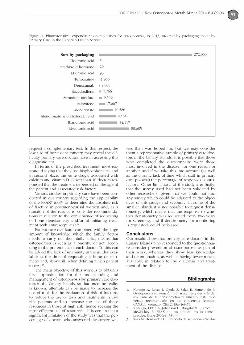
The duration of treatment with bisphosphonateswas checked in 85.20% of cases, while 12% of thoseresponding do not do so (item 12). Lastly, in rela-tion to the data obtained from the service for therational use of medicines8, in the year 2013 theexpenditure on medicines for osteoporosis in theCanarian health service8 totalled € 7,684,393.61.
In primary care the expenditure was €7,265,491.06, the top drug prescribed being rise-dronic acid, followed by ibandronic acid (Figures1 and 2). It should be clarified that included withinthe figure for expenditure in primary care is theexpenditure originating both in prescriptions fromprimary care, as well as those from specialisedcare. With reference to drugs which should beused as first choice for the treatment with osteo-porosis, in primary care these represented 13.71%.
DiscussionThe family doctor is an essential pillar of support forthe care of osteoporosis in all its aspects: preventa-tive, educative and therapeutic. For this reason theyneed to be capable of identifying the population athighest risk of osteoporotic fracture in the earlysilent phase, before the first fracture appears9.
Osteoporosis is an asymptomatic disease whichis difficult to diagnose in the absence of a fractu-re10,11. And even if there are fractures, these often donot produce symptoms. A number of authors haveindicated that is it important that the doctor has ade-quate diagnostic methods at their disposal, but it isalso necessary that they have the correct informa-tion regarding the treatment of this disease12-15. InSpain, until the publication of the first guide to oste-oporosis by the semFYC16, the study of this diseasein primary care was not well documented, neitherwas it included in the programme of preventative
and health promotion activities. In the ABOPAP2000 study carried out in Spain17, the approach toosteoporosis in primary care was studied. Notableamong the results of this study was the fact thataround a quarter of doctors had access to bone den-sitometry, whereas around 50% said that they conti-nued to study patients suspected of having osteopo-rosis. It is also interesting that screening for risk fac-tors is lower than expected in certain risk situations,such as family history of osteoporosis or hip fractu-re, chronic treatment with glucocorticoids, etc. Aswas expected, those doctors who had available thebest diagnostic tools also carried out greater scree-ning for risk factors18.
Another study published in Spain19, whichanalysed approaches to osteoporosis in a primaryhealth care centre concluded that the family doc-tors rarely complied with directives emanatingfrom guides to diagnosis and treatment.
In this study, according to the results from thedoctors surveyed, we are able to say that, in gene-ral, what primary care doctors in the Canariescarry out best is prevention, which is shown in thehigh percentage of those who responded whotook into account risk factors (96.40%), ensured agood intake of calcium and vitamin D (83.70%)and routinely applied non-pharmacological mea-sures to the general population (90%).
Even so, it is notable that only 61.40% said thatthey used risk factor scales to a greater or lesserextent. Given the simplicity of the tests, they couldbe of general use in primary care. It is possiblethat these staff are not yet convinced by theassessment of risk factors, perhaps due to the riskscales not being completely accepted consen-sually among researchers.
On the other hand, 71.60% of the doctors whoresponded did not measure patients’ heights in theirclinic. This is contradictory since this data is neces-sary when using the scales. We raise the question asto whether doctors really take into account height ornot, since this parameter is measured by the nurse,and in the survey we do not ask who measured it.This may also reflect a lack of knowledge on thepart of many staff of the significance of loss ofheight as an indicator of vertebral fracture.
Table 1. Distribution of primary care physicians (inpercentage) who answered surveys on each island ofthe Canary Islands
Tenerife 53.54%
Fuerteventura 40.74%
Gran Canaria 3.74%
Hierro 87.5%
Lanzarote 22.78%
La Gomera 35.29%
La Palma 8.16%
ORIGINALS / Rev Osteoporos Metab Miner 2014 6;4:89-9691
ORIGINALS / Rev Osteoporos Metab Miner 2014 6;4:89-9692
Another point of note is that 91% of doctors donot request a vitamin D test in patients with oste-oporosis or at risk of fracture, so we believe eitherthat there is a lack of knowledge of the usefulnessof such a test, or that there is an administrative dif-ficulty in requesting one, although the fact that83.70% said that they ensured a sufficient supplyof vitamin D to their patients inclines us to thesecond explanation. In terms of the diagnosis, it isnotable that nearly 50% used X-rays, occasionally
or routinely, to diagnose osteoporosis. And, if areduction in height is detected, only 50% requesta spinal X-ray. This leads us to suspect that thereis an under-diagnosis of possible vertebral fractu-res, and in some way corroborates the possibilityof a lack of knowledge about the loss of height asbeing indicative of vertebral fracture as has beenpointed out before. Furthermore, according to theresults, if a doctor were to detect a fragility fractu-re 28.70% would not, or would only occasionally,
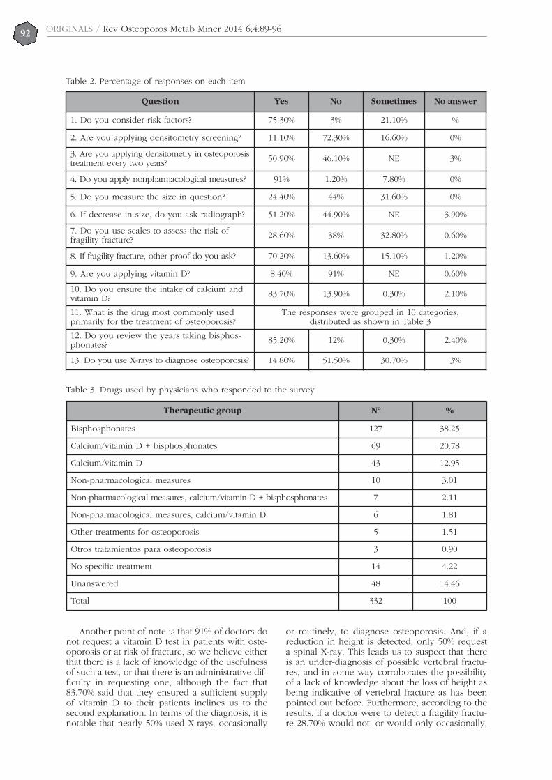
Table 2. Percentage of responses on each item
Question Yes No Sometimes No answer
1. Do you consider risk factors? 75.30% 3% 21.10% %
2. Are you applying densitometry screening? 11.10% 72.30% 16.60% 0%
3. Are you applying densitometry in osteoporosistreatment every two years? 50.90% 46.10% NE 3%
4. Do you apply nonpharmacological measures? 91% 1.20% 7.80% 0%
5. Do you measure the size in question? 24.40% 44% 31.60% 0%
6. If decrease in size, do you ask radiograph? 51.20% 44.90% NE 3.90%
7. Do you use scales to assess the risk offragility fracture? 28.60% 38% 32.80% 0.60%
8. If fragility fracture, other proof do you ask? 70.20% 13.60% 15.10% 1.20%
9. Are you applying vitamin D? 8.40% 91% NE 0.60%
10. Do you ensure the intake of calcium andvitamin D? 83.70% 13.90% 0.30% 2.10%
11. What is the drug most commonly usedprimarily for the treatment of osteoporosis?
The responses were grouped in 10 categories,distributed as shown in Table 3
12. Do you review the years taking bisphos-phonates? 85.20% 12% 0.30% 2.40%
13. Do you use X-rays to diagnose osteoporosis? 14.80% 51.50% 30.70% 3%
Table 3. Drugs used by physicians who responded to the survey
Therapeutic group Nº %
Bisphosphonates 127 38.25
Calcium/vitamin D + bisphosphonates 69 20.78
Calcium/vitamin D 43 12.95
Non-pharmacological measures 10 3.01
Non-pharmacological measures, calcium/vitamin D + bisphosphonates 7 2.11
Non-pharmacological measures, calcium/vitamin D 6 1.81
Other treatments for osteoporosis 5 1.51
Otros tratamientos para osteoporosis 3 0.90
No specific treatment 14 4.22
Unanswered 48 14.46
Total 332 100
ORIGINALS / Rev Osteoporos Metab Miner 2014 6;4:89-9693
request a complementary test. In this respect, thelow use of bone densitometry may reveal the dif-ficulty primary care doctors have in accessing thisdiagnostic test.
In terms of the prescribed treatment, most res-ponded saying that they use bisphosphonates, andin second place, the same drugs, associated withcalcium and vitamin D. Fewer than 10 doctors res-ponded that the treatment depended on the age ofthe patient and associated risk factors.
Various studies in primary care have been con-ducted in our country regarding the applicabilityof the FRAX© tool2,3 to determine the absolute riskof fracture in postmenopausal women and, as afunction of the results, to consider recommenda-tions in relation to the convenience of requestingof bone densitometry and/or of initiating treat-ment with antiresorptives20,21.
Patient care overload, combined with the largeamount of knowledge which the family doctorneeds to carry out their daily tasks, means thatosteoporosis is seen as a priority, or not, accor-ding to the preferences of each doctor. To this canbe added the lack of unanimity in the guides avai-lable at the time of requesting a bone densito-metry and, above all, when defining which patientto treat17.
The main objective of this work is to obtain afirst approximation for the understanding andmanagement of osteoporosis by primary care doc-tors in the Canary Islands, so that once the realityis known, attempts can be made to increase theuse of tools for the evaluation of risk of fracture,to reduce the use of tests and treatments in lowrisk patients and to increase the use of theseresources in those at high risk, hence seeking themost efficient use of resources. It is certain that asignificant limitation of the study was that the per-centage of doctors who answered the survey was
less than was hoped for, but we may considerthem a representative sample of primary care doc-tors in the Canary Islands. It is possible that thosewho completed the questionnaire were thosemost involved in the disease, for one reason oranother, and if we take this into account (as wellas the chronic lack of time which staff in primarycare possess) the percentage of responses is satis-factory. Other limitations of the study are: firstly,that the survey used had not been validated byother researchers, given that we could not findany survey which could be adjusted to the objec-tives of this study; and secondly, in some of thesmaller islands it is not possible to request densi-tometry, which means that the response to whe-ther densitometry was requested every two yearsfor screening, and if densitometry for monitoringis requested, could be biased.
ConclusionsOur results show that primary care doctors in theCanary Islands who responded to the questionnai-re consider prevention of osteoporosis as part oftheir work, whereas they show less knowledgeand determination, as well as having fewer meansavailable, in relation to the diagnosis and treat-ment of the disease.
Bibliography
1. Naranjo A, Rosas J, Ojeda S, Salas E. Manejo de laOsteoporosis en atención primaria antes y después delresultado de la densitometría:tratamiento instauradoversus recomendado en los consensos (estudioCANAL). Reumatol Clin 2013;9:269-73.
2. Kanis JA, Oden A, Johanson H, Borgstrom F, Strom O,McCloskey E. FRAX and its applications to clinicalpractice. Bone 2009;44:734-43.
3. Sosa M, Hernández D. Protocolo de actuación ante dos
Figure 1. Pharmaceutical expenditure on medicines for osteoporosis, in 2013, ordered by packaging made byPrimary Care in the Canarian Health Service
Sort by packaging
Clodronic acid
Parathyroid hormone
Etidronic acid
Teriparatide
Denosumab
Bazedoxifene
Strontium ranelate
Raloxifene
Alendronate
Alendronate and cholecalciferol
Ibandronic acid
Risedronic acid
272.900
5
29
90
1.966
2.868
7.766
9.509
17.667
40.586
49.612
54.117
88.685
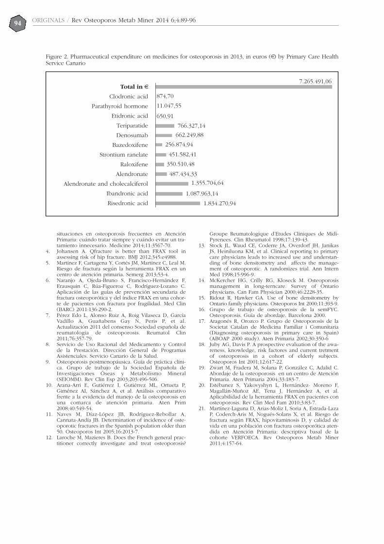
ORIGINALS / Rev Osteoporos Metab Miner 2014 6;4:89-9694
situaciones en osteoporosis frecuentes en AtenciónPrimaria: cuándo tratar siempre y cuándo evitar un tra-tamiento innecesario. Medicine 2014;11:3567-70.
4. Johansen A. Qfracture is better than FRAX tool inassessing risk of hip fracture. BMJ 2012;345:e4988.
5. Martínez F, Cartagena Y, Cortés JM, Martínez C, Leal M.Riesgo de fractura según la herramienta FRAX en uncentro de atención primaria. Semerg 2013;53-4.
6. Naranjo A, Ojeda-Bruno S, Francisco-Hernández F,Erausquin C, Rúa-Figueroa C, Rodríguez-Lozano C.Aplicación de las guías de prevención secundaria defractura osteoporótica y del índice FRAX en una cohor-te de pacientes con fractura por fragilidad. Med Clin(BARC) 2011:136:290-2.
7. Pérez Edo L, Alonso Ruiz A, Roig Vilaseca D, GarcíaVadillo A, Guañabens Gay N, Peris P, et al.Actualización 2011 del consenso Sociedad española dereumatología de osteoporosis. Reumatol Clin2011;76:357-79.
8. Servicio de Uso Racional del Medicamento y Controlde la Prestación. Dirección General de ProgramasAsistenciales. Servicio Canario de la Salud.
9. Osteoporosis postmenopáusica. Guía de práctica clíni-ca. Grupo de trabajo de la Sociedad Española deInvestigaciones Óseas y Metabolismo Mineral(SEIOMM). Rev Clin Esp 2003;203:496-506.
10. Arana-Arri E, Gutiérrez I, Gutiérrez ML, Ortueta P,Giménez AI, Sánchez A, et al. Análisis comparativofrente a la evidencia del manejo de la osteoporosis enuna comarca de atención primaria. Aten Prim2008;40:549-54.
11. Naves M, Díaz-López JB, Rodríguez-Rebollar A,Cannata-Andía JB. Determination of incidence of oste-oporotic fractures in the Spanish population older than50. Osteoporos Int 2005;16:2013-7.
12. Laroche M, Mazieres B. Does the French general prac-titioner correctly investigate and treat osteoporosis?
Groupe Reumatologique d’Etudes Cliniques de Midi-Pyrenees. Clin Rheumatol 1998;17:139-43.
13. Stock JL, Waud CE, Coderre JA, Overdorf JH, JanikasJS, Heiniluona KM, et al. Clinical reporting to primarycare physicians leads to increased use and understan-ding of bone densitometry and affects the manage-ment of osteoporotic. A randomizes trial. Ann InternMed 1998;15:996-9.
14. McKercher HG, Crilly RG, Kloseck M. Osteoporosismanagement in long-terncare. Survey of Ontariophysicians. Can Fam Physician 2000;46:2228-35.
15. Ridout R, Hawker GA. Use of bone densitometry byOntario family physicians. Osteoporos Int 2000;11:393-9.
16. Grupo de trabajo de osteoporosis de la semFYC.Osteoporosis. Guía de abordaje. Barcelona 2000.
17. Aragonés R, Orozco P. Grupo de Osteoporosis de laSocietat Catalan de Medicina Familiar i Comunitaria(Diagnosing osteoporosis in primary care in Spain)(ABOAP 2000 study). Aten Primaria 2002;30:350-6
18. Juby AG, Davis P. A prospective evaluation of the awa-reness, knowledge, risk factores and current tretmentof osteoporosis in a cohort of elderly subjects.Osteoporos Int 2001;12:617-22.
19. Zwart M, Fradera M, Solana P, González C, Adalid C.Abordaje de la osteoporosis en un centro de AtenciónPrimaria. Aten Primaria 2004;33:183-7.
20. Estébanez S, Yakovyshyn L, Hernández- Moreno F,Magallán-Muñoz AE, Tena J, Hernández A, et al.Aplicabilidad de la herramienta FRAX en pacientes conosteoporosis. Rev Clin Med Fam 2010;3:83-7.
21. Martínez-Laguna D, Arias-Moliz I, Soria A, Estrada-LazaP, Coderch-Aris M, Nogués-Solans X, et al. Riesgo defractura según FRAX, hipovitaminosis D, y calidad devida en una población con fractura osteoporótica aten-dida en Atención Primaria: descriptiva basal de lacohorte VERFOECA. Rev Osteoporos Metab Miner2011;4:157-64.
Figure 2. Pharmaceutical expenditure on medicines for osteoporosis in 2013, in euros (€) by Primary Care HealthService Canario
Total in €
Clodronic acid
Parathyroid hormone
Etidronic acid
Teriparatide
Denosumab
Bazedoxifene
Strontium ranelate
Raloxifene
Alendronate
Alendronate and cholecalciferol
Ibandronic acid
Risedronic acid
7.265.491,06
874,70
11.047,55
650,91
766.327,14
662.249,88
256.874,94
451.582,41
350.510,48
1.087.963,14
487.434,33
1.355.704,64
1.834.270,94

95ORIGINALS / Rev Osteoporos Metab Miner 2014 6;4:89-96
1) Do you consider all risk factors for fracture: age, personal history of fracture, risk of falls anduse of corticosteroids, as usual in the query?
2) Are you applying routinely densitometry, screening for all postmenopausal women?
3) Are you applying control densitometry every two years in patients with osteoporosis treated?
4) Do you apply nonpharmacological measures the general population routinely: no smoking,no drinking, physical exercise regularly, diet rich in calcium?
5) Do you measure regularly in consultation up to their patients?
6) In case of decreasing size do you ask x-thoracic and lumbar spine?
7) Do you use any scale (FRAX, QFracture) to assess the risk of fragility fracture?
8) In case you are facing a fragility fracture patient with some complementary test do you ask?
9) Are you applying vitamin D levels routinely?
10) Do you ensure good calcium intake as needed (depending on age, sex, ...) and vitamin D?
11) What treatment for osteoporosis using home?
12) In patients taking bisphosphonates Do you regularly check how many years have you been taking it?
13) Do you use radiography as a method to diagnose osteoporosis?
Annex I. Survey data collection
Knowledge about osteoporotic fractures in primary care professionals
YES No SOMETIMES
YES No
YES No
YES No
YES No
YES No
YES No SOMETIMES
YES No SOMETIMES
YES No SOMETIMES
YES No SOMETIMES
YES No SOMETIMES
YES No SOMETIMES

ORIGINALS / Rev Osteoporos Metab Miner 2014 6;4:89-9696
Gifre L1, Vidal J2, Ruiz-Gaspà S3, Portell E2, Monegal A1, Muxi A4, Guañabens N1,3, Peris P1,3
1 Servicio de Reumatología - Unidad de Patología Metabólica Ósea - Hospital Clínico de Barcelona2 Unidad de Lesionados Medulares - Instituto de Neurorrehabilitación Guttmann - Universidad Autónoma de Barcelona - Badalona3 CIBERehd4 Servicio de Medicina Nuclear - Hospital Clínico de Barcelona
ORIGINALS / Rev Osteoporos Metab Miner 2014 6;4:97-10297
Effect of spinal cord injury recentlyin bone turnover and in bone massevolution of complete motor.Preliminary findings
Correspondence: Laia Gifre - Servicio de Reumatología - Hospital Clínic - Carrer Villarroel, 170 - 08036 Barcelona (Spain)e-mail: [email protected]
Date of receipt: 11/07/2014Date of acceptance: 24/10/2014
Work scholarship from the SEIOMM to attend the 35th Congress of the ASBMR (Baltimore, 2013).
SummaryBackground and aim: Spinal cord injury (SCI) has been associated with a marked increase in bone lossand a higher incidence of skeletal fractures, however the pathogenesis and clinical management of thiscondition remains unclear. The aim of this study was to analyze the bone mineral density (BMD) evolu-tion in patients with complete SCI and its relationship with parameters of bone metabolism and bone tur-nover markers.Methods: Patients with a recent complete motor SCI (ASIA A)(<6 months) were prospectively included. Bonemetabolism parameters (calcium, phosphate, PTH and 25-OHD), bone turnover markers (bone formation: pro-collagen type 1 aminoterminal propeptide -P1NP-, bone alkaline phosphatase -bone AP-, osteocalcin -OC-;bone resorption: C-telopeptides of type I collagen -CTx-) and BMD were assessed in all patients at baselineand at 6 months. The results were compared with a control group.Results: 23 men with complete SCI (ASIA A) and a mean age of 38±15 years were included at 102±33 daysof SCI onset. 52% had paraplegia. 12 patients were assessed at 6 months of follow-up. Patients with SCIshowed a significant increase in bone turnover markers, especially P1NP and CTx, compared to controls(P1NP: 191±90 vs 51±19 ng/ml, p<0.001; CTx: 1.37±0.49 vs 0.51±0.23 ng/ml, p<0.001). At 6 months, boneturnover markers decreased (P1NP: -34%, p=0.005 and CTx: -26%, p=0.002) and BMD had a mean decrea-se of 12% at total femur (p=0.002) compared to baseline, with osteoporosis development in 50% of patients.Bone markers (bone AP, P1NP and OC) were negatively correlated with total femur BMD values.Conclusions: Patients with complete SCI show a marked increase in bone turnover and bone loss, espe-cially at the proximal femur, with the development of osteoporosis being observed in 50% of thesepatients at 6 months of follow-up. These findings indicate the need to implement preventive measureswithin the therapeutic approach in these patients.
Key words: osteoporosis, spinal cord injury, bone metabolism, bone turnover markers.
IntroductionAn absence of mechanical load on the skeleton isassociated with a marked loss of bone mass whichmay result in the development of osteoporosis andfractures. Spinal cord injury (SCI), especially whenthey are complete, are a common cause and anarchetypal example of an absence of load on theskeleton. So, a marked loss of bone mineral density(BMD) after an SCI has been reported, of the orderof 35% after two years from its occurrence, and thedevelopment of osteoporosis and fractures in morethan 50% of patients1-5. Although the physiopatho-logy of this process is not well understood, after anSCI there has also been observed a marked increa-se in bone turnover, especially during the first yearafter the SCI6-10. While the absence of load is themain factor related to this finding, the regulatorymechanism for this process is not clear. This fact,together with the absence of guidelines aimed at theprevention and treatment of osteoporosis after anSCI, could be the cause of defective treatment forthese patients. Indeed, a study recently carried outin our unit found evidence that after a complete SCIfewer than 10% of patients had obtained anti-osteo-porotic treatment, even after having had fragilityfractures2, a fact which has also been observed inother studies11. It is important to remember that theindividuals who have a complete SCI are usuallyyoung people, which means that the risk of deve-loping fractures over their life time is very high,clearly increasing at 3-5 years after the SCI2, whichindicates the necessity of adopting preventativemeasures in these patients.
The objective of this study is to analyse thedevelopment of BMD and bone turnover in patientswith recent SCI, and the factors relating to the lossof bone mass in this process. This preliminaryanalysis shows the development of the BMD andthe markers for bone turnover in the short-term, inthe first 6 months of monitoring.
Patients and methodsPopulation of the studyProspective study in which were included patientswith recently occurring (<6 months) SCI of traumaticorigin and severe in character (complete motor SCI[ASIA scale: A or B]). The patients were recruitedconsecutively (from August 2010 to January 2012) atthe Guttman Institute for Neurorehabilitation andthen referred to the bone metabolism pathology unitof the rheumatology service of the Clinical Hospitalof Barcelona.
Those included were patients over 18 years ofage, while those having diseases or processeswhich affected bone metabolism (Paget’s disease ofbone, rheumatoid arthritis, hyperparathyroidism,hypercortisolism, malabsorption syndrome, malig-nant tumours, transplants, recent pregnancy or bre-astfeeding) and/or who were following treatmentwith drugs which would interfere with bone meta-bolism (bisphosphonates, strontium ranelate, selec-tive estrogen receptor modulators, calcitonin, hor-mone therapy, denosumab and teriparatide,amongst others) were excluded.
In all the patients the risk factors for osteopo-rosis were evaluated, including: family history offemoral fractures, personal history of fractures,tobacco and alcohol consumption, dietary intakeof calcium (mg/day) and history of renal lithiasis.In addition the cause, level (tetraplegia/paraple-gia), severity and type (spastic/flaccid) of SCI andassociated complications were analysed.
The severity of the SCI was evaluated usingthe ASIA (American Spinal Injury Association)scale which, classifies MLs into 5 categories accor-ding to motor function and residual sensitivity: A:complete motor and sensory loss; B: completemotor and partial sensory loss; C and D: partialmotor and sensory loss; E: without motor or sen-sory lesion12.
The results were compared with a healthy con-trol group of the similar age and sex.
The study was carried out with the approval ofethics committee of the hospital and adjusted inaccordance with directives pertaining to researchin humans. All the patients signed their informedconsent for inclusion.Analytical testsBlood was taken from all patients at between 8and 10 in the morning after overnight fasting. Abiochemistry profile was performed which inclu-ded calcium, phosphate and creatinine, determi-ned by standard techniques, and levels of 25-hydroxyvitamin D (25-OHD) and parathyroid hor-mone (PTH) were assessed using automated che-moluminescence (Liaison, Diasorin and AdviaCentaur XP, Siemens, respectively). In addition,the following biochemical markers for bone for-mation were determined: bone alkaline phospha-tase (Bone AP, IDS, Vitro); osteocalcin (OC,radioimmunoassay, Elsa-Osteo-Cis, Gif-sur-Yvette,France) and amino-terminal propeptide of colla-gen type 1 P1NP, Cobas e411 automated method,Roche), and for bone resorption: carboxyl-termi-nal telopeptide of collagen type 1 (CTx, Cobase411 automated method, Roche).Bone mineral densityThe BMD in the lumbar spine, proximal femur(femoral neck and total femur) and in the lowerlimbs (EI) were determined in all patients bymeans of double X-ray absorptiometry (DXA;Lunar Prodigy, Radiation Corporation Madison,WI, US). The densitometric risk categories (normalBMD, osteopenia and osteoporosis were definedaccording to the criteria of the WHO13.Statistical analysisThe results are expressed as the mean ± standarddeviation of the mean (SD). The differences bet-ween the means of the continuous variables wereanalysed using the Mann-Whitney nonparametricU test, and the differences between proportions,by the Fisher test. For the comparison of pairedvariables the Wilcoxon test was used. To evaluatethe association between variables the Pearsoncorrelation coefficient was used. A value p<0.05was considered statistically significant. The statisti-cal analysis of the data was carried out using theSPSS program (version 18.0, Chicago, US).
ORIGINALS / Rev Osteoporos Metab Miner 2014 6;4:97-10298
ResultsThe clinical characteristics of the patients includedin the study are shown in Table 1.
The study included 23 males with an averageage of 38±15 years (range: 18-64) at an average of102±33 days from suffering the SCI. All thepatients had a severe SCI (ASIA A); 48% had tetra-plegia and 52% paraplegia. The majority ofpatients (83%) had an SCI of the spastic type. Allthe patients had a severe residual functional affec-tation: 3 patients (13%) remained totally immobili-zed in bed, the rest (87%) required a wheelchairfor mobility. The main cause of the SCI was a traf-fic accident (57%). The rest of the patients had anSCI due to falling (17%), diving into shallow water(13%), a sporting accident (9%) or domestic acci-dent (4%). 12 of the 23 patients (7 with tetraplegiaand 5 with paraplegia) were newly assessed after6 months of follow up.
After the SCI a marked increase in markers forbone turnover (OC, P1NP and CTx) were obser-ved compared with the control group (Table 2).No significant differences were observed in thevalue of bone markers as a function of the degreeof lesion (patients with tetraplegia vs those withparaplegia). Also, those patients with SCI had asignificant increase in levels of phosphate in theblood, and a reduction in values of PTH compa-red with the control group (Table 2). 39% of thepatients had vitamin D deficit (<20 ng/ml), howe-ver, no differences were observed in levels of 25-OHD or in values of calcium in the blood compa-red with the control group.
At 6 months of follow up a significant reduc-tion in markers for bone turnover (P1NP: -34%,p=0.005 and CTx: -26%, p=0.002) were observed,although they remained higher with respect to thecontrol group, and there was a normalisation ofthe parameters for phosphorous-calcium metabo-lism (baseline phosphate: 4.4±0.4 mg/dl vs 3.9±0.4mg/dl in follow up, p=0.011; baseline PTH: 32±20pg/ml vs 40.8±22.9 pg/ml in follow up, p=0.09).
The BMD in the proximal femur and in the EIreduced significantly at 6 months follow up (totalfemur: -12.3+4.9%, p=0.002; femoral neck: -12.8+6.7%,p=0.002; EI: -7.7+3.7%, p=0.003) compared with base-line values (Figure 1). No significant changes wereobserved in the development of the lumbar BMD(1.214±0.2 g/cm2 baseline vs 1.224±.2 g/cm2 at 6months, p=n.s). 50% of the patients had criteria fordensitometric osteoporosis after 6 months followup. No significant differences were observed inthe development of BMD as a function of the levelof SCI (tetraplegia vs paraplegia) or the type oflesion (spastic vs flaccid). None of the patientshad skeletal fractures during the first 6 months ofmonitoring.
A negative correlation was observed betweenthe values of BMD in the total femur and markersfor bone turnover (Bone AP: r=-0.63, p=0.001;P1NP: r=-0.459, p=0.028; OC: r=-0.454, p=0.051).The values of CTx were not related to the valuesof BMD. The change in BMD at 6 months was notrelated to the change in markers for bone turnover.
DiscussionThe results of this study show that after a comple-te motor SCI a marked increase in bone turnoverand in loss of bone mass occurs, especially in theproximal femur, which leads to the developmentof osteoporosis in half of the patients, a complica-tion which is already observed after 6 months offollow up, and which indicates the need to adoptpreventative measures in the therapeutic approachwith these patients.
Hence, over a period of only 6 months thepatients included in this study had a loss of BMDin the proximal femur of 12% after an SCI and 50%developed densitometric osteoporosis. The BMDin the lumbar spine, however, remained stableduring the follow up. These results coincide withthose of earlier studies which indicate that the lossof bone mass after an SCI occurs early, alreadybeing evident at 6 weeks from the lesion1,14 and ofgreatest magnitude during the first two years afterthe SCI, with loss of BMD which varies between 8and 35%, depending on it location and the timeover which it had developed1,4,6,15,16, which leads tothe development of osteoporosis and fractures inmore than 50% of patients4. One of the main cha-racteristics of the loss of bone mass associatedwith an SCI is its location, since, as seen in ourstudy, it occurs below the level of the lesion,affecting, above all, the lower limbs1,17,18. This factappears to be associated with the absence ofmechanical load in the said location and whichexplains, furthermore, the high incidence of frac-tures in lower limbs which is observed in thesepatients, especially in the femur and tibia2,19,20.Also, even though it has been suggested that thereis a greater loss of trabecular bone following anSCI15, other studies show that the bone loss occu-rring in this process takes place in various sec-tions. Hence, a study which analysed the develop-ment of bone mass using peripheral quantitativecomputerised tomography (pQCT) in the proximalfemur of patients with a recent SCI, describes cor-tical bone loss and an alteration in bone microar-chitecture and strength after SCI, which would bemeasured by an increase in trabecular and endos-teal bone resorption14. The results of our studyalso suggest an early affectation in both bone sec-tions; thus, the magnitude of the bone loss in thedifferent sections of the proximal femur, femoralneck and total femur, was similar after 6 monthsof follow up, of the order of 12% in both type oflocation. However, no significant changes wereseen in the lumbar BMD after 6 months of followup, a finding which has also been reported inother studies1,4, and which confirms the determi-ning effect which the absence of mechanical loadhas in the lower limbs on bone loss associatedwith this process.
The majority of studies, both in experimentaland human studies, report a marked increase inbone turnover after an SCI1,6.8-10, especially during thefirst year of the lesion, a finding which we alsoobserved in our patients. In fact, the patients inclu-ded in this study had an increase in markers for
ORIGINALS / Rev Osteoporos Metab Miner 2014 6;4:97-10299
bone turnover, especially P1NP and CTx in theblood, after an SCI of the order of 2 to 3 times hig-her than the control group, which, in addition –ashas been observed in the study of Sabour et al.21–are negatively correlated with values of BMD in theproximal femur. This increase in the markers forbone turnover diminishes progressively, such that at6 months, although they remain slightly higher, theirvalues have reduced significantly. Furthermore, inthose patients it was also observed that there was areduction in the values of PTH and a secondaryincrease in phosphate, two findings previously des-cribed in patients with recent SCI and which hadbeen attributed to the marked increase in in boneturnover which occurred after the lesion6,8,9,22. Bothparameters, PTH and phosphate, were normalisedafter 6 months follow up. Even though a high pre-valence of hypovitaminosis D has been seen inpatients affected by SCI23, this has been observedmostly in patients with a longstanding SCI. Ourpatients had recent MLs, less than 6 months earlier,a fact which may explain the absence of differencein values of vitamin D compared to the controlgroup. Furthermore, although the increase in bone
turnover which is observed after arecent SCI is a finding reported in mostof the studies, the magnitude of thisincrease, and above all, its long-termdevelopment are aspects which are lesswell-documented. Hence, although theincrease in turnover is particularly evi-dent during the first few months afterthe SCI, some authors indicate the per-sistence of this increase some years afterthe SCI. Thus, Zehnder et al.6 observedthat around 30% of patients had anincrease in bone resorption, evaluatedby determining levels of free deoxypy-ridinoline ten years on from the SCI.This is a fact which coincides with thepersistence in BMD loss, although at alower level, which patients have afterseveral years with an SCI6,24, and whichconfirms the necessity of adopting pre-ventative measures for the monitoringand treatment of this process.
The physiopathology of the altera-tions in bone turnover and loss ofbone mass associated with an SCI isunclear, nevertheless, the absence ofmechanical charge appears to be thedetermining factor for the loss of bonemass associated with this process1.Other factors such as the denervationwhich occurs after an SCI could alsocontribute as an additional factor inthis loss. In this vein, experimental stu-dies indicate a higher loss of bone inmice with an SCI than in mice subjectto load on their extremities16,25.
One of the main limitations of thisstudy is the loss of subjects during thefollow up. However, this is an initialanalysis which includes a highly homo-
geneous sample of patients, all having had a com-plete motor SCI in the past 6 months, which there-fore allows the application of the results to otherpatients with similar characteristics. Furthermore, itis remarkable in featuring a prospective cohortwhich includes one of the highest numbers ofpatients with SCI.
In summary, the results of this preliminarystudy show that after a complete motor SCI thereis a marked increase in bone turnover and loss ofbone mass below the level of the lesion, whichleads to the development of osteoporosis in halfthe patients during the first year of follow up.These results confirm the necessity of establishingpreventative measures against the development ofosteoporosis as part of the therapeutic approachtaken with these patients.
Conflict of interest: There are no conflicts ofinterest on the part of the authors.
This work is funded by grants from the ClinicHospital of Barcelona and the La MaratóFoundation of TV3.
ORIGINALS / Rev Osteoporos Metab Miner 2014 6;4:97-102100
Table 1. Clinical characteristics of the patients include in the study
Patients with SCI(n=23)
Age (years) 38±15
Males (n) 23
Risk factors for osteoporosis:
BMI (kg/m2) 24±5
History of renal lithiasis (%) 13
Active smoker (%) 30
Daily alcohol consumption (%) 13
Family history:
Femoral fracture (%) 13
SCI characteristics:
Development period of SCI (days) 102±33
Paraplegia/tetraplegia (%) 52/48
Spasticity (%) 83
Causes of SCI:
-Traffic accident 57
-Fall from a height 17
-Diving into shallow water 13
-Sporting accident 9
-Direct axial trauma 4
SCI: spinal cord injury; BMI: body mass index.
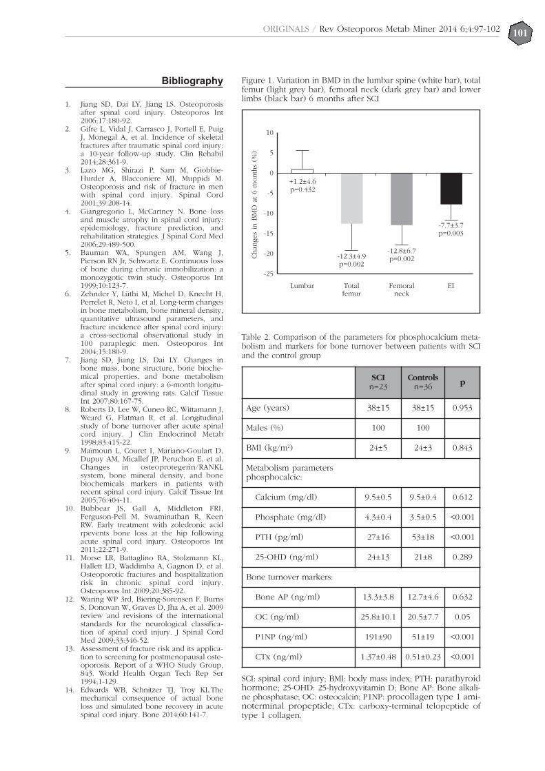
Bibliography
1. Jiang SD, Dai LY, Jiang LS. Osteoporosisafter spinal cord injury. Osteoporos Int2006;17:180-92.
2. Gifre L, Vidal J, Carrasco J, Portell E, PuigJ, Monegal A, et al. Incidence of skeletalfractures after traumatic spinal cord injury:a 10-year follow-up study. Clin Rehabil2014;28:361-9.
3. Lazo MG, Shirazi P, Sam M, Giobbie-Hurder A, Blacconiere MJ, Muppidi M.Osteoporosis and risk of fracture in menwith spinal cord injury. Spinal Cord2001;39:208-14.
4. Giangregorio L, McCartney N. Bone lossand muscle atrophy in spinal cord injury:epidemiology, fracture prediction, andrehabilitation strategies. J Spinal Cord Med2006;29:489-500.
5. Bauman WA, Spungen AM, Wang J,Pierson RN Jr, Schwartz E. Continuous lossof bone during chronic immobilization: amonozygotic twin study. Osteoporos Int1999;10:123-7.
6. Zehnder Y, Lüthi M, Michel D, Knecht H,Perrelet R, Neto I, et al. Long-term changesin bone metabolism, bone mineral density,quantitative ultrasound parameters, andfracture incidence after spinal cord injury:a cross-sectional observational study in100 paraplegic men. Osteoporos Int2004;15:180-9.
7. Jiang SD, Jiang LS, Dai LY. Changes inbone mass, bone structure, bone bioche-mical properties, and bone metabolismafter spinal cord injury: a 6-month longitu-dinal study in growing rats. Calcif TissueInt 2007;80:167-75.
8. Roberts D, Lee W, Cuneo RC, Wittamann J,Weard G, Flatman R, et al. Longitudinalstudy of bone turnover after acute spinalcord injury. J Clin Endocrinol Metab1998;83:415-22.
9. Maïmoun L, Couret I, Mariano-Goulart D,Dupuy AM, Micallef JP, Peruchon E, et al.Changes in osteoprotegerin/RANKLsystem, bone mineral density, and bonebiochemicals markers in patients withrecent spinal cord injury. Calcif Tissue Int2005;76:404-11.
10. Bubbear JS, Gall A, Middleton FRI,Ferguson-Pell M, Swaminathan R, KeenRW. Early treatment with zoledronic acidrpevents bone loss at the hip followingacute spinal cord injury. Osteoporos Int2011;22:271-9.
11. Morse LR, Battaglino RA, Stolzmann KL,Hallett LD, Waddimba A, Gagnon D, et al.Osteoporotic fractures and hospitalizationrisk in chronic spinal cord injury.Osteoporos Int 2009;20:385-92.
12. Waring WP 3rd, Biering-Sorensen F, BurnsS, Donovan W, Graves D, Jha A, et al. 2009review and revisions of the internationalstandards for the neurological classifica-tion of spinal cord injury. J Spinal CordMed 2009;33:346-52.
13. Assessment of fracture risk and its applica-tion to screening for postmenopausal oste-oporosis. Report of a WHO Study Group,843. World Health Organ Tech Rep Ser1994;1-129.
14. Edwards WB, Schnitzer TJ, Troy KL.Themechanical consequence of actual boneloss and simulated bone recovery in acutespinal cord injury. Bone 2014;60:141-7.
ORIGINALS / Rev Osteoporos Metab Miner 2014 6;4:97-102101
SCIn=23
Controlsn=36 p
Age (years) 38±15 38±15 0.953
Males (%) 100 100
BMI (kg/m2) 24±5 24±3 0.843
Metabolism parametersphosphocalcic:
Calcium (mg/dl) 9.5±0.5 9.5±0.4 0.612
Phosphate (mg/dl) 4.3±0.4 3.5±0.5 <0.001
PTH (pg/ml) 27±16 53±18 <0.001
25-OHD (ng/ml) 24±13 21±8 0.289
Bone turnover markers:
Bone AP (ng/ml) 13.3±3.8 12.7±4.6 0.632
OC (ng/ml) 25.8±10.1 20.5±7.7 0.05
P1NP (ng/ml) 191±90 51±19 <0.001
CTx (ng/ml) 1.37±0.48 0.51±0.23 <0.001
Table 2. Comparison of the parameters for phosphocalcium meta-bolism and markers for bone turnover between patients with SCIand the control group
SCI: spinal cord injury; BMI: body mass index; PTH: parathyroidhormone; 25-OHD: 25-hydroxyvitamin D; Bone AP: Bone alkali-ne phosphatase; OC: osteocalcin; P1NP: procollagen type 1 ami-noterminal propeptide; CTx: carboxy-terminal telopeptide oftype 1 collagen.
Figure 1. Variation in BMD in the lumbar spine (white bar), totalfemur (light grey bar), femoral neck (dark grey bar) and lowerlimbs (black bar) 6 months after SCI
Chan
ges
in B
MD
at 6
month
s (%
)
10
5
0
-5
-10
-15
-20
-25
Lumbar
+1.2±4.6p=0.432
-12.3±4.9p=0.002
-12.8±6.7p=0.002
-7.7±3.7p=0.003
Totalfemur
Femoralneck
EI
15. Frey-Rindova P, de Bruin ED, Stüssi E, Dambacher MA,Dietz V. Bone mineral density in upper and lowerextremities during 12 months after spinal cord injurymeasured by peripheral quantitative computed tomo-graphy. Spinal Cord 2000;38:26-32.
16. de Bruin ED, Dietz V, Dambacher MA, Stüssi E.Longitudinal changes in bone in men with spinal cordinjury. Clin Rehabil 2000;14:145-52.
17. Liu D, Zhao CQ, Li H, Jiang SD, Jiang LS, Dai LY.Effects of spinal cord injury and hindlimb immobiliza-tion on sublesional and supralesional bones in younggrowing rats. Bone 2008;43:119-25.
18. Goemaere S, Van Laere M, De Neve P, Kaufman JM.Bone mineral status in paraplegic patients who do or donot perform standing. Osteoporos Int 1994;4:138-43.
19. Lala D, Craven BC, Thabane L, Papaioannou A, AdachiJD, Popovic MR, et al. Exploring the determinants offracture risk among individuals with spinal cord injury.Osteoporos Int 2014;25:177-85.
20. Craven BC, Robertson CF, McGillivray CF, Adachi J.D.Detection and treatment of sublesional osteoporosis
among patients with chronic spinal cord injury: proposedparadigms. Top Spinal Cord Inj Rehabil 2009;14:1-22.
21. Sabour H, Javidan AN, Latifi S, Larijani B, Shidfar F, VafaMR, et al. Bone biomarkers in patients with chronictraumatic spinal cord injury. Spine J 2014;14:1132-8.
22. Maïmoun L, Fattal C, Micallef JP, Peruchon E, RabischongP. Bone loss in spinal cord-injured patients: from physio-pathology to therapy. Spinal Cord 2006;44:203-10.
23. Rivero González L, Méndez Suárez JL, MirandaCalderín G, Bárbara Bataller E, Sánchez Enríquez J,Sosa Henríquez M. Prevalencia de la hipovitaminosisD e hiperparatiroidismo secundario en la Unidad deLesionados Medulares de Gran Canaria. Estudio preli-minar. Rev Osteoporos Metab Miner 2013;5:67-72.
24. Garland DE, Adkins RH, Stewart CA. Five-year longitudi-nal bone evaluations in individuals with chronic comple-te spinal cord injury. J Spinal Cord Med 2008;31:543-50.
25. He JY .Jiang LS, Dai LY. The roles of the sympatheticnervous system in osteoporotic diseases: A review ofexperimental and clinical studies. Ageing Res Rev2011;10:253-63.
102ORIGINALS / Rev Osteoporos Metab Miner 2014 6;4:97-102
Delgado-Calle J1, Pérez-Campo FM2, Riancho JA2
1 Departamento de Anatomía y Biología Celular - Facultad de Medicina de Indiana - Centro Médico de Administración de Veteranos Roudebush -Indianapolis (EE.UU. )2 Departamento de Medicina Interna - Hospital Universitario Marqués de Valdecilla - Universidad de Cantabria - IDIVAL - Santander (España)
103REVIEWS / Rev Osteoporos Metab Miner 2014 6;4:103-108
Advances in the study of the mechanismsinvolved in the modulation of the expressionof sclerostin in human cells
Correspondence: José A. Riancho Moral - Departamento de Medicina Interna - Hospital U. Marqués de Valdecilla -39011 Santander (Spain)e-mail: [email protected]
Date of receipt: 18/08/2014Date of acceptance: 15/10/2014
Work rewarded with the scholarship investigation FEIOMM 2011.
SummarySclerostin plays an important role in the regulation of bone metabolism, as is shown in the dramatic chan-ges in bone mass which occur when its activity is inhibited by means of monoclonal antibodies. However,the mechanisms which regulate its expression are still not well-understood. Various studies have shownan association between polymorphisms of the SOST gene promoter (which codes for sclerostin) and bonemineral density. Also, the degree of methylation of a CpG island near the start of the transcription is asso-ciated with marked changes in the expression of the gene. Therefore, it appears that the production ofsclerostin is influenced by both genetic and epigenetic mechanisms, in addition to other hormonal andmechanical factors. A greater knowledge of these mechanisms would not only contribute to a betterunderstanding of bone biology, but could open up new therapeutic opportunities.
Key words: sclerostin, SOST, methylation, epigenetic.
IntroductionSclerostin is a protein coded by the SOST gene.This protein is secreted specifically by the osteocy-tes and has a negative effect on bone formation,through the inhibition of the Wnt canonical path-way1. The inhibition of this pathway has profoundconsequences for the activity of the osteoblasts;specifically, their differentiation is inhibited andtheir apoptosis induced2,3. The importance of scle-rostin in bone biology has been seen in the des-cription of cases of mutations of the SOST gene inhumans which provoke an altered bone phenoty-pe, with an increased bone mass4,5. On the otherhand, the inhibition of sclerostin through the useof neutralising antibodies has been demonstratedto have a powerful anabolic effect in bone, bothin animals and in humans6,7.Although the significance of sclerostin in bonehomeostasis appears indubitable, there are variousaspects of its biology which still remain unknown.Some of the least-known aspects are the factorswhich regulate the expression of sclerostin andthe mechanisms involved. For example, it is notknown why solely the osteocytes, and not otherosteoblast line cells, are capable of expressingsclerostin. Possibly even more intriguing is the factthat there are osteocytes within the bone produ-cing sclerostin, while others located within a fewmicrons of them do not8.
In some experimental models various effectorshave been identified which are capable of regula-ting levels of sclerostin. On the one hand, amongthe positive effectors are the bone morphogeneticproteins (BMPs)9 or the combined action of thetumor necrosis factor (TNF) and the tumour necro-sis factor-like weak inducer of apoptosis(TWEAK)10. On the other, notable among thenegative regulators are parathyroid hormone(PTH)11,12, prostaglandin E2 (PGE2)12 and mechani-cal load, this last factor being of special significan-ce due to the role which this type of stimulus hason bone homeostasis13. Unfortunately, many ofthese experiments have been carried out in muri-ne models and it remains to be seen to whatextent they are transferable to human bone.Furthermore, although the effects of these factorshave been described, the molecular mechanismswhich underlie their effects on the expression ofSOST have hardly been identified.
One of the obstacles which researchers areencountering when studying the regulation ofsclerostin production is the absence of systems inwhich this gene is actively expressed. Currently,there are no human osteocyte lines available. Thegeneration of some murine lines has been repor-ted, but in spite of the fact that these show someof the characteristic phenotypes of osteocytes,their production of sclerostin is barely detectable.It would therefore of great interest to find a goodsystem in which the factors involved in the regu-lation of the expression of this gene may be iden-tified.
Curiously, not the entire promoter sequencefor the SOST gene is preserved between species,
which suggests that the regulation may differ as afunction of the species. This is still more evidenceof the necessity of developing human models.Some works suggest that region 5’ of the genewould have two sections: one, close to the start ofthe transcription, which shows marked transcrip-tional activity, and the other, situated at some1,000 base pairs’ distance from the start of thetranscription, which could have an inhibitoryeffect14. On the other hand, it is interesting to notethat various groups, including ours, have shownan association between some polymorphismslocated in the 5’ region of the gene and bonemineral density (BMD)15,16. In the same vein, ingenome-wide association studies (GWAS) somepolymorphisms of a nucleotide (SNPs) have beenfound near this gene associated with BMD17. Thissuggests that these polymorphisms may have afunctional impact and modulate the expression ofthe gene, but actually, it is not known if this reallyis the case or what would be the molecularmechanisms involved.
Given the importance attributed to sclerostin inbone formation, the identification of the molecu-lar mechanisms which regulate its levels couldopen new areas of investigation in bone biology,and perhaps help to identify new therapeutic tar-gets related to the inhibition of its production,which would have an anabolic effect on bone.Furthermore, the validation of new models of cellsof human origin in which it would be possible tostudy these mechanisms could be crucial for theadvancement of the understanding of the regula-tion of sclerostin. In this article we briefly reviewsome recent results from our laboratory and thoseof other researchers to shed some light on thesequestions.
DNA methylation and the regulation ofgene expressionA good number of the cytosines of mammalianDNA are methylated, especially when they arefollowed by a guanine, which is to say, when for-ming CG dinucleotides (often also known asCpGs, the “p” indicating the phosphate groupwhich links the two bases). It is supposed that themethylation brings stability to the DNA and avoids“transcriptional noise” in the background. Thereare zones of DNA, called “CpG islands”, whichhave a particular behaviour. These islands consistof regions of a few hundred nucleotides which areespecially rich in CpG and which are found fre-quently in the promoter regions of many genes. Inrecent years it has been shown that the level ofmethylation in these CpG islands (and in the adja-cent regions, called “CpG island shores”) plays animportant role in the regulation of the expressionof many genes. In general, when the CpG of thepromoter regions is highly methylated, the trans-cription of DNA to RNA is repressed, and, as aconsequence, the levels of the protein for whichthe gene codes are reduced. Inversely, thedemethylation of the promoter tends to be asso-ciated with the active transcription of the gene.
104REVIEWS / Rev Osteoporos Metab Miner 2014 6;4:103-108
There are many molecular mechanisms invol-ved in the regulation of gene expression throughchanges in methylation, of which only some areknown. Thus, for example, the methylation ofDNA may impede its bonding with some activa-ting transcription factors. On the other hand, themethylated regions attract some proteins whichbond specifically to these methylated regions. Thisis the case with MeCP2, the binding protein formethylated CpG18. However, it should also betaken into account that the methylation of DNAacts in combination with other epigenetic mecha-nisms, specifically with the postranslational modi-fications of the histones. In fact, when MeCP2bonds to DNA, it recruits other proteins, such asHDACs (histone deacetylases) which modify thetails of the histones near this region. Together,these modifications contribute to the modulationof gene expression. For example, the highestlevels of histone acetylation are usually associatedwith an activation of transcription; while, to thecontrary, the methylation of certain lysines presentin the histones is associated with gene repres-sion19.
The patterns of DNA methylation are transmit-ted through mitosis, which means that they areinherited from the cell which divides into twodaughter cells. In this process an essential role isplayed by a family of enzymes called DNA-methyltransferases (DNMTs), in particular type120,21.
DNA methylation can be a passive phenome-non, which is to say, it may appear during somecell divisions if the DNMTs do not perform theirfunction of the remethylation the DNA daughterchains. But demethylation may also be an activeprocess. This is to suggest that it is possible thatsome regions of DNA are demethylated withoutthe necessity of cell division and the consequentreplication of DNA having taken place. The pro-cess by which active demethylation occurs is notwell understood, but the enzyme GADD45 andthe conversion of the methylcytosines to hydro-methylcytosines appears to play a special role init19,22,23. In addition, its true significance in tissuehomeostasis is also not well known. Nevertheless,it has been suggested that this process could beinvolved in osteoblast differentiation24.
Our group has demonstrated that methylationand demethylation of some genes plays an essen-tial role in variations in the patterns of geneexpression which occur during the different stagesof the differentiation of the osteoblast-lineagecells. For example, using the technique of laserassisted microdissection and subsequent DNAanalysis of the cells thus captured, we have con-firmed that during the step from osteoblasts toosteocytes there occurs a marked reduction in themethylation of the SOST gene promoter.Differently from that which occurs in the case ofosteoblasts, this is a necessary requisite for osteo-clasts to be able to synthesise sclerostin25. Othergenes involved in the biology of the skeleton arealso regulated, in part, through the level of methy-
lation of their promoters. This is the case, forexample, with osteoprotogerin, the ligand ofRANK (RANKL), alkaline phosphatase, osterix orestrogen receptor26-28.
DNA demethylation as experimental toolThe changes in the methylation of the CpG islandsare very powerful regulation mechanisms. Theyare possibly not involved in the fine regulation ofgene expression, but act as a type of molecular“interrupter” which starts and stops gene transcrip-tion. Once the demethylation allows transcription,other mechanisms, (humoral, physical, etc.) willbe responsible for adjusting precisely the geneexpression, in response to what is required at thatmoment29.
The regulatory power of the mechanisms lin-ked to methylation are shown in certain experi-ments in which DNA methylation is pharmacolo-gically induced. For this, nucleotide analogs areoften used, such as azacytidine and deoxy-azacy-tidine (or decytabine) which inhibit the activity ofthe DNMTs. Thus we have been able to demons-trate that the incubation of different types of cellwith decytabine strongly induces the expressionof sclerostin even when in normal conditionsthese cells do not express the gene25.
This phenomenon has, furthermore, an interes-ting practical repercussion, in that it facilitates thestudy of the mechanisms which modulate theexpression of sclerostin. Given that there are nohuman osteocyte lineages, or techniques to isola-te viable osteocytes from human bone, it is com-plicated to explore the regulatory mechanisms forthis gene in humans. Although there are differentimmortalised osteoblast lineages, and it is relati-vely easy to obtain osteoblasts from bone biop-sies, these cells do not express sclerostin.However, the demethylation of its promoter withdecytabine induces the expression of this gene,thus functioning, at least in theory, as an experi-mental model to analyse the physical and chemi-cal factors involved in its regulation. But for thismodel to be really useful the osteoblasts shouldhave a pattern of response to different stimulisimilar to that of the osteocytes in in vivo animalexperimental models30,31.
In fact, this appears to be the case. The resultswhich we have obtained with this model of osteo-blasts treated with decytabine have confirmed theinhibitory effect of PTH and the stimulatory effectof the BMPs (bone morphogenetic proteins) on theexpression of SOST32. We have also been able toconfirm that the osteoblasts treated with decytabi-ne maintain their response not only to humoralfactors but also to mechanical stimuli. When thesecells are subject to a pulsating flow of the culturemedium (which simulates the stimulus of the oste-ocyte membranes by the liquid present in the lacu-nae and canaliculi of the bone) a series of bioche-mical responses is induced, notable among whichis the induction of nitric oxide synthase (NOS),with its consequent accumulation in the medium.This response is maintained in the osteoblasts pre-
105REVIEWS / Rev Osteoporos Metab Miner 2014 6;4:103-108
treated with decytabine. Furthermore, in these cul-tures it is possible to confirm that mechanical sti-mulus induces a reduction in the expression ofsclerostin, in line with that demonstrated in in vivoexperimental models13,33. The later experimentswith nitric oxide inhibitors and donors have ena-bled the confirmation that nitric oxide synthesisreally is involved in the inhibitor effect of SOSTinduced by mechanical stimulation34.
SOST gene promoter, sclerostin andbone massVarious studies of candidate genes and also thoseof genome association (GWAS) have found poly-morphisms of the SOST gene associated with bonemineral density15,35. Hence, we have proved thatwomen who are homozygous for the minor allele(G) of SNP rs851054, situated in promoter region5’ of the gene, have a BMD significantly lowerthan women with other genotypes. This suggestthat this polymorphism may provoke differencesin transcriptional activity as a function of the alle-le which is present. To explore further the mecha-nisms involved, we cloned the entire SOST pro-moter region (positions -1440/+30 in relation tothe transcription start site or TSS) and confirmedits transcriptional activity in a luciferase reportervector, after its transfection in different types ofcell. The cloning of various regions of this frag-ment allowed us to confirm that the most activeregion appears to be in the first 500 nucleotides.In fact, the transcriptional activity of the vectorswith insertion in the region -580/+30 is somewhatgreater than that of the complete regions(-1440/+30). Contrarily, the most distal region(-1440/1030) is not active, while the intermediateregion (-1032/-571) has a certain degree of acti-vity, although clearly lower than that of the com-plete region or the region nearest to the TSS.Also, we found that BMP2 increases the transcrip-tional activity of these constructions, while PTHhas no effect, which is in accordance with thosestudies which show that the effect of this hormo-ne is mediated by an enhancer region located inseveral thousand base pairs32.
On the other hand, from the genomic DNA ofindividuals for various polymorphisms frequent inthe SOST promoter (rs801054 and rs801056), wecloned the promoter regions with each of thepossible alleles for these polymorphisms in luci-ferase reporter vectors. We then analyzed itstranscriptional activity after transfection intovarious osteoblast-type lineages. However, thedifferences in activity of the difference alleleswere small (data not published). This suggeststhat the demonstrated association of these alleleswith bone mineral density should be measured byindirect mechanisms which are not reproduced inthese experimental models. Among these shouldbe considered: certain factors, physical or humo-ral, present in vivo but not in vitro; the interactionof other cell elements in the bone microenviron-ment; or complex actions which involve the threedimensional structure of chromatin and the invol-
vement of other distant regions of the DNA. Anobvious candidate is called the Van Buchemregion, situated several thousand gene basesaway, and in which have been described regula-tory regions (such as that called ECR5), whichappear to mediate the response to some factors,PTH in particular36,37.
ConclusionSclerostin plays a significant role in the regulationof bone metabolism, as is demonstrated in the dra-matic changes in bone mass which occur when itsactivity is inhibited by means of monoclonal anti-bodies38,39. However, knowledge of the mecha-nisms which regulate their expression is stillincomplete. Nevertheless, in recent years newdata have been generated which allow one tosketch out, albeit schematically, some of the fac-tors and pathways involved (Figure 1).
This work and the experiments which are men-tioned have also been carried out with the help ofresearch grants from ISCIII (PI12/615) and withfinancial support from IFIMAV-IDIVAL.
The authors have no conflicts of interest inrelation to this work.
Bibliography
1. van Bezooijen RL, Roelen BA, Visser A, Wee-Pals L, deWilt E, Karperien M, et al. Sclerostin is an osteocyte-expressed negative regulator of bone formation, butnot a classical BMP antagonist. J Exp Med2004;199:805-14.
2. Sutherland MK, Geoghegan JC, Yu C, Turcott E,Skonier JE, Winkler DG, et al. Sclerostin promotes theapoptosis of human osteoblastic cells: a novel regula-tion of bone formation. Bone 2004;35:828-35.
3. Poole KE, van Bezooijen RL, Loveridge N, HamersmaH, Papapoulos SE, Lowik CW, et al. Sclerostin is adelayed secreted product of osteocytes that inhibitsbone formation. FASEB J 2005;19(13):1842-4.
4. Balemans W, Ebeling M, Patel N, Van Hul E, Olson P,Dioszegi M, et al. Increased bone density in scleroste-osis is due to the deficiency of a novel secreted pro-tein (SOST). Hum Mol Genet 2001;10:537-43.
5. Brunkow ME, Gardner JC, Van Ness J, Paeper BW,Kovacevich BR, Proll S, et al. Bone dysplasia scleros-teosis results from loss of the SOST gene product, anovel cystine knot-containing protein. Am J HumGenet 2001;68:577-89.
6. Padhi D, Jang G, Stouch B, Fang L, Posvar E. Single-dose, placebo-controlled, randomized study of AMG785, a sclerostin monoclonal antibody. J Bone MinerRes 2011;26:19-26.
7. Agholme F, Li X, Isaksson H, Ke HZ, Aspenberg P.Sclerostin antibody treatment enhances metaphysealbone healing in rats. J Bone Miner Res 2010;25:2412-8.
8. Delgado-Calle J, Arozamena J, Garcia-Renedo R,Garcia-Ibarbia C, Pascual-Carra MA, Gonzalez-MaciasJ, et al. Osteocyte deficiency in hip fractures. CalcifTissue Int 2011;89:327-34.
9. Kamiya N, Kobayashi T, Mochida Y, Yu PB, YamauchiM, Kronenberg HM, et al. Wnt inhibitors Dkk1 andSost are downstream targets of BMP signaling throughthe type IA receptor (BMPRIA) in osteoblasts. J BoneMiner Res 2010;25:200-10.
10. Vincent C, Findlay DM, Welldon KJ, Wijenayaka AR,Zheng TS, Haynes DR, et al. Pro-inflammatory cytoki-nes TNF-related weak inducer of apoptosis (TWEAK)and TNFalpha induce the mitogen-activated protein
106REVIEWS / Rev Osteoporos Metab Miner 2014 6;4:103-108
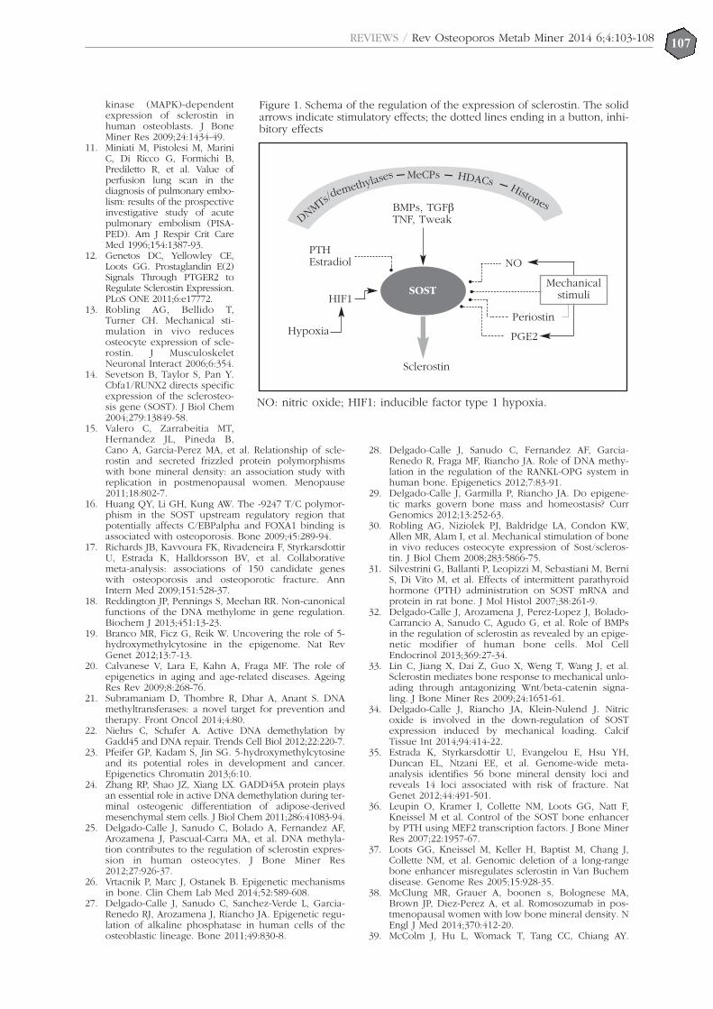
kinase (MAPK)-dependentexpression of sclerostin inhuman osteoblasts. J BoneMiner Res 2009;24:1434-49.
11. Miniati M, Pistolesi M, MariniC, Di Ricco G, Formichi B,Prediletto R, et al. Value ofperfusion lung scan in thediagnosis of pulmonary embo-lism: results of the prospectiveinvestigative study of acutepulmonary embolism (PISA-PED). Am J Respir Crit CareMed 1996;154:1387-93.
12. Genetos DC, Yellowley CE,Loots GG. Prostaglandin E(2)Signals Through PTGER2 toRegulate Sclerostin Expression.PLoS ONE 2011;6:e17772.
13. Robling AG, Bellido T,Turner CH. Mechanical sti-mulation in vivo reducesosteocyte expression of scle-rostin. J MusculoskeletNeuronal Interact 2006;6:354.
14. Sevetson B, Taylor S, Pan Y.Cbfa1/RUNX2 directs specificexpression of the sclerosteo-sis gene (SOST). J Biol Chem2004;279:13849-58.
15. Valero C, Zarrabeitia MT,Hernandez JL, Pineda B,Cano A, Garcia-Perez MA, et al. Relationship of scle-rostin and secreted frizzled protein polymorphismswith bone mineral density: an association study withreplication in postmenopausal women. Menopause2011;18:802-7.
16. Huang QY, Li GH, Kung AW. The -9247 T/C polymor-phism in the SOST upstream regulatory region thatpotentially affects C/EBPalpha and FOXA1 binding isassociated with osteoporosis. Bone 2009;45:289-94.
17. Richards JB, Kavvoura FK, Rivadeneira F, StyrkarsdottirU, Estrada K, Halldorsson BV, et al. Collaborativemeta-analysis: associations of 150 candidate geneswith osteoporosis and osteoporotic fracture. AnnIntern Med 2009;151:528-37.
18. Reddington JP, Pennings S, Meehan RR. Non-canonicalfunctions of the DNA methylome in gene regulation.Biochem J 2013;451:13-23.
19. Branco MR, Ficz G, Reik W. Uncovering the role of 5-hydroxymethylcytosine in the epigenome. Nat RevGenet 2012;13:7-13.
20. Calvanese V, Lara E, Kahn A, Fraga MF. The role ofepigenetics in aging and age-related diseases. AgeingRes Rev 2009;8:268-76.
21. Subramaniam D, Thombre R, Dhar A, Anant S. DNAmethyltransferases: a novel target for prevention andtherapy. Front Oncol 2014;4:80.
22. Niehrs C, Schafer A. Active DNA demethylation byGadd45 and DNA repair. Trends Cell Biol 2012;22:220-7.
23. Pfeifer GP, Kadam S, Jin SG. 5-hydroxymethylcytosineand its potential roles in development and cancer.Epigenetics Chromatin 2013;6:10.
24. Zhang RP, Shao JZ, Xiang LX. GADD45A protein playsan essential role in active DNA demethylation during ter-minal osteogenic differentiation of adipose-derivedmesenchymal stem cells. J Biol Chem 2011;286:41083-94.
25. Delgado-Calle J, Sanudo C, Bolado A, Fernandez AF,Arozamena J, Pascual-Carra MA, et al. DNA methyla-tion contributes to the regulation of sclerostin expres-sion in human osteocytes. J Bone Miner Res2012;27:926-37.
26. Vrtacnik P, Marc J, Ostanek B. Epigenetic mechanismsin bone. Clin Chem Lab Med 2014;52:589-608.
27. Delgado-Calle J, Sanudo C, Sanchez-Verde L, Garcia-Renedo RJ, Arozamena J, Riancho JA. Epigenetic regu-lation of alkaline phosphatase in human cells of theosteoblastic lineage. Bone 2011;49:830-8.
28. Delgado-Calle J, Sanudo C, Fernandez AF, Garcia-Renedo R, Fraga MF, Riancho JA. Role of DNA methy-lation in the regulation of the RANKL-OPG system inhuman bone. Epigenetics 2012;7:83-91.
29. Delgado-Calle J, Garmilla P, Riancho JA. Do epigene-tic marks govern bone mass and homeostasis? CurrGenomics 2012;13:252-63.
30. Robling AG, Niziolek PJ, Baldridge LA, Condon KW,Allen MR, Alam I, et al. Mechanical stimulation of bonein vivo reduces osteocyte expression of Sost/scleros-tin. J Biol Chem 2008;283:5866-75.
31. Silvestrini G, Ballanti P, Leopizzi M, Sebastiani M, BerniS, Di Vito M, et al. Effects of intermittent parathyroidhormone (PTH) administration on SOST mRNA andprotein in rat bone. J Mol Histol 2007;38:261-9.
32. Delgado-Calle J, Arozamena J, Perez-Lopez J, Bolado-Carrancio A, Sanudo C, Agudo G, et al. Role of BMPsin the regulation of sclerostin as revealed by an epige-netic modifier of human bone cells. Mol CellEndocrinol 2013;369:27-34.
33. Lin C, Jiang X, Dai Z, Guo X, Weng T, Wang J, et al.Sclerostin mediates bone response to mechanical unlo-ading through antagonizing Wnt/beta-catenin signa-ling. J Bone Miner Res 2009;24:1651-61.
34. Delgado-Calle J, Riancho JA, Klein-Nulend J. Nitricoxide is involved in the down-regulation of SOSTexpression induced by mechanical loading. CalcifTissue Int 2014;94:414-22.
35. Estrada K, Styrkarsdottir U, Evangelou E, Hsu YH,Duncan EL, Ntzani EE, et al. Genome-wide meta-analysis identifies 56 bone mineral density loci andreveals 14 loci associated with risk of fracture. NatGenet 2012;44:491-501.
36. Leupin O, Kramer I, Collette NM, Loots GG, Natt F,Kneissel M et al. Control of the SOST bone enhancerby PTH using MEF2 transcription factors. J Bone MinerRes 2007;22:1957-67.
37. Loots GG, Kneissel M, Keller H, Baptist M, Chang J,Collette NM, et al. Genomic deletion of a long-rangebone enhancer misregulates sclerostin in Van Buchemdisease. Genome Res 2005;15:928-35.
38. McClung MR, Grauer A, boonen s, Bolognese MA,Brown JP, Diez-Perez A, et al. Romosozumab in pos-tmenopausal women with low bone mineral density. NEngl J Med 2014;370:412-20.
39. McColm J, Hu L, Womack T, Tang CC, Chiang AY.
107REVIEWS / Rev Osteoporos Metab Miner 2014 6;4:103-108
••••
•SOST
BMPs, TGFβTNF, Tweak
PTHEstradiol
HIF1
NO
Hypoxia
Sclerostin
Periostin
PGE2
Mechanicalstimuli
Figure 1. Schema of the regulation of the expression of sclerostin. The solidarrows indicate stimulatory effects; the dotted lines ending in a button, inhi-bitory effects
NO: nitric oxide; HIF1: inducible factor type 1 hypoxia.
MeCPs
DNMTs/demethylase
s HDACs Histones
– ––

REVIEWS / Rev Osteoporos Metab Miner 2014 6;4:103-108108
Single- and multiple-dose randomized studies of blo-sozumab, a monoclonal antibody against sclerostin, in
healthy postmenopausal women. J Bone Miner Res2014;29:935-43.
REVIEWS / Rev Osteoporos Metab Miner 2014 6;4:109-121109
Arboleya L1, Castañeda S2
1 Hospital Universitario Central de Asturias - Oviedo2 Hospital Universitario de la Princesa - IIS-Princesa - Madrid
Osteoclasts: much more than boneremodelling cells
Correspondence: Luis Arboleya - Servicio de Reumatología - Hospital Universitario Central de Asturias - Avda. deRoma, s/n - 33011 Oviedo (Spain)e-mail: [email protected]
Date of receipt: 07/06/2014Date of acceptance: 07/10/2014
SummaryThe osteoclast has been considered classically as a cell with the exclusive function of bone remodelling,with a gregarious behaviour.However, advances which have been made in recent years have changed this concept drastically, and wenow know that this multinuclear cell is subject to complex biological regulation, necessary for it to exerta multifunctional role of unknown dimensions. In addition to its participation as the only cell capable of reabsorbing the calcified bone matrix, the osteoclastis one of the cellular elements effective in the immune system, a function still little-known but expected,given its belonging to the monocyte-macrophage lineage. Its role in other processes, both local, such asas a collaborative element in osteoformation and hematopoietic stem cell niche maintenance, and syste-mic, is also beginning to be understood.In this review the most significant findings contributing to our understanding of the biology of the osteoclastare analysed, with an eminently practical content and an approach aimed at understanding the possiblemolecular targets which will allow a better therapeutic treatment of such important diseases as osteopo-rosis, arthritis or cancer.
Key words: osteoclasts, osteoporosis, arthritis, RANKL.
IntroductionOsteoclasts (OCs), as the only cells capable ofextracting the calcified bone matrix, are the protago-nists in the delicate task of dissolving the crystals ofcalcium phosphate and digesting the collagen, bymeans of highly specialised structures1. Their patho-genic role in the induction of excessive boneresorption observed in pathological processes suchas osteoporosis2, arthritis3, or cancer4, is fundamen-tal. The notable advances which have occurredsince the start of this new century have allowed usto understand the intimate mechanisms which regu-late the formation, activity and survival of OC, ope-ning new possibilities for the design of drugs withmore specific actions than those that already exist.
In recent years, the scientific effort dedicated tounderstanding the complex resorptive mechanismshas grown exponentially, with great advancesbeing made through three main lines of research:1) the study of a series of genetic diseases, relatingthe phenotypes observed to the dysfunction detec-ted; 2) experimental studies based on the creationof animal models in which a determined gene isannulled or overexpressed; and 3) by obtainingprecursors and mature cells in culture and analy-sing their responses to various stimuli. Taking intoaccount the fundamental importance of OCs in thepathogeny of such significant diseases as arthritis,osteoporosis and cancer, along with the enormousquantity of information which has emerged in thelast five years, we consider it necessary to carry outa review to update our knowledge in this impor-tant area of research.
General characteristics of osteoclastsOCs are located on the internal surfaces of theHaversian canals of the cortical bone, in the trabe-culae large than 200 microns and in the externalwalls of the bone, beneath the periosteum.Although potential precursors may be found in theperipheral blood, spleen and bone marrow, themature cells are very rarely found away from thebone surfaces, except in pathological situations,such as in giant cell tumours. In the absence of thespecific situation of high levels of remodelling,such as occurs at the metaphysis of the long bonesduring growth or in diseases such as primaryhyperparathyroidism, OCs are scarce in the skele-ton since they only comprise 1-2% of bone cells.They have a half-life of two weeks, and in normalconditions, after this period, undergo apoptosis5.
In spite of their rarity in samples of non-decalcifiedtissue, their morphology is characteristic when acti-vated, which enables them to be easily recognisedas strongly polarised multinucleated structures,with a basal region for the interchange of externalsignals and a zone joined to the calcified matrix bya structure called the brush border. The OCsmove, by means of podosomes, over the calcifiedsurfaces, on which a single cell can form consecu-tively a number of Howship’s lacunae. They havea number of immunohistochemical characteristicswhich facilitate their identification, among whichare the expression of tartrate-resistant acid phos-
phatase (TRAP). Although TRAP mRNA has beenidentified in other tissues, such as the kidney,intestine and lung, as well as in activated macro-phages, this enzyme continues to be an essentialosteoclast marker whose expression appears veryearly, immediately before the mononuclear OCinitiates the fusion mechanisms, increasing pro-gressively through the different post-fusion stagesuntil maturity is reached.
The OCs belong to the monocyte-dendritic-macrophage lineage, although, differently fromother members of its progeny, it has the capacity tobond to bone by means of the αvβ3 integrins,which are expressed in the surface of the podoso-mes and which have the property of interacting withthe proteins of the matrix, such as osteopontin andvitronectin. Following the primary activation signal,the multinuclear OC is polarised and is stuck to thebone surface by means of specialised structureknown as the brush border, at the ends of which arefound the integrins which become bonded to thematrix producing a hermetic seal with the lacuna, anessential step for the interchange of ions and prote-ases necessary for proper bone resorption.
The basolateral zone of the membrane doesnot undergo significant morphological changes,but will play a role, which is poorly-understood,in cell communication and in the transport of ions.In the osteoblast cytoplasm there is a high level ofcarbonic anhydrase II activity which causes a disso-ciation of the cytosolic carbonic acid into protons(H+) and bicarbonate (HCO3−), the latter interchan-ged with chloride (Cl−) by means of a specific chan-nel, which allows the conservation of the intra-cellu-lar isoelectric state. The proton is directed to thebrush border, where a proton pump dependent ona specific ATPase (H+-ATPase) transports it to thelacuna. In the vicinity of this pump is situated an ionchannel (chloride 7 channel, ClC7) which is a sim-ple ion interchanger which uses voltage gradient toobtain the energy necessary to transport themthrough the membrane. Specifically, this channelinterchanges 2 Cl− for 1H+, and its function is highlyimportant in the processes of lysosome acidificationin general6 and in bone resorption in particular.
The loss of function of the ClC7 is one of themost common causes of osteopetrosis7 and is, toge-ther with the proton pump, an interesting therapeu-tic target8, but limited, at the moment, due to theconsequences of its extra-skeletal actions, aboveall, the risk of production of lysosomal diseases9. Inthe lacunae, through the union of these two ions,hydrochloric acid is formed, which acidifies theenvironment causing the hydroxyapatite to dissol-ve, liberating calcium and phosphate, while at thesame time maintaining the cytoplasmic ionic char-ge in equilibrium. Lastly, through the lysosomes, acysteine protease, cathepsin K, and a series ofmetalloproteases are secreted which, finally, causethe dissolution of the organic matrix. The resultingdegradation products enter the OC by endocytosisand are transported to the basolateral region invesicles rich in TRAP and released to the exterior byexocytosis.
110REVIEWS / Rev Osteoporos Metab Miner 2014 6;4:109-121
111REVIEWS / Rev Osteoporos Metab Miner 2014 6;4:109-121
Formation and activation of osteoclastsThe osteoblasts (OB) of mesenchymal origin resi-de, essentially, in the bone tissue and the adjacentbone marrow. However, the OCs and their precur-sors are a highly dynamic population, and themechanisms which control their migration andarrival at the bone surfaces have recently emergedas essential elements of the homeostasis of theskeleton. OCs derive from hematopoietic stemcells, which will lead, through myeloid progeni-tors, to circulating monocytes and tissue macro-phages10. The target organ will define the finalcharacteristics of these cell populations, emittingdifferent signals which will determine their diffe-rent morphological and functional qualities:Küpffer cells in the liver, alveolar macrophages inthe lungs, microglia in the central nervous system,histiocytes in the connective tissue, dendritic cellsand macrophages in the lymphoid organs, andOCs in the bone. In spite of the fact that many ofthe properties of these differentiated myeloidcells, essentially their structure and function in thetissues, are known, there is still very little knownof the intimate mechanisms which govern theirdifferentiation and dynamics.
Migration of the precursorsMononuclear lineage cells with the capability ofdifferentiating into osteoclasts have been found inthe bone marrow and in the bloodstream11,12.Although it is not known if there is a mononuclearprecursor population specific to OCs, it is knownthat certain sub-classes of circulating monocytesand dendritic cells, as well as progenitor cells ofmonocyte-macrophage lineage resident in thebone marrow, have the capability of being trans-formed into OCs if they are subject to certain spe-cific signals13. Using innovative fluorescence tech-niques which allow the visualisation of the beha-viour of cells in vivo, Kotani et al. have recentlyshown that the mature OCs situated in the resorp-tion surfaces come from the circulating monocyteswhich migrate to these regions of the bone wherethey undergo fusion, polarisation and develop-ment of the elements of the cytoskeleton whichcharacterise active OCs14.
The signals which attract the circulating precur-sor population towards the bone surfaces are star-ting to become understood, constituting an inte-resting group of molecules of potential therapeu-tic interest. These cells, which should expressRANK in their membranes, become attracted tothe bone marrow or the quiescent surfaces where,after receiving the RANKL signal, they are transfor-med into mature, polarised OCs with the charac-teristic cytoskeleton. This main signal comes fromthe mesenchymal cells of the bone marrow, fromlining cells or from the osteocytes situated in thedepths of the calcified matrix.
The RANKL signal is essential for the final acti-vation of the OCs, although it is probably onlyexecuted in the target organ, there being signalswhich we could consider to be “anterior” whichprovoke the migration of the precursors from the
circulation system. To date, various recruitmentsignals have been identified, notable amongwhich is chemokine CXCL12, strongly expressedin stromal cells located in the perivascular regionsof the bone marrow. The osteoclast precursorsexpress the receptor of chemokine CXCR4, whoseunion with CXCL12 promotes the recruitment andsurvival of the OCs15. The CXCL12/CXCR4 axis hasbecome a target of great interest in oncology16,17
due to its key role in the migratory behaviour oftumour cells, although, taking into account theabove, it is highly probable that it also participatesin functions such as accelerated bone remodellingwhich occurs in postmenopausal osteoporosis, orin the different forms of bone destruction whichcharacterise rheumatoid arthritis.
Another chemokine axis of interest is that fea-turing CXCL1 (fractalkine), expressed in osteo-blasts, and its receptor, CX3CR1, expressed in OCswhose action could also be important in therecruitment of precursors18. Nevertheless, thedesign of small molecules with activity inhibitoryto chemokines19 is encountering a number of diffi-culties due to the toxicity caused by their poorspecificity.
Another group of molecules with recruitingaction are the bioactive sphingolipids. Known fortheir structural role in cell membranes, they haveacquired additional importance due to their beingprecursors of molecules with a strong chemotacticcapacity, such as sphingosine-1-phosphate (S1P)and ceramide-1-phosphate (C1P)20,21. The latter,with significant roles in the function and dynamicsof other myeloid populations22, does not appear tointervene in the migration of the OCs, with, todate, no receptors associated with these cellshaving been identified.
S1P is the product of the phosphorylation ofsphingosine by two kinases, sphingosine-kinase 1and 2, a reaction which is activated in response toa number of mediators which include variouscytokines and hormones. After its synthesis it maybe activated in the intracellular environment butalso be released into the bloodstream, where itinteracts with at least five G protein-coupledreceptors, of whichS1PR1 and S1PR2 have beenidentified in osteoclast precursors23,24. After thebonding of S1P to its receptor, this is rapidly inter-nalised in a way very similar to that which hap-pens with the bonding of the ligand to CXCR4,and, at the present time, this is considered to be ahighly significant factor in the dynamics of hema-topoietic progenitor cells and in the traffic ofimmune cells between the lymphoid organs andthe peripheral tissues. Its role in bone diseases isbeginning to be understood, it having been obser-ved that low concentrations of S1P are chemotac-tic for the osteoclast precursors, while high con-centrations have the opposite effect. S1PR2-nulmice develop osteopetrosis, while in ovariectomi-sed rats, the S1PR2 antagonist, JTE013, slows oste-oporosis, reducing the number of OCs24.Contrarily, the ablation of osteoclast S1PR1 causesosteoporosis25.
These facts suggest the existence of a fine con-trol of osteoclast migration dependent on the gra-dient of S1P26, which may be summarised as follows:in the bloodstream there is a high concentration ofS1P, while in the bone tissue it is lower. The skele-tal OCs, after the activation of the S1PR1, migratetowards the circulation system, while the activationof S1PR2 exerts an opposite effect, inducing migra-tion in the opposite direction, with OCs accumula-ting in the bone. We are, therefore, looking at amolecular system of therapeutic interest27-29, sincethe stimulus of S1PR1 or the blocking of S1PR2 cau-ses an antiresorptive effect notable in murinemodels in, respectively, provoking the departure orslowing the arrival of OCs to the resorption sites.
Regulation of osteoclast differentiationOsteoclast differentiation is a strongly regulated pro-cess whose study has been limited due to the neces-sity of using mixed cultures of osteoblasts and OCsto obtain mature cells30. Since the discovery ofRANKL, the advance in the knowledge of thesemechanisms has been enormous by making possi-ble the culture of isolated osteoclast precursors inthe presence of RANKL without the need for theinteraction of other cells31. It is widely known thatthe mature OCs are the only cells in an organismcapable of reabsorbing bone32. Nevertheless, toachieve the development of their complete resorpti-ve mechanism the osteoclasts have to undergo aprofound transformation after their arrival in theproximity of the mineralised surfaces, which startswith the initial intervention of M-CSF and theexpression in its membrane of RANK (Figure 1). Atpresent, the mechanism by which a sub-group ofmultipotential mononuclear precursors begin toexpress RANK in their membranes, and as a conse-quence, follow the path to differentiation as osteo-clasts after being exposed to RANKL33, is not known.
a) M-CSF signalAfter the initial expression of PU-1, a transcriptionfactor required for the generation of the progenitorsof the lymphoid and granulocyte-macrophageseries, which acts in the very early phases of mye-loid differentiation, the expression of c-Fms occurs,the receptor of M-CSF which will characterise thepopulation of the primitive osteoclast precursors13,34.After its union with the ligand, the c-Fms, as withother members of the super-family of tyrosine-kinase receptors to which it belongs, is phos-phorylated and activated to ERK (extracellular sig-nal-regulated kinase) through GRB-2 (growth fac-tor receptor bound protein 2) and to AKT throughPl3K (phosphoinositide 3-kinase), provoking cellproliferation signals. In addition, through the acti-vation of MITF (microphthalmia-associated trans-cription factor) the expression of Bcl-2 (anti-apop-totic B-cell leukaemia/lymphoma-associated gene2) an essential factor for survival, is induced35-38.Lastly, the expression of RANK occurs in the mem-brane of the precursors, which will enable theaction of RANKL on these cells and their final diffe-rentiation into mature OCs.
b) RANKL signalRANK lacks intrinsic enzyme activity in its interce-llular domain and needs to transduce the signalfrom the ligand through the recruitment of adap-tor molecules, among them TRAF-6, GAB-2 (Grb-2-associated binder-2) and phospholipase C. Thelast two of these are not indispensable in theinitial phase but are necessary in a subsequentamplification phase39. However, TRAF-6 is essen-tial to activate the distal signal, in which NFkB,AP-1 and various MAPKs (mitogen-activated kina-ses), above all JNK (Jun N-terminal kinase), p38and ERK, are involved.
The activation of NF-κB is one of the earliestand most crucial molecular events which occurafter the union of the ligand to RANK. NF-κBbelongs to a family of dimeric transcription factorswhich, in the non-activated cell, stays captive in thecytoplasm due to being bonded with inhibitoryproteins called IkB (inhibitors of κB kinase). TheRANKL/RANK/TRAF6 signal provokes the proteoly-sis of these inhibitors, which allows the translationto the nucleus of free NFkB, where it bonds withDNA response elements, inducing the transcriptionof the target genes40. This intracellular signallingpathway participates in the regulation of variousgenes involved in immune and inflammatory res-ponses, which produce cytokines such as IL-1, IL-2,IL-6, IL-7 and TNF, chemokines, interferons andanti-apoptotic proteins, such as BIRC2, BIRC3 andBCL2L1. In humans, the deregulation of NF-κB isassociated with various diseases such as diabetesmellitus, Alzheimer’s, autoimmune diseases, osteo-porosis and arthrosis, and is a potential therapeutictarget, partly limited by its non-specificity41.
RANK also induces the activation of NFATc1(nuclear factor of activated T-cells, cytoplasmic 1),currently considered to be the master regulator forosteoclast activation42. NFATc1 belongs to the familyof NFAT transcription factors, identified initially innuclear extracts of activated T-lymphocytes43. In sub-sequent studies it was shown that its role in osteo-clast activation was significant when it was observedthat the monocyte-macrophage precursor cells inbone marrow stimulated by RANKL had a selectiveand marked overexpression of NFATc144. The activa-tion of this factor is dependent on NFkB and c-Fms,probably in this order45.
c) Co-stimulation and amplification of the RANKLsignalCoordinated with the RANKL signal other trans-duction pathways for inductor signals for NFATc1have been observed in the OC (Figure 2), whoserole could be decisive in pathological states46. Atleast two Ig-like receptors are known: OSCAR47
(osteoclast-associated receptor) and TREM-248
(triggering receptor expressed in myeloid cells).Both are associated with adaptor proteins whichcontain ITAM (immunoreceptor tyrosine-basedactivation motifs) motifs such as DAP-12 (DNAX-activation protein 12) or FcRγ (Fc receptor com-mon γsubunit). Although the ligand for thesereceptors is not known with any certainty
112REVIEWS / Rev Osteoporos Metab Miner 2014 6;4:109-121
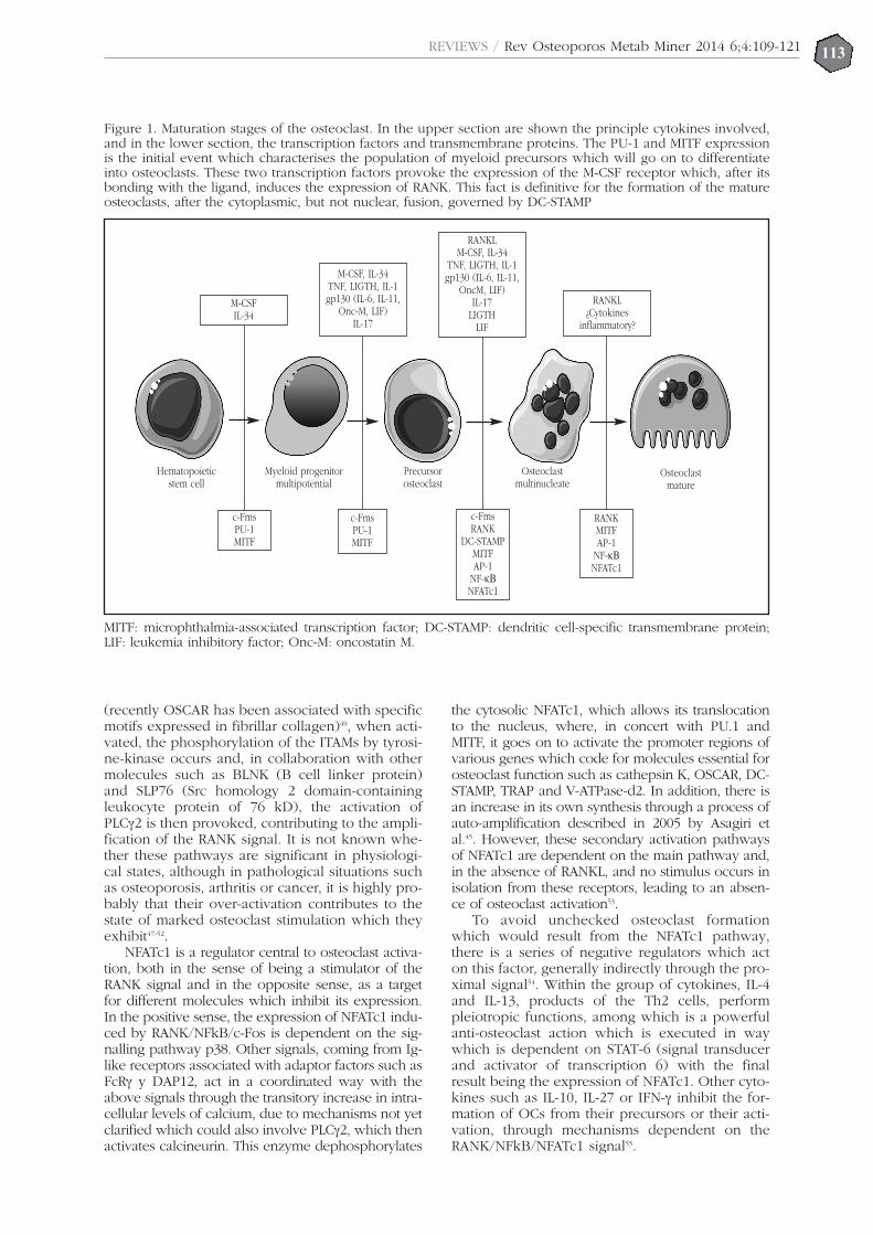
REVIEWS / Rev Osteoporos Metab Miner 2014 6;4:109-121113
(recently OSCAR has been associated with specificmotifs expressed in fibrillar collagen)49, when acti-vated, the phosphorylation of the ITAMs by tyrosi-ne-kinase occurs and, in collaboration with othermolecules such as BLNK (B cell linker protein)and SLP76 (Src homology 2 domain-containingleukocyte protein of 76 kD), the activation ofPLCγ2 is then provoked, contributing to the ampli-fication of the RANK signal. It is not known whe-ther these pathways are significant in physiologi-cal states, although in pathological situations suchas osteoporosis, arthritis or cancer, it is highly pro-bably that their over-activation contributes to thestate of marked osteoclast stimulation which theyexhibit47-52.
NFATc1 is a regulator central to osteoclast activa-tion, both in the sense of being a stimulator of theRANK signal and in the opposite sense, as a targetfor different molecules which inhibit its expression.In the positive sense, the expression of NFATc1 indu-ced by RANK/NFkB/c-Fos is dependent on the sig-nalling pathway p38. Other signals, coming from Ig-like receptors associated with adaptor factors such asFcRγ y DAP12, act in a coordinated way with theabove signals through the transitory increase in intra-cellular levels of calcium, due to mechanisms not yetclarified which could also involve PLCγ2, which thenactivates calcineurin. This enzyme dephosphorylates
the cytosolic NFATc1, which allows its translocationto the nucleus, where, in concert with PU.1 andMITF, it goes on to activate the promoter regions ofvarious genes which code for molecules essential forosteoclast function such as cathepsin K, OSCAR, DC-STAMP, TRAP and V-ATPase-d2. In addition, there isan increase in its own synthesis through a process ofauto-amplification described in 2005 by Asagiri etal.45. However, these secondary activation pathwaysof NFATc1 are dependent on the main pathway and,in the absence of RANKL, and no stimulus occurs inisolation from these receptors, leading to an absen-ce of osteoclast activation53.
To avoid unchecked osteoclast formationwhich would result from the NFATc1 pathway,there is a series of negative regulators which acton this factor, generally indirectly through the pro-ximal signal54. Within the group of cytokines, IL-4and IL-13, products of the Th2 cells, performpleiotropic functions, among which is a powerfulanti-osteoclast action which is executed in waywhich is dependent on STAT-6 (signal transducerand activator of transcription 6) with the finalresult being the expression of NFATc1. Other cyto-kines such as IL-10, IL-27 or IFN-γ inhibit the for-mation of OCs from their precursors or their acti-vation, through mechanisms dependent on theRANK/NFkB/NFATc1 signal55.
Figure 1. Maturation stages of the osteoclast. In the upper section are shown the principle cytokines involved,and in the lower section, the transcription factors and transmembrane proteins. The PU-1 and MITF expressionis the initial event which characterises the population of myeloid precursors which will go on to differentiateinto osteoclasts. These two transcription factors provoke the expression of the M-CSF receptor which, after itsbonding with the ligand, induces the expression of RANK. This fact is definitive for the formation of the matureosteoclasts, after the cytoplasmic, but not nuclear, fusion, governed by DC-STAMP
Hematopoieticstem cell
Myeloid progenitormultipotential
Precursorosteoclast
Osteoclastmultinucleate
Osteoclastmature
M-CSFIL-34
RANKL¿Cytokines
inflammatory?
M-CSF, IL-34TNF, LIGTH, IL-1gp130 (IL-6, IL-11,
Onc-M, LIF)IL-17
RANKLM-CSF, IL-34
TNF, LIGTH, IL-1gp130 (IL-6, IL-11,
OncM, LIF)IL-17
LIGTHLIF
c-FmsPU-1MITF
c-FmsPU-1MITF
c-FmsRANK
DC-STAMPMITFAP-1
NF-κΒNFATc1
RANKMITFAP-1
NF-κΒNFATc1
MITF: microphthalmia-associated transcription factor; DC-STAMP: dendritic cell-specific transmembrane protein;LIF: leukemia inhibitory factor; Onc-M: oncostatin M.
The activation of various TLRs (toll like recep-tors) reduces the rate of formation of mature OCsinduced by RANKL through IFN-β-dependentmechanisms, although independent mechanismshave also been observed. On the other hand, theactivation of TLRs is one of the most powerfulinductors of inflammatory cytokines, such as TNFand IL-1, which act synergistically with RANKL inthe production of inflammatory osteolysis in disea-ses such as rheumatoid arthritis or periodontaldisease56.
In brief, we may intuit that the TLRs, as keyelements in the innate immune system, have anantagonistic role strongly dependent on context.On the one hand, by initiating the inflammatoryresponse, the transformation of precursors intoOCs would reduce, which would increase thepool of cells available for transformation intomacrophages. However, in a more advancedstage, if their activation persisted in a sustainedway, they would act as inductors for osteoclasto-genesis, indirectly by means of inflammatory cyto-kines. The confirmation of this attractive hypothe-sis would constitute one more element to supportthe idea of the OCs’ significant participation in theimmune response.
There are other factors which inhibit the for-mation or activation of the OCs in addition tothose already cited: cytokines such as TRAIL57
(TNF-related apoptosis inducing ligand), IL-12 andIL-1858, different intracellular signalling moleculessuch as SHIP159 (Src homology 2-containing inosi-tol-5-phosphatase 1), NF-κB p10060 and somecomponents of the Notch pathway61, various trans-criptional repressors such as MafB (v-maf muscu-loaponeurotic fibrosarcoma oncogene family pro-tein B)62, C/EBPβ (CCAATenhancer-binding pro-tein β)63, IRF-8 (Interferon regulatory factor)64, andBcL6 (B cell lymphoma)65. All these molecules arepotential targets of therapeutic interest, but theirdetailed analysis is beyond the scope of thisreview.
d) Osteoclast activation pathways independent ofRANKLThe RANKL signal is the most important osteoclastactivation pathway and its annulment in murinemodels results in the complete disappearance ofthe OCs, which means that the role of pathwaysindependent of activation appear, in theory, to beunimportant. However, in 2005 Kim et al. demons-trated that the presence of cofactors such as TGF-β, the hematopoietic precursors in mice null forRANKL, RANK and TRAF-6 would succeed inbeing differentiated into OCs66. It is evident thatthe interest in this topic is enormous, since therecould be, at least in pathological circumstances,non-canonical osteoclast activation pathwayswhich could be modulated to achieve differenttherapeutic responses to the complete annulmentof OCs.
Within the TNF superfamily, given the structuralhomology between its members, various ligandsand receptors have been investigated. One of the
most interesting is LIGHT (also known as TNFSF14and CD258). This type II transmembrane protein isexpressed primarily in activated T-cells, NK cells,dendritic cells and macrophages, performing keybiological functions in the innate and adaptiveimmune responses through the homeostasis, diffe-rentiation and activation of the T-lymphocytes67. Itjoins three receptors which share a structural simila-rity in their cytoplasmic stem: TNFRSF14/HVEM(herpes virus entry mediator), LT-βR (lymphotoxin βreceptor) and DcR3 (decoy receptor 3)68. Althoughthe role of LIGHT in bone resorption is notknown, it has been observed that it causes apowerful osteoclastogenetic action independent ofRANK and OPG, through AKT, NFkB and JNK inhuman and murine monocytes, using TRAF-2 andTRAF-5. Its function in bone diseases has not beenclarified, but it is, without a doubt, an interestingtarget of potential therapeutic interest69,70.
Two other members of the TNF superfamilyhave shown an osteoclastogenetic capability inde-pendent of RANKL. APRIL (a proliferation indu-cing ligand, TNFSF13) and BAFF (cell activatingfactor belonging to the TNF, also known as BLySand TNFSF 13b), are capable, in in vitro culture,of inducing cells with the osteoclast phenotypefrom mononuclear precursors, although of a sma-ller size and with a lower number of nuclei andresorptive capacity than those induced by RANKLor LIGHT71.
e) Origin of RANKL in osteoclast activationAlthough the origin of RANKL which is involved inbone remodelling is classically thought to be theOBs, there have been a number of experimentalfindings which have cast doubt on this idea. In apioneering study, Corral et al.72 showed that theablation of osteoblast progenitors by the adminis-tration of ganciclovir in mice bearing a thymidine-kinase transgene under the control of the osteocal-cin promoter, did not cause any effects on theosteoclastic surfaces or on the markers for resorp-tion, even after several weeks of follow up, inthose in which the population of osteoblasts haddisappeared from the bone surfaces. Morerecently, and using a similar transgenic murinemodel, Galli et al. observed that the absence ofosteoblasts did not affect the levels at the baseli-ne, or after being stimulated by PTH, of RANKLmRNA73. These studies indicate that the classicparadigm, which is that the RANKL which governsosteoclast activation comes from OBs or their pre-cursors, should be revised74.
OCs are formed in different locations in theskeleton with different purposes and with avariety of support cells charged with synthesisingthe RANKL necessary for their activation. Forexample, the femurs of mice which lack osteocy-tic RANKL develop a normal morphology, whichindicates that the cortical modelling of the longbones is controlled by cells other than the oste-ocytes; whereas during chondral ossification, themain source of RANKL, which enables the reab-sorptive action of the osteoclasts on the calcified
114REVIEWS / Rev Osteoporos Metab Miner 2014 6;4:109-121
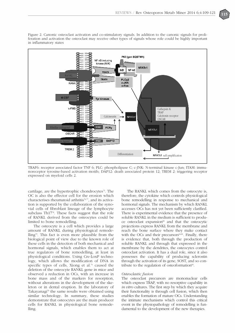
115REVIEWS / Rev Osteoporos Metab Miner 2014 6;4:109-121
cartilage, are the hypertrophic chondrocytes75. TheOC is also the effector cell for the erosion whichcharacterises rheumatoid arthritis76,77, and its activa-tion is supported by the collaboration of the syno-vial cells of fibroblast lineage of the lymphocytesubclass Th1778. These facts suggest that the roleof RANKL derived from the osteocytes could belimited to bone remodelling.
The osteocyte is a cell which provides a largeamount of RANKL during physiological remode-lling79. This fact is even more plausible from thebiological point of view due to the known role ofthese cells in the detection of both mechanical andhormonal signals, which enables them to act attrue regulators of bone remodelling, at least inphysiological conditions. Using Cre-LoxP techno-logy, which allows the modification of DNA inspecific types of cells, Xiong et al.74 caused thedeletion of the osteocyte RANKL gene in mice andobserved a reduction in OCs, with an increase inbone mass and of the markers for resorption,without alterations in the development of the ske-leton or in dental eruption. In the laboratory ofTakayanagi79 the same results were obtained usingsimilar technology. In summary, these studiesdemonstrate that osteocytes are the main producercells for RANKL in physiological bone remode-lling.
The RANKL which comes from the osteocyte is,therefore, the cytokine which controls physiologicalbone remodelling in response to mechanical andhormonal signals. The mechanism by which RANKLaccesses OCs has not yet been sufficiently clarified.There is experimental evidence that the presence ofsoluble RANKL in the medium is sufficient to produ-ce osteoclast expansion80 and that the osteocyticprojections express RANKL from the membrane andreach the bone surface where they make contactwith the OCs and their precursors64,81. Finally, thereis evidence that, both through the production ofsoluble RANKL and through that expressed in themembrane by the dendrites, the osteocytes controlosteoclast activation. It has a dual role, since it alsopossesses the capability of producing sclerostinthrough the activation of its gene, SOST, and so con-tribute to the regulation of osteoformation82.
Osteoclastic fusionThe osteoclast precursors are mononuclear cellswhich express TRAP, with no resorptive capability inin vitro cultures. The first step by which they acquiretheir functionality is through cell fusion, which thenenables the formation of mature OCs. Understandingthe intimate mechanisms which control this criticalevent in the physiopathology of remodelling is fun-damental to the development of the new therapies.
Figure 2. Canonic osteoclast activation and co-stimulatory signals. In addition to the canonic signals for proli-feration and activation the osteoclast may receive other types of signals whose role could be highly importantin inflammatory states
TRAF6: receptor associated factor TNF 6; PLC: phospholipase C; c-JNK: N-terminal kinase c-Jun; ITAM: immu-noreceptor tyrosine-based activation motifs; DAP12: death associated protein 12; TREM 2: triggering receptorexpressed on myeloid cells 2.
RANKL
Fibrillar collagens?
Unknown ligand
Calcineurin
Differentiation self-amplification
Cathepsin KTRAPCalcitonin receptorOSCAR
REVIEWS / Rev Osteoporos Metab Miner 2014 6;4:109-121116
In physiological conditions, the pre-OC TRAPcells + and the mature OCs are only found on thebone surfaces, which indicates that the fusionoccurs in these locations. Using techniques ofDNA subtraction in precursor cells stimulated byisolated M-CSF or M-CSF and RANKL, it was obser-ved that DC-STAMP (dendritic cell-specific trans-membrane protein) is an essential molecule forthe fusion of mononuclear cells as a first step forthe formation of active mature OCs. This trans-membrane protein, discovered in 200083, is alsoexpressed in dendritic cells and macrophages84. Itsannulment in murine models provoked osteopo-rosis associated with a complete absence of fusedmononuclear OCs as well as foreign-body giantcells. In these mice there persisted a moderatedegree of resorptive activity in the mature cells,which indicates that their fundamental role is per-formed at fusion85. The regulation of DC-STAMP iscomplex and depends not only on theRANKL/RANK pathway but also on other indepen-dent factors, such as IL-3286, Tal1 (T-cell acutelymphocytic leukemia 1)87, LDLR (low-densitylipoprotein receptor)88, CCN2/CTGF (CCN family2/connective tissue growth factor)89 and vitaminE90, among others, whose role is even less wellknown but which could be future targets of thera-peutic interest.
OC fusion is promoted by other moleculessuch as the inflammatory cytokines. Among these,in addition to the actions already mentioned ofRANKL, both TNF-α and LPS (lipopolysaccharide)are capable of inducing OC fusion under certaincircumstances. For example, the action of TNF-αis specifically blocked by Ac anti-TNF-α, while theeffect of LPS is partly blocked by these drugs, andcompletely blocked by polymyxin B91. The activa-tion of these pathways is accompanied by intrace-llular signals dependent on kinases, and wheninhibitors are used specific to these pathways OCfusion is reduced, while levels of DC-STAMP arenot altered. These findings indicate that there arealternative pathways which regulate OC fusionindependently of DC-STAMP, although it is notknown if they exert physiological functions oronly interfere with pathological processes92.
Additional roles for osteoclastsIn addition to their function as the only cells capa-ble of reabsorbing calcified bone matrix, OCs par-ticipate in other processes which we summarisebelow.
1. Stimulation of bone formationBone remodelling is a coupled process in whichthe osteoclast activity is followed by the action ofthe osteoblasts. The pharmacological inhibition ofthe former provokes a reduction in the latter, whilethe osteoforming stimulus is followed by a secon-dary increase in resorption. In principle, the modelwould appear to be simple, attributing to factorsreleased from the matrix reabsorbed by the OCs arole in the recruitment of osteoblast93,94. However, ina study published in 2001, the Molecular Biology
Group of the University of Hamburg demonstratedthat, in some murine models of osteopetrosis andin a patient with the malignant infantile form, inspite of a functional alteration in the resorptivemechanism in the presence of a normal number ofOCs, such as is produced with the annulment of thechloride channels ClC-7 C, there was normal boneformation7. This fact suggests that there are factorsindependent of the matrix reabsorbed by the OCswhose role in the coupling is probably more signi-ficant.
Among the mechanisms in which OCs interve-ne directly stimulating osteoformation, the follo-wing have been proposed95: on the one hand,ephrin B2, expressed in the osteoclast membrane,is capable of provoking an activation signal bybonding with its osteoblast receptor EphB4; also,sphingosine-1-phosphate is capable of causing therecruitment of osteoblast precursors to the remo-delling sites96, although treatment with analoguesof this molecule has not shown significant resultsin the mending of fractures97. OC expresses, inaddition, regulatory factors negative to osteoblasts,such as Atp6v0d2 (a subunit of the V-ATPase pro-ton pump)98. Even though the physiological role ofthese molecular signals is not known, the findingswhich have been commented on suggest that theintervention of the OCs in remodelling is not limi-ted to bone resorption, but that they also play asignificant role in the coupling through molecularsignals which participate in the recruitment, acti-vation and inhibition of the osteoblasts.
2. Immune cellsBoth OCs and OBs have the capability of respon-ding to a wide variety of cytokines produced bythe cells of the innate and adaptive immunesystems78,99-101. The OCs contain all the mechanismsnecessary for endocytosis and the processing ofexogenous proteins coming from the materialgenerated during resorption and in pathologicalsituations such as osteomyelitis. In 2009, Kiesel etal.102 demonstrated that the OCs could recruit TCD8+ FoxP3+ cells and present their antigens.These cells would play a regulatory role, whosefunction in non-inflammatory situations is unk-nown. A very attractive but non-proven hypothe-sis relates this capacity of the OCs as presenters ofantigens to the existence of a large reservoir ofCD8+ central memory T-lymphocytes in the bonemarrow, the former participating in the latter’srecruitment and maintenance103.
The extraction of necrotic bone during a bac-terial infection is another of the mechanisms inwhich OCs play a part in the immune response. Infact, in an elegant study in which murine modelswhich emulated the biology of osteomyelitis andof periodontal implants were used, Li et al.104
demonstrated that the functional inhibition of theOCs by bisphosphonates and by osteoprotogerinwas associated with an increase in the quantity ofnecrotic cortical bone around the implant whichacted as nests for the bacterial colonisation, whileat the same time reducing the size of the drainage
REVIEWS / Rev Osteoporos Metab Miner 2014 6;4:109-121117
orifice through which the opsonised bacteria wereexpelled to the exterior of the lesion. These dataare highly significant since they suggest that thepharmacological inhibition of osteoclasts could becontraindicated in bone infections, as well as inthe pathogenesis of osteonecrosis of the jaw,where bacterial colonisation is very important, andwhere OCs would play a key role, at least in itsinitial phases.
3. Articular cartilageIn those process in which the destruction of hyali-ne articular cartilage occurs, giant multinucleatedcells have been observed which express the osteo-clast phenotype (TRAP+, cathepsin K+, MMP9+,CD14−, HLA-DR−, CD45+, CD51+ and CD68+).These cells, called “chondroclasts” in some publica-tions, have the capability of reabsorbing the cartila-ginous matrix and have been implicated in thepathogeny of diseases such as rheumatoid arthritisor arthrosis105. Their specific role has not been esta-blished with any certainty, although there is variousindirect evidence to suggest that they may play asignificant role on articular damage. It is knownthat 30% of the total RANKL which is produced inarthritic joints is synthesised in the cartilage, essen-tially through the chondrocytes106. The soluble partof this cytokine acting like a paracrine, may partici-pate, through osteoclast activation in locations ofchondral-sinovial contact, in the pathology of ero-sion and of juxtaarticular osteopenia, which charac-terise rheumatoid lesions. Furthermore, eventhough it has not been demonstrated with sufficientcertainty, chondrocytic RANKL may contribute tothe transformation and activation of the mononu-clear precursors, resulting in chondroclasts capa-ble of degrading the cartilage. The mechanismsthrough which this action would occur is not yetknown, but there is, undoubtedly, an interestingquestion to be asked based around about the possi-ble therapeutic role of the inhibitors of RANKL inprocesses such as arthrosis.
4. Energy metabolismOsteocalcin, a small peptide produced by osteo-blasts, stimulates the secretion of insulin by thebeta pancreatic cells, a finding of enormousimportance in decisively implicating bone tissue inthe hormonal control of energy metabolism107. Thismolecule has a number of the characteristics of ahormone: it is a specifically cellular product,synthesised in a pre-propeptide form and secretedinto the circulation after a process of vitamin K-dependent gamma-carboxylation. This factexplains its great affinity for the bone matrix,which causes it to be released during bone resorp-tion and converted into its active form after expo-sure to the acid pH of the resorption lacuna. Intransgenic mice which lack V-ATPase activity,hypoinsulinemia and glucose intolerance associa-ted with reduced levels of osteocalcin are obser-ved108. A study which analysed the effects of alen-dronate in a small sample of patients showedreduced levels of infra-carboxylated osteocalcin
which is associated inversely with an increase inbody weight and of fat mass109. However, a reviewof the results of the FIT, HORIZON and FREEDOMstudies did not show any alteration in these para-meters, nor in glucose metabolism110. In summary,while animal models suggest a role for boneremodelling in the control of energy metabolism,the studies carried out in humans show discordantresults which need to be clarified in the future111.
ConclusionsThe OC has been considered classically to be a cellwhose function is exclusively that of bone remode-lling, and which exhibits gregarious behaviour.However, in the last decade experimental findingshave drastically transformed this over-simplisticview. The OC shares common origins with thecells of the immune system, both in the myeloidand the lymphoid series. Its role in articular inflam-matory diseases such as rheumatoid arthritis is pro-bably highly significant, since, to its well-knownfunction as the only cell capable of dissolving thecalcified bone matrix, are added new roles due toits capacity to secrete cytokines and as an antigenpresenter cell. OCs, as extraordinarily dynamiccells, are therapeutic targets of enormous interest(Table 1) due to their participation in processessuch as osteoporosis, arthrosis or cancer.
Bibliography
1. Seeman E. Modelling and remodelling. En: Bilezikian J,Raisz LG, Martin TJ, editores. Principles of bone biology(Third Edition). Filadelfia: Elsevier Inc; 2008;p.3-28.
2. Schett G. Biology, physiology and morphology of bone.En: Firestein GS, Budd RC, Gabriel SE, McInnes IB,O'Dell JR, editores. Kelley's Textbook of Rheumatology(Ninth Edition). Filadelfia: Saunders; 2013;p.61-6.
3. Goldring SR, Schett G. The role of the immune systemin the bone loss of inflammatory artritis. En: Lorenzo J,Horowitz M, Choi Y, Schett G, Takayanagi H, editores.Osteoimmunology. Londres: Elsevier; 2011;p.301-22.
4. Olechnowicz SW, Edwards CM. Contributions of thehost microenvironment to cancer-induced bone disea-se. Cancer Res 2014;74:1625-31.
5. Väänänen HK, Zhao H. Osteoclast function: biologyand mechanisms En: Bilezikian JP, Raisz LG, Martin TJ.Principles of Bone Biology (Third Edition). Filadelfia:Elsevier Inc; 2008;p:193-209.
6. Graves AR, Curran PK, Smith C, Mindell JA. The Cl-/H+antiporter ClC-7 is the primary chloride permeationpathway in lysosomes. Nature 2008;453:788-92.
7. Kornak U, Kasper D, Bösl MR, Kaiser E, Schweizer M,Schulz A, et al. Loss of the ClC-7 chloride channel leadsto osteopetrosis in mice and man. Cell 2001;104:205-15.
8. Schaller S, Henriksen K, Sveigaard C, Heegaard AM,Hélix N, Stahlhut M, et al. The chloride channel inhi-bitor NS3736 prevents bone resorption in ovariectomi-zed rats without changing bone formation. J BoneMiner Res 2004;19:1144-53.
9. Kasper D, Planells-Cases R, Fuhrmann JC, Scheel O,Zeitz O, Ruether K, et al. Loss of the chloride channelClC-7 leads to lysosomal storage disease and neurode-generation. EMBO J 2005;24:1079-91.
10. Kraft-Terry SD, Gendelman HE. Proteomic biosignatu-res for monocyte-macrophage differentiation. CellImmunol 2011;271:239-55.
11. Teitelbaum SL, Ross FP. Genetic regulation of osteoclastdevelopment and function. Nat Rev Genet 2003;4:638-49.
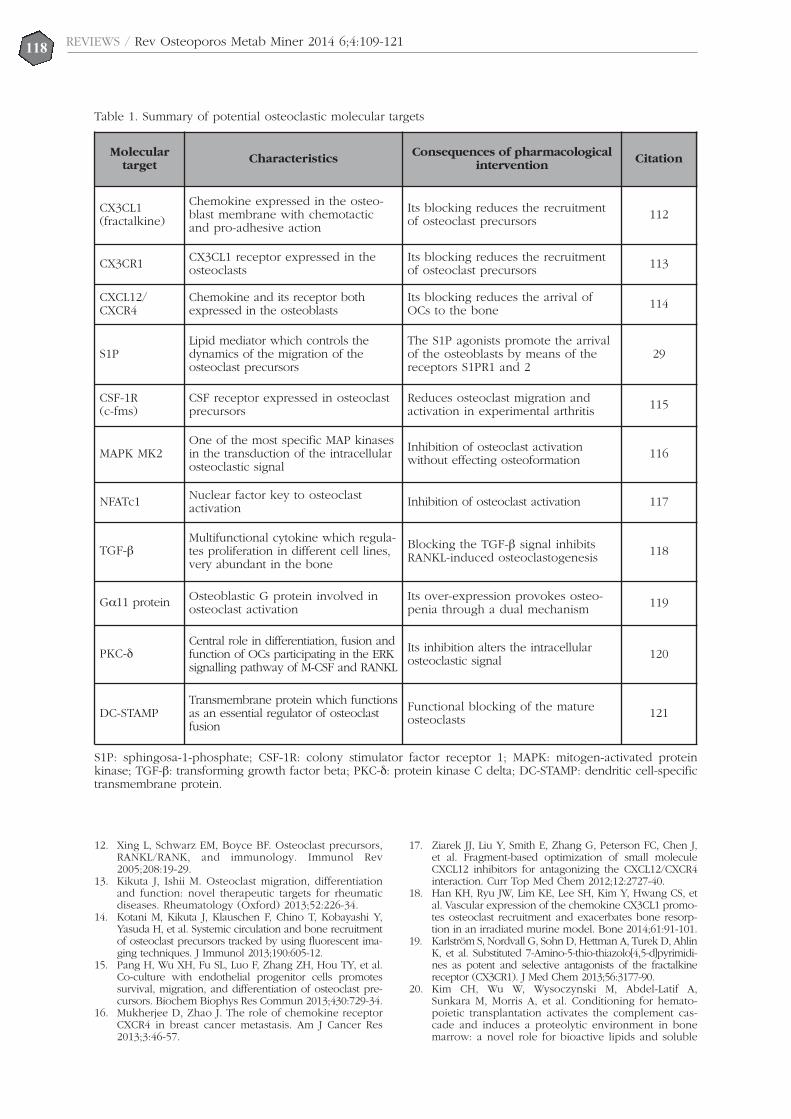
12. Xing L, Schwarz EM, Boyce BF. Osteoclast precursors,RANKL/RANK, and immunology. Immunol Rev2005;208:19-29.
13. Kikuta J, Ishii M. Osteoclast migration, differentiationand function: novel therapeutic targets for rheumaticdiseases. Rheumatology (Oxford) 2013;52:226-34.
14. Kotani M, Kikuta J, Klauschen F, Chino T, Kobayashi Y,Yasuda H, et al. Systemic circulation and bone recruitmentof osteoclast precursors tracked by using fluorescent ima-ging techniques. J Immunol 2013;190:605-12.
15. Pang H, Wu XH, Fu SL, Luo F, Zhang ZH, Hou TY, et al.Co-culture with endothelial progenitor cells promotessurvival, migration, and differentiation of osteoclast pre-cursors. Biochem Biophys Res Commun 2013;430:729-34.
16. Mukherjee D, Zhao J. The role of chemokine receptorCXCR4 in breast cancer metastasis. Am J Cancer Res2013;3:46-57.
17. Ziarek JJ, Liu Y, Smith E, Zhang G, Peterson FC, Chen J,et al. Fragment-based optimization of small moleculeCXCL12 inhibitors for antagonizing the CXCL12/CXCR4interaction. Curr Top Med Chem 2012;12:2727-40.
18. Han KH, Ryu JW, Lim KE, Lee SH, Kim Y, Hwang CS, etal. Vascular expression of the chemokine CX3CL1 promo-tes osteoclast recruitment and exacerbates bone resorp-tion in an irradiated murine model. Bone 2014;61:91-101.
19. Karlström S, Nordvall G, Sohn D, Hettman A, Turek D, AhlinK, et al. Substituted 7-Amino-5-thio-thiazolo[4,5-d]pyrimidi-nes as potent and selective antagonists of the fractalkinereceptor (CX3CR1). J Med Chem 2013;56:3177-90.
20. Kim CH, Wu W, Wysoczynski M, Abdel-Latif A,Sunkara M, Morris A, et al. Conditioning for hemato-poietic transplantation activates the complement cas-cade and induces a proteolytic environment in bonemarrow: a novel role for bioactive lipids and soluble
118REVIEWS / Rev Osteoporos Metab Miner 2014 6;4:109-121
Table 1. Summary of potential osteoclastic molecular targets
Moleculartarget Characteristics Consequences of pharmacological
intervention Citation
CX3CL1(fractalkine)
Chemokine expressed in the osteo-blast membrane with chemotacticand pro-adhesive action
Its blocking reduces the recruitmentof osteoclast precursors 112
CX3CR1 CX3CL1 receptor expressed in theosteoclasts
Its blocking reduces the recruitmentof osteoclast precursors 113
CXCL12/CXCR4
Chemokine and its receptor bothexpressed in the osteoblasts
Its blocking reduces the arrival ofOCs to the bone 114
S1PLipid mediator which controls thedynamics of the migration of theosteoclast precursors
The S1P agonists promote the arrivalof the osteoblasts by means of thereceptors S1PR1 and 2
29
CSF-1R(c-fms)
CSF receptor expressed in osteoclastprecursors
Reduces osteoclast migration andactivation in experimental arthritis 115
MAPK MK2One of the most specific MAP kinasesin the transduction of the intracellularosteoclastic signal
Inhibition of osteoclast activationwithout effecting osteoformation 116
NFATc1 Nuclear factor key to osteoclastactivation Inhibition of osteoclast activation 117
TGF-βMultifunctional cytokine which regula-tes proliferation in different cell lines,very abundant in the bone
Blocking the TGF-β signal inhibitsRANKL-induced osteoclastogenesis 118
Gα11 protein Osteoblastic G protein involved inosteoclast activation
Its over-expression provokes osteo-penia through a dual mechanism 119
PKC-δCentral role in differentiation, fusion andfunction of OCs participating in the ERKsignalling pathway of M-CSF and RANKL
Its inhibition alters the intracellularosteoclastic signal 120
DC-STAMPTransmembrane protein which functionsas an essential regulator of osteoclastfusion
Functional blocking of the matureosteoclasts 121
S1P: sphingosa-1-phosphate; CSF-1R: colony stimulator factor receptor 1; MAPK: mitogen-activated proteinkinase; TGF-β: transforming growth factor beta; PKC-δ: protein kinase C delta; DC-STAMP: dendritic cell-specifictransmembrane protein.
119REVIEWS / Rev Osteoporos Metab Miner 2014 6;4:109-121
C5b-C9 as homing factors. Leukemia 2012;26:106-16. 21. Ratajczak MZ, Kim C, Janowska-Wieczorek A, Ratajczak J.
The expanding family of bone marrow homing factors forhematopoietic stem cells: Stromal Derived Factor 1 Is notthe only player in the game. Sci World J 2012; 2012:758512.
22. Gangoiti P, Arana L, Ouro A, Granado MH, Trueba M,Gómez-Muñoz A. Activation of mTOR and RhoA is amajor mechanism by which Ceramide 1-phosphate stimu-lates macrophage proliferation. Cell Signal 2011;1:27-34.
23. Ishii M, Egen JG, Klauschen F, Meier-Schellersheim M,Saeki Y, Vacher J, et al. Sphingosine-1-phosphatemobilizes osteoclast precursors and regulates bonehomeostasis. Nature 2009;458:524-8.
24. Ishii M, Kikuta J, Shimazu Y, Meier-Schellersheim M,Germain RN. Chemorepulsion by blood S1P regulatesosteoclast precursor mobilization and bone remode-ling in vivo. J Exp Med 2010;207:2793-8.
25. Maceyka M, Harikumar KB, Milstien S, Spiegel S.Sphingosine-1-phosphate signaling and its role in dise-ase. Trends Cell Biol 2012;1:50-60.
26. Kikuta J, Kawamura S, Okiji F, Shirazaki M, Sakai S,Saito H, et al. Sphingosine-1-phosphate-mediated oste-oclast precursor monocyte migration is a critical pointof control in antibone-resorptive action of active vita-min D. Proc Natl Acad Sci USA 2013;110:7009-13.
27. Boyce BF. Sphingosine-1 phosphate: a new player inosteoimmunology. Dev Cell 2009;3:323-4.
28. Ishii M, Kikuta J. Sphingosine-1-phosphate signalingcontrolling osteoclasts and bone homeostasis. BiochimBiophys Acta 2013;1831:223-7.
29. Quint P, Ruan M, Pederson L, Kassem M, WestendorfJJ, Khosla S, et al. Sphingosine 1-phosphate (S1P)receptors 1 and 2 coordinately induce mesenchymalcell migration through S1P activation of complemen-tary kinase pathways. J Biol Chem 2013;288:5398-406.
30. Takahashi N, Yamana H, Yoshiki S, Roodman GD,Mundy GR, Jones SJ, et al. Osteoclast-like cell formationand its regulation by osteotropic hormones in mousebone marrow cultures. Endocrinology 1988;122:1373-82.
31. Arai F, Miyamoto T, Ohneda O, Inada T, Sudo T, BraselK, et al. Commitment and differentiation of osteoclastprecursor cells by the sequential expression of c-Fmsand receptor activator of nuclear factor kappaB(RANK) receptors. J Exp Med 1999;190:1741-54.
32. Asagiri M, Takayanagi H. The molecular understandingof osteoclast differentiation. Bone 2007;40:251-64.
33. González Macías J, Olmos Martínez JM. Fisiopatologíade la osteoporosis y mecanismo de acción de la PTH.Rev Osteoporos Metab Miner 2010;2 (Suppl 2);5-17.
34. Horowitz MC, Lorenzo JA. Immunologic regulation ofbone development. Adv Exp Med Biol 2007;602:47-56.
35. Chai RC, Kouspou MM, Lang BJ, Nguyen CH, van derKraan AG, Vieusseux JL, et al. Molecular stress inducingcompounds increase osteoclast formation in a Heat ShockFactor 1 dependent manner. J Biol Chem 2014; Apr 1.
36. Asai K, Funaba M, Murakami M. Enhancement ofRANKL-induced MITF-E expression and osteoclastoge-nesis by TGF-β. Cell Biochem Funct 2014; Feb 12. doi:10.1002/cbf.3028.
37. Matsumoto T, Nagase Y, Iwasawa M, Yasui T, MasudaH, Kadono Y, et al. Distinguishing the proapoptoticand antiresorptive functions of risedronate in murineosteoclasts: role of the Akt pathway and the ERK/Bimaxis. Arthritis Rheum 2011;12:3908-17.
38. Matsumoto T, Nagase Y, Hirose J, Tokuyama N, YasuiT, Kadono Y, et al. Regulation of bone resorption andsealing zone formation in osteoclasts occurs throughprotein kinase b-mediated microtubule stabilization. JBone Miner Res 2013;5:1191-202.
39. Mao D, Epple H, Uthgenannt B, Novack DV, Faccio R.PLCgamma2 regulates osteoclastogenesis via its inter-action with ITAM proteins and GAB2. J Clin Invest2006;116:2869-79.
40. Hayden MS, Ghosh S. Shared principles in NF-kB sig-naling. Cell 2008;132:344-62.
41. Mantovani A. Molecular pathways linking inflamma-tion and cancer. Curr Mol Med 2010;4:369-73.
42. Nakashima T, Hayashi M, Takayanagi H. New insights
into osteoclastogenic signaling mechanisms. TrendsEndocrinol Metab 2012;23:582-90.
43. Shaw JP, Utz PJ, Durand DB, Toole JJ, Emmel EA,Crabtree GR. Identification of a putative regulator ofearly T cell activation genes. Science 1998;241:202-5.
44. Takayanagi H, Kim S, Koga T, Nishina H, Isshiki M,Yoshida H, et al. Induction and activation of the trans-cription factor NFATc1 (NFAT2) integrate RANKL signa-ling in terminal differentiation of osteoclasts. Dev Cell2002;6:889-901.
45. Asagiri M, Sato K, Usami T, Ochi S, Nishina H, YoshidaH, et al. Autoamplification of NFATc1 expression deter-mines its essential role in bone homeostasis. J ExpMed 2005;202:1261-9.
46. Kuroda Y, Matsuo K. Molecular mechanisms of trigge-ring, amplifying and targeting RANK signaling in oste-oclasts. World J Orthop 2012;3:167-74.
47. Barrow AD, Raynal N, Andersen TL, Slatter DA, BihanD, Pugh N, et al. OSCAR is a collagen receptor thatcostimulates osteoclastogenesis in DAP12-deficienthumans and mice. J Clin Invest 2011;121:3505-16.
48. Paradowska-Gorycka A, Jurkowska M. Structure, expres-sion pattern and biological activity of molecular complexTREM-2/DAP12. Human Immunol 2013;74:730-7.
49. Nemeth K, Schoppet M, Al-Fakhri N, Helas S, JessbergerR, Hofbauer LC, et al. The role of osteoclast-associatedreceptor in osteoimmunology. J Immunol 2011;186:13-8.
50. Pelham CJ, Agrawal DK. Emerging roles for triggeringreceptor expressed on myeloid cells receptor familysignaling in inflammatory diseases. Expert Rev ClinImmunol 2014;10:243-56.
51. Colonna M, Turnbull I, Klesney-Tait J. The enigmaticfunction of TREM-2 in osteoclastogenesis. Adv ExpMed Biol 2007;602:97-105.
52. Takahashi N, Maeda K, Ishihara A, Uehara S, KobayashiY. Regulatory mechanism of osteoclastogenesis byRANKL and Wnt signals. Front Biosci 2011;16:21-30.
53. Otero K, Shinohara M, Zhao H, Cella M, Gilfillan S,Colucci A, et al. TREM2 and β-catenin regulate bonehomeostasis by controlling the rate of osteoclastogene-sis. J Immunol 2012;188:2612-21.
54. Takayanagi H. The role of NFAT in osteoclast forma-tion. Ann N Y Acad Sci 2007;1116:227-37.
55. Ivashkiv LB, Donlin LT. Regulation of type I interferonresponses. Nat Rev Immunol 2014;14:36-49.
56. Meng S, Zhang L, Tang Y, Tu Q, Zheng L, Yu L, et al.BET inhibitor JQ1 blocks inflammation and bone des-truction. J Dent Res 2014;93:657-62.
57. Yen ML, Hsu PN, Liao HJ, Lee BH, Tsai HF. TRAF-6dependent signaling pathway is essential for TNF-rela-ted apoptosis-inducing ligand (TRAIL) induces osteo-clast differentiation. PLoS One 2012;7:e38048.
58. Kitaura H, Kimura K, Ishida M, Sugisawa H, Kohara H,Yoshimatsu M, et al. Effect of cytokines on osteoclastformation and bone resorption during mechanicalforce loading of the periodontal membrane. ScientificWorld Journal 2014; Jan 19. doi:10.1155/2014/617032.
59. Iyer S, Margulies BS, Kerr WG. Role of SHIP1 in bonebiology. Ann N Y Acad Sci 2013;1280:11-4.
60. Taniguchi R, Fukushima H, Osawa K, Maruyama T,Yasuda H, Weih F, et al. RelB-induced expression of Cot,a MAP3K family member, rescues RANKL-induced osteo-clastogenesis in alymphoplasia mice by promoting NF-κB2 processing by IKKα. J Biol Chem 2014;289:7349-61.
61. Canalis E, Adams DJ, Boskey A, Parker K, Kranz L,Zanotti S. Notch signaling in osteocytes differentiallyregulates cancellous and cortical bone remodeling. JBiol Chem 2013;288:25614-25.
62. Smink JJ, Bégay V, Schoenmaker T, Sterneck E, deVries TJ, Leutz A. Transcription factor C/EBPbeta iso-form ratio regulates osteoclastogenesis through MafB.EMBO J 2009;28:1769-81.
63. Fu SL, Pang H, Xu JZ, Wu XH. C/EBPβMediates OsteoclastRecruitment by Regulating Endothelial Progenitor CellExpression of SDF-1α. PLoS One 2014;9:e91217.
64. Zhao B, Takami M, Yamada A, Wang X, Koga T, Hu X,et al. Interferon regulatory factor-8 regulates bonemetabolism by suppressing osteoclastogenesis. Nat
Med 2009;15:1066-71.65. Park-Min KH, Lee EY, Moskowitz NK, Lim E, Lee SK,
Lorenzo JA, et al. Negative regulation of osteoclast pre-cursor differentiation by CD11b and β2 integrin-B-celllymphoma 6 signaling. J Bone Miner Res 2013;28:135-49.
66. Kim N, Kadono Y, Takami M, Lee J, Lee SH, Okada F, etal. Osteoclast differentiation independent of the TRAN-CE–RANK–TRAF6 axis. J Exper Med 2005; 202:589-95.
67. Mellis DJ, Itzstein C, Helfrich MH, Crockett JC. Theskeleton: a multi-functional complex organ. The roleof key signalling pathways in osteoclast differentiationand in bone resorption. J Endocrinol 2011;211:131-43.
68. Ware CF. Targeting lymphocyte activation through thelymphotoxin and LIGHT pathways. Immunol Rev2008;223:186-201.
69. Ware CF, Sedy J. TNF superfamily networks: bidirectionaland interference pathways of the Herpesvirus EntryMediator (TNFSF14). Curr Opin Immunol 2011;23:627-31.
70. Hemingway F, Kashima TG, Knowles HJ, Athanasou NA.Investigation of osteoclastogenic signalling of the RANKLsubstitute LIGHT. Exper Mol Pathol 2013;94:380-5.
71. Hemingway F, Taylor R, Knowles HJ, Athanasou NA.RANKL-independent human osteoclast formation withAPRIL, BAFF, NGF, IGF I 2 and IGF II. Bone2011;48:938-44.
72. Corral DA, Amling M, Priemel M, Loyer E, Fuchs S,Ducy P. Dissociation between bone resorption andbone formation in osteopenic transgenic mice. ProcNatl Acad Sci USA 1998;95:13835-40.
73. Galli C, Fu Q, Wang W, Olsen BR, Manolagas SC, JilkaRL, et al. Commitment to the osteoblast lineage is notrequired for RANKL gene expression. J Biol Chem2009;284:12654-62.
74. Xiong J, O’Brien CA. Osteocyte RANKL: New Insightsinto the control of bone remodeling. J Bone Miner Res2012;27:499-505.
75. Gravallese EM, Harada Y, Wang JT, Gorn AH, ThornhillTS, Goldring SR. Identification of cell types responsiblefor bone resorption in rheumatoid arthritis and juvenilerheumatoid arthritis. Am J Pathol 1998;152:943-51.
76. Pettit AR, Ji H, von Stechow D, Müller R, Goldring SR,Choi Y, et al. TRANCE/RANKL knockout mice are pro-tected from bone erosion in a serum transfer model ofarthritis. Am J Pathol 2001;159:1689-99.
77. Goldring SR, Purdue PE, Crotti TN, Shen Z, FlanneryMR, Binder NB, et al. Bone remodelling in inflamma-tory arthritis. Ann Rheum Dis 2013;72:52-55.
78. Arboleya L, Castañeda S. Osteoimmunology. ReumatolClin 2013;9:303-15.
79. Nakashima T, Hayashi M, Fukunaga T, Kurata K, Oh-Hora M, Feng JQ, et al. Evidence for osteocyte regula-tion of bone homeostasis through RANKL expression.Nat Med 2011;17:1231-4.
80. Zhao S, Kato Y, Zhang Y, Harris S, Ahuja SS, BonewaldLF. MLO-Y4 osteocyte-like cells support osteoclast forma-tion and activation. J Bone Miner Res 2002;17:2068-79.
81. Kurata K, Heino TJ, Higaki H, Vaananen HK. Bonemarrow cell differentiation induced by mechanicallydamaged osteocytes in 3D gel-embedded culture. JBone Miner Res 2006;21:616-25.
82. Van Bezooijen RL, Roelen BAJ, Visser A, Wee-Pals L, deWilt E, Karperien M, et al. Sclerostin is an osteocyte-expressed negative regulador of bone formation, but nota classical BMP antagonist. J Exp Med 2004;199:805-14.
83. Hartgers FC, Vissers JL, Looman MW, van Zoelen C,Huffine C, Figdor CG, et al. DC-STAMP, a novel multi-membrane-spanning molecule preferentially expres-sed by dendritic cells. Eur J Immunol 2000;30:3585-90.
84. Xing L, Xiu Y, Boyce BF. Osteoclast fusion and regu-lation by RANKL-dependent and independent factors.World J Orthop 2012;3:212-22.
85. Yagi M, Ninomiya K, Fujita N, Suzuki T, Iwasaki R,Morita K, et al. Induction of DC-STAMP by alternativeactivation and downstream signaling mechanisms. JBone Miner Res 2007;22:992-1001.
86. Kim YG, So MW, Koo BS, Chang EJ, Song SJ, Lee CK,et al. The influence of interleukin-32γ on osteoclasto-genesis with a focus on fusion-related genes. J Clin
Immunol 2012;32:201-6. 87. Courtial N, Smink JJ, Kuvardina ON, Leutz A, Göthert
JR, Lausen J. Tal1 regulates osteoclast differentiationthrough suppression of the master regulator of cellfusion DC-STAMP. FASEB J 2012;26:523-32.
88. Okayasu M, Nakayachi M, Hayashida C, Ito J, Kaneda T,Masuhara M, et al. Low-density lipoprotein receptordeficiency causes impaired osteoclastogenesis andincreased bone mass in mice because of defect in oste-oclastic cell-cell fusion. J Biol Chem 2012;287:19229-41.
89. Nishida T, Emura K, Kubota S, Lyons KM, Takigawa M.CCN family 2/connective tissue growth factor(CCN2/CTGF) promotes osteoclastogenesis via inductionof and interaction with dendritic cell-specific transmembra-ne protein (DC-STAMP). J Bone Miner Res 2011;26:351-63.
90. Fujita K, Iwasaki M, Ochi H, Fukuda T, Ma C,Miyamoto T, et al. Vitamin E decreases bone mass bystimulating osteoclast fusion. Nat Med 2012;18:589-94.
91. Hotokezaka H, Sakai E, Ohara N, Hotokezaka Y,Gonzales C, Matsuo K, et al. Molecular analysis ofRANKL-independent cell fusion of osteoclast-like cellsinduced by TNF-alpha, lipopolysaccharide, or pepti-doglycan. J Cell Biochem 2007;101:122-34.
92. Zhu M, Van Dyke TE, Gyurko R. Resolvin E1 regulatesosteoclast fusion via DC-STAMP and NFATc1. FASEB J2013;27:3344-53.
93. Bonewald LF, Mundy GR. Role of transforming growthfactor-beta in bone remodeling. Clin Orthop Relat Res1990;250:261-76.
94. Mohan S, Baylink DJ. Insulin-like growth factor systemcomponents and the coupling of bone formation toresorption. Horm Res 1996;45(Suppl 1):59-62.
95. Tamma R, Zallone A. Osteoblast and osteoclast cross-talks: from OAF to Ephrin. Inflamm Allergy DrugTargets 2012;11:196-200.
96. Boyce BF. Advances in osteoclast biology revealpotential new drug targets and new roles for osteo-clasts. J Bone Miner Res 2013;28:711-22.
97. Heilmann A, Schinke T, Bindl R, Wehner T, Rapp A,Haffner-Luntzer M, et al. Systemic treatment with thesphingosine-1-phosphate analog FTY720 does notimprove fracture healing in mice. J Orthop Res 2013Jul 1.doi: 10.1002/jor.22426.
98. Lee SH, Rho J, Jeong D, Sul JY, Kim T, Kim N, et al. v-ATPase V0 subunit d2-deficient mice exhibit impairedosteoclast fusion and increased bone formation. NatMed 2006;12:1403-9.
99. Jones D, Glimcher LH, Aliprantis AO. Osteoimmunologyat the nexus of arthritis, osteoporosis, cancer, and infec-tion. J Clin Invest 2011;121:2534-42.
100. Manilay JO, Zouali M. Tight relationships between Blymphocytes and the skeletal system. Trends Mol Med2014;Apr 10.doi: 10.1016/j.molmed.2014.03.003.
101. Feng W, Xia W, Ye Q, Wu W. Osteoclastogenesis andosteoimmunology. Front Biosci 2014;19:758-6.
102. Kiesel JR, Buchwald ZS, Aurora R. Cross-presentationby osteoclasts induces FoxP3 in CD8+ T cells. JImmunol 2009;182:5477-87.
103. Mazo IB, Honczarenko M, Leung H, Cavanagh LL,Bonasio R, Weninger W. Bone marrow is a major reser-voir and site of recruitment for central memory CD8+T cells. Immunity 2005;22:259-70.
104. Li D, Gromov K, Proulx ST, Xie C, Li J, Crane DP, et al.Effects of antiresorptive agents on osteomyelitis: novelinsights into the pathogenesis of osteonecrosis of thejaw. Ann N Y Acad Sci 2010;1192:84-94.
105. Knowles HJ, Moskovsky L, Thompson MS, Grunhen J,Cheng X, Kashima TG, et al. Chondroclasts are matu-re osteoclasts which are capable of cartilage matrixresorption. Virchows Arch 2012;461:205-10.
106. Martínez-Calatrava MJ, Prieto-Potín I, Roman-Blas JA,Tardio L, Largo R, Herrero-Beaumont G. RANKL synthesi-zed by articular chondrocytes contributes to juxta-articularbone loss in chronic artritis. Arthritis Res Ther 2012;14:R149.
107. Lee NK, Sowa H, Hinoi E, Ferron M, Ahn JD, ConfavreuxC, et al. Endocrine regulation of energy metabolism by theskeleton. Cell 2007;130:456-69.
108. Ferron M, Wei J, Yoshizawa T, Del Fattore A, De Pinho RA,
120REVIEWS / Rev Osteoporos Metab Miner 2014 6;4:109-121
121REVIEWS / Rev Osteoporos Metab Miner 2014 6;4:109-121
Teti A, et al. Insulin signaling in osteoblasts integrates boneremodeling and energy metabolism. Cell 2010;142:296-308.
109. Schafer AL, Sellmeyer DE, Schwartz AV, Rosen CJ,Vittinghoff E, Palermo L, et al. Change in undercar-boxylated osteocalcin is associated with changes inbody weight, fat mass, and adiponectin: parathyroidhormone (1-84) or alendronate therapy in postmeno-pausal women with osteoporosis (the PaTH study). JClin Endocrinol Metab 2011;96:1982-9.
110. Schwartz AV, Schafer AL, Grey A, Vittinghoff E, PalermoL, Lui LY, et al. Effects of antiresorptive therapies on glu-cose metabolism: results from the FIT, HORIZON-PFT,and FREEDOM trials. J Bone Miner Res 2013;28:1348-54.
111. Karsenty G, Ferron M. The contribution of bone towhole-organism physiology. Nature 2012;481:314-20.
112. Koizumi K, Saitoh Y, Minami T, Takeno N, TsuneyamaK, Miyahara T, et al. Role of CX3CL1/fractalkine inosteoclast differentiation and bone resorption. JImmunol 2009;183:7825-31.
113. Hoshino A, Ueha S, Hanada S, Imai T, Ito M, Yamamoto K,et al. Roles of chemokine receptor CX3CR1 in maintainingmurine bone homeostasis through the regulation of bothosteoblasts and osteoclasts. J Cell Sci 2013;126:1032-45.
114. Shahnazari M, Chu V, Wronski TJ, Nissenson RA,Halloran BP. CXCL12/CXCR4 signaling in the osteo-blast regulates the mesenchymal stem cell and osteo-clast lineage populations. FASEB J 2013;27:3505-13.
115. Toh ML, Bonnefoy JY, Accart N, Cochin S, Pohle S,Haegel H, et al. A CSF-1 Receptor monoclonal anti-body has potent bone and cartilage protective effectsin experimental arthritis. Arthritis Rheumatol 2014;Mar12. doi: 10.1002/art.38624.
116. Braun T, Lepper J, Ruiz Heiland G, Hofstetter W,Siegrist M, Lezuo P, et al. Mitogen-activated proteinkinase 2 regulates physiological and pathological boneturnover. J Bone Miner Res 2013;28:936-47.
117. Intini G, Katsuragi Y, Kirkwood KL, Yang S. Alveolarbone loss: mechanisms, potential therapeutic targets,and interventions. Adv Dent Res 2014;26:38-46.
118. Yasui T, Kadono Y, Nakamura M, et al. Regulation ofRANKL-induced osteoclastogenesis by TGF-betathrough molecular interaction between Smad3 andTraf6. J Bone Miner Res 2011;26:1447-56.
119. De la Cruz A, Mattocks M, Sugamori KS, Grynpas MD,Mitchell J. Reduced trabecular bone mass and strengthin mice overexpressing Gα11 protein in cells of theosteoblast lineage. Bone 2014;59:211-22.
120. Khor EC, Abel T, Tickner J, Chim SM, Wang C, ChengT,et al. Loss of protein kinase C-δ protects against LPS-induced osteolysis owing to an intrinsic defect in oste-oclastic bone resorption. PLoS One 2013;8:e70815.
121. Zhang C, Dou C, Xu J, Dong S. DC-STAMP, the keyfusion-mediating molecule in osteoclastogenesis. J CellPhysiol 2014;doi: 10.1002/jcp.24553.
Robaina Bordón JM1, Morales Castellano E1, López Rodríguez JF1, Sosa Henríquez M1,2
1 Universidad de Las Palmas de Gran Canaria - Grupo de Investigación en Osteoporosis y Metabolismo Mineral - Las Palmas de Gran Canaria2 Hospital Universitario Insular - Servicio de Medicina Interna - Unidad Metabólica Ósea - Las Palmas de Gran Canaria
122SPECIAL ARTICLE/ Rev Osteoporos Metab Miner 2014 6;4:122-126
Doctors handwritingCorrespondence: Manuel Sosa Henríquez - c/Espronceda, 1 - 35005 Las Palmas de Gran Canaria (Spain)e-mail: [email protected]
IntroductionDoctors are famous for their poor handwriting. Itis not for nothing that the expression “doctor’shandwriting” exists, referring to handwritingwhich is almost illegible, and which, in all cases,only the art and sagacity of the pharmacist candecipher. In fact, if one looks for the definition of“legibility” in some online dictionaries phrasessuch as: “the legibility of this prescription is nil”are given as an examples1. Popular culture consi-ders this fact as an almost inherent quality of themedical profession.
However, from a legislative point of view,Royal Decree 1718/2010 of 17th December, regar-ding medical prescriptions and dispensary orders,states: “All the data and instructions given onmedical prescriptions should be clearly legible”2.Doctors therefore have a duty to write their pres-criptions clearly.
Is there any truth to all this? It may be that doc-tors’ handwriting is as legible as that of the rest ofthe population and that what we have here is anurban legend. Or if it is indeed the case that doc-tors have worse, often illegible writing, whatimpact could this fact have on the health of theirpatients? These questions have led us to investiga-te what has been published about this matter inthe scientific literature, with the aim of findingfirm answers.
Materials and methodsIn order to research this article we carried out abibliographic search in the following databases:
a) In Spanish: Google Academic, SciELO,Dialnet, Freemedicaljournals and Latindex, usingdifferent combinations with the following keywords: letra, médico, legible, ilegible, legibilidad,prescripción, doctor,
b) In English: PuMed, Google Scholar, DOAJ,Freemedicaljournals, Open J-Gate, ElectronicJournals Library, EBSCO, EMCARE and AcademicKeys. The terms used were: Writing, medical, ille-gible, legibility, prescription, doctor.
ResultsHistoryThere have been references to the handwriting ofdoctors since the time of Molière. Hence, in hisplay “The Doctor in Spite of Himself” (in Spanish,“El médico a palos”) the author satirised doctorswho wrote in a Latin which was illegible to all butthemselves3. A century ago, in The Lancet, inJanuary 1915, an editorial condemned poor hand-writing, reproducing “the most atrociously illegibleprescription ever seen”, as well as the arbitraryway in which it was interpreted by the pharmacist.They concluded that “unless there may be anunderstanding or private code between the pres-criber and the pharmacist, the only thing thatcould be said of this prescription is that the doc-tor wrote it should have been ashamed of them-selves”4. Forty years later, the topic appears againin a letter from J.J. Conybeare, who comes to thedefence of bad handwriting, saying that he consi-dered it to be a mistake to penalise an examineefor poor handwriting, maintaining that “trying todecipher handwriting was a question of honour,and that deliberate penalisation should be avoi-ded”5. A debate then ensued between defendersand detractors of the illegibility of doctors’ hand-writing. Hence, a month later, a letter from W.WKaye supported the opposite position, expressingthe view that it was not enough to penalise examsor written tests by reducing marks for poor hand-writing, but even proposing that disapprovalshould be shown. He considered that “poor hand-writing could only be considered to be no less anexample of bad manners than presenting oneselfat a social occasion poorly-dressed, dirty and withmuddy boots”6. One week later, the debate hadturned to whether or not the defect could becorrected or not, and E.W. Playfair held that “yes,this was possible up to the age of 60, and that hehad achieved it at 22…”. And his elegant signatu-re appears as proof of this. The author was anno-yed with his complacent colleagues and agreedwith the view that “poor handwriting appears to
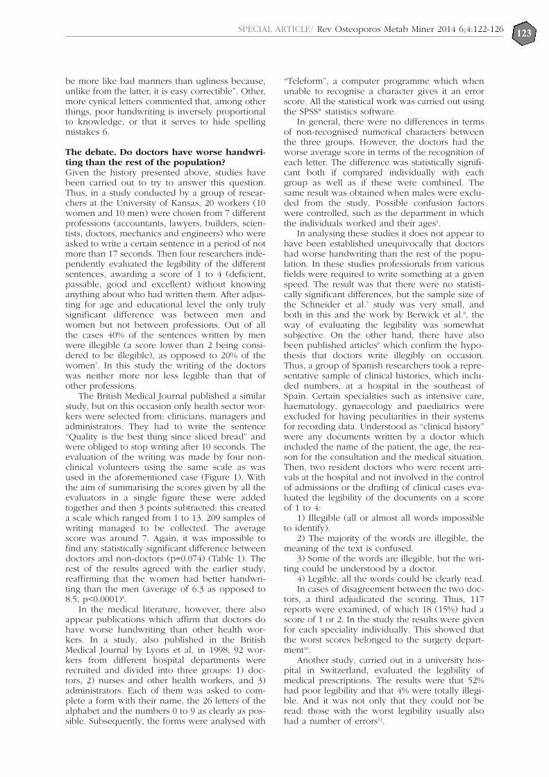
SPECIAL ARTICLE/ Rev Osteoporos Metab Miner 2014 6;4:122-126123
be more like bad manners than ugliness because,unlike from the latter, it is easy correctible”. Other,more cynical letters commented that, among otherthings, poor handwriting is inversely proportionalto knowledge, or that it serves to hide spellingmistakes 6.
The debate. Do doctors have worse handwri-ting than the rest of the population?Given the history presented above, studies havebeen carried out to try to answer this question.Thus, in a study conducted by a group of resear-chers at the University of Kansas, 20 workers (10women and 10 men) were chosen from 7 differentprofessions (accountants, lawyers, builders, scien-tists, doctors, mechanics and engineers) who wereasked to write a certain sentence in a period of notmore than 17 seconds. Then four researchers inde-pendently evaluated the legibility of the differentsentences, awarding a score of 1 to 4 (deficient,passable, good and excellent) without knowinganything about who had written them. After adjus-ting for age and educational level the only trulysignificant difference was between men andwomen but not between professions. Out of allthe cases 40% of the sentences written by menwere illegible (a score lower than 2 being consi-dered to be illegible), as opposed to 20% of thewomen7. In this study the writing of the doctorswas neither more nor less legible than that ofother professions.
The British Medical Journal published a similarstudy, but on this occasion only health sector wor-kers were selected from: clinicians, managers andadministrators. They had to write the sentence“Quality is the best thing since sliced bread” andwere obliged to stop writing after 10 seconds. Theevaluation of the writing was made by four non-clinical volunteers using the same scale as wasused in the aforementioned case (Figure 1). Withthe aim of summarising the scores given by all theevaluators in a single figure these were addedtogether and then 3 points subtracted: this createda scale which ranged from 1 to 13. 209 samples ofwriting managed to be collected. The averagescore was around 7. Again, it was impossible tofind any statistically significant difference betweendoctors and non-doctors (p=0.074) (Table 1). Therest of the results agreed with the earlier study,reaffirming that the women had better handwri-ting than the men (average of 6.3 as opposed to8.5, p<0.0001)8.
In the medical literature, however, there alsoappear publications which affirm that doctors dohave worse handwriting than other health wor-kers. In a study, also published in the BritishMedical Journal by Lyons et al. in 1998, 92 wor-kers from different hospital departments wererecruited and divided into three groups: 1) doc-tors, 2) nurses and other health workers, and 3)administrators. Each of them was asked to com-plete a form with their name, the 26 letters of thealphabet and the numbers 0 to 9 as clearly as pos-sible. Subsequently, the forms were analysed with
“Teleform”, a computer programme which whenunable to recognise a character gives it an errorscore. All the statistical work was carried out usingthe SPSS® statistics software.
In general, there were no differences in termsof non-recognised numerical characters betweenthe three groups. However, the doctors had theworse average score in terms of the recognition ofeach letter. The difference was statistically signifi-cant both if compared individually with eachgroup as well as if these were combined. Thesame result was obtained when males were exclu-ded from the study. Possible confusion factorswere controlled, such as the department in whichthe individuals worked and their ages9.
In analysing these studies it does not appear tohave been established unequivocally that doctorshad worse handwriting than the rest of the popu-lation. In these studies professionals from variousfields were required to write something at a givenspeed. The result was that there were no statisti-cally significant differences, but the sample size ofthe Schneider et al.7 study was very small, andboth in this and the work by Berwick et al.8, theway of evaluating the legibility was somewhatsubjective. On the other hand, there have alsobeen published articles9 which confirm the hypo-thesis that doctors write illegibly on occasion.Thus, a group of Spanish researchers took a repre-sentative sample of clinical histories, which inclu-ded numbers, at a hospital in the southeast ofSpain. Certain specialities such as intensive care,haematology, gynaecology and paediatrics wereexcluded for having peculiarities in their systemsfor recording data. Understood as “clinical history”were any documents written by a doctor whichincluded the name of the patient, the age, the rea-son for the consultation and the medical situation.Then, two resident doctors who were recent arri-vals at the hospital and not involved in the controlof admissions or the drafting of clinical cases eva-luated the legibility of the documents on a scoreof 1 to 4:
1) Illegible (all or almost all words impossibleto identify).
2) The majority of the words are illegible, themeaning of the text is confused.
3) Some of the words are illegible, but the wri-ting could be understood by a doctor.
4) Legible, all the words could be clearly read.In cases of disagreement between the two doc-
tors, a third adjudicated the scoring. Thus, 117reports were examined, of which 18 (15%) had ascore of 1 or 2. In the study the results were givenfor each speciality individually. This showed thatthe worst scores belonged to the surgery depart-ment10.
Another study, carried out in a university hos-pital in Switzerland, evaluated the legibility ofmedical prescriptions. The results were that 52%had poor legibility and that 4% were totally illegi-ble. And it was not only that they could not beread: those with the worst legibility usually alsohad a number of errors11.
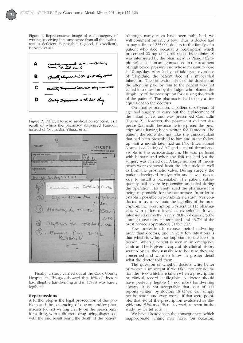
Finally, a study carried out at the Cook CountyHospital in Chicago showed that 16% of doctorshad illegible handwriting and in 17% it was barelylegible12.
RepercussionsA further step is the legal prosecution of this pro-blem and the sentencing of doctors and/or phar-macists for not writing clearly on the prescriptionfor a drug, with a different drug being dispensed,with the end result being the death of the patient.
Although many cases have been published, wewill comment on only a few. Thus, a doctor hadto pay a fine of 225,000 dollars to the family of apatient who died because a prescription whichprescribed 20 mg of Isordil (isosorbide dinitrate)was interpreted by the pharmacist as Plendil (felo-pidine), a calcium antagonist used in the treatmentof high blood pressure and whose maximum doseis 10 mg/day. After 6 days of taking an overdoseof felopidine, the patient died of a myocardialinfarction. The professionalism of the doctor andthe attention paid by him to the patient was notcalled into question by the judge, who blamed theillegibility of the prescription for causing the deathof the patient13. The pharmacist had to pay a fineequivalent to the doctor’s.
On another occasion, a patient of 65 years ofage had surgery to carry out the replacement ofthe mitral valve, and was prescribed Coumadin(Figure 2). However, the pharmacist did not dis-pense Coumadin because he interpreted the pres-cription as having been written for Famodin. Thepatient therefore did not take the anticoagulantthat had been prescribed to him and in the followup visit a month later had an INR (InternationalNormalised Ratio) of 0.7 and a mitral thrombosisvisible in the echocardiogram. He was perfusedwith heparin and when the INR reached 3.6 thesurgery was carried out. A large number of throm-boses were extracted from the left auricle as wellas from the prosthetic valve. During surgery thepatient developed bradycardia and it was neces-sary to install a pacemaker. The patient subse-quently had severe hypotension and died duringthe operation. His family sued the pharmacist forbeing responsible for the occurrence. In order toestablish possible responsibilities a study was con-ducted to try to evaluate the legibility of the pres-cription: the prescription was sent to 113 pharma-cists with different levels of experience. It wasinterpreted correctly in only 70.8% of cases (75.6%among those most experienced and 43.7% of themost novice apprentices) (Table 2)14.
Few professionals expose their handwritingmore than doctors, and in very few situations isthat which is written so important to the life of aperson. When a patient is seen in an emergencyclinic and he is given a copy of his clinical historywritten by us, they usually read because they areconcerned and want to know in greater detailwhat the doctor told them.
The question of whether doctors write betteror worse is important if we take into considera-tion the risks which are taken when a prescriptionor clinical record is illegible. A doctor shouldhave perfectly legible (if not nice) handwritingalways. It is not acceptable that, out of 117reports written by doctors 18 (15%) can simplynot be read10, and even worse, if that were possi-ble, that 4% of the prescription evaluated as ille-gible and 52% as difficult to read, as seen in thestudy by Hartel et al.11.
We have already seen the consequences whichinappropriate writing may have. On occasion,
124SPECIAL ARTICLE/ Rev Osteoporos Metab Miner 2014 6;4:122-126
Figure 1. Representative image of each category ofwriting (receiving the same score from all the evalua-tors. A deficient, B passable, C good, D excellent).Berwick et al.8
Figure 2. Difficult to read medical prescription, as aresult of which the pharmacy dispensed Famodininstead of Coumadin. Yilmaz et al.14
125SPECIAL ARTICLE/ Rev Osteoporos Metab Miner 2014 6;4:122-126
patients die because of this type of negligence.The pharmacist should not be having to guesswhat it was the doctor wanted to write. In the finalstudy14, around 30% of the pharmacists end upproviding an incorrect drug due to the ambiguityof the prescription.
Why, it should therefore be asked, does thistype of thing happen? Too much work and too lit-tle time in which to carry it out could explain it,but it could also be related to a lack of awarenessof this issue. Socially, it does not appear to besomething which is worth addressing, but ratheras seen as an amusing aspect of doctor’s writing.
Be that as it may, handwriting which involvesthe health of patients should be able to be readwithout any difficulty, however long it takes to bewritten. Having said all this, there is no excuse fornot writing correctly. Therefore, this requires acommitment and an awareness of the risks which
are taken when notes are written unclearly and ina hurry. In the end it is the patients who suffer.
Possible solutionsOne solution which, at least in the field of medicalprescriptions appears to be happening, is the compu-terisation of documentation. This possibility has alre-ady been suggested in many of the aforementionedarticles. The same is happening with clinical records.The electronic prescription may be an important aidin facilitating the legibility of prescriptions as well asclinical reports. However, this also runs the risk that,again with the excuse of being in a hurry and havingtoo much work, computerised jargon will start to beused based on abbreviations and invented words,plagued with spelling mistakes, above all withaccents, which could eventually make the text legiblebut difficult to understand or interpret correctly. Anexample could be that shown in Table 3.
Table 1. Average number of handwriting errors by group and character type. Berwick et al.8
Table 2. Percentage of pharmacists according to level of experience who interpreted the same prescription,associating it with different medications. Yilmaz et al.14
Median errors Value of p
Doctors
Nursesand
other healthprofessions
Administrative AllMedicalversus
the rest
All N=38 N=32 N=22
Letters*
Numbers#
7 (0-10)
1 (0-1)
3 (1-6)
1 (0-2)
4 (2-5)
0 (0-1)
0.006
0.15
0.001
0.60
Only women N=13 N=28 N=16
Letters*
Numbers#
6 (3-10)
1 (0-1)
3 (1-6)
1 (0-1)
3 (1-5)
0 (0-1)
0.10
0.29
0.036
0.82
* Maximum possible error in the alphabet = 26# Possible error numbers = 10
Pharmacist Apprentice withexperience
Rookieapprentice Total
Business name N % N % N % N %
Coumadin 34 75.6 39 75 7 43.7 80 70.8
Famodin 10 22.2 13 25 9 56.3 32 28.3
Famoser 1 2.2 - - - - 1 0.9
ConclusionsIt seems clear that a significant percentage of doc-tors have illegible handwriting which, on the onehand, results in significant difficulties in unders-tanding medical reports written by hand, above allfor people not related to the health sector, and onthe other, is the cause of the dispensing and admi-nistering of the wrong medication.
Conflicts of interest: The authors declare thatthey do not have conflicts of interest of anykind.
Bibliography
1. Dictionaries. WcOL. http://www.wordreference.com/definicion/legibilidad. Consultado el 9 de septiembrede 2014.
2. Boletín Oficial del Estado. Real Decreto 1718/2010 de17 de diciembre, sobre receta médica y órdenes dedispensación. BOE núm. 17, de 20 de enero de 2011,capítulo II artículo 3 apartado 4 Consultado el 9 deseptiembre de 2014.http://www.boe.es/diario_boe/txt.php?id=BOE-A-2011-1013.
3. Moliere. El médico a palos. Ediciones ESEBE. 1969.4. Notes, short comments, and answers to correspon-
dents. Lancet 1915;185(4766):55.5. Conybeare JJ. The illegible candidate. Lancet 1953;261
(6768):1001.6. Paz RA. Mala letra. Medicina (Buenos Aires) 2001;61:
495-6.7. Schneider KA, Murray CW, Shadduck RD, Meyers DG.
Legibility of doctors' handwriting is as good (or bad)as everyone else's. Qual Saf Health Care 2006;15:445.
8. Berwick DM, Winickoff DE. The truth about doctors'handwriting: a prospective study. BMJ 1996;313(7072):1657-8.
9. Lyons R, Payne C, McCabe M, Fielder C. Legibility ofdoctors' handwriting: quantitative comparative study.BMJ 1998;317(7162):863-4.
10. Rodríguez-Vera FJ, Marín Y, Sánchez A, Borrachero C,Pujol E. Illegible handwriting in medical records. J RoySoc Med 2002;95:545-6.
11. Hartel MJ, Staub LP, Roder C, Eggli S. High incidenceof medication documentation errors in a Swiss univer-sity hospital due to the handwritten prescription pro-cess. BMC Health Ser Res 2011;11:199.
12. Dunea G. Beastly handwriting. BMJ 1999;319(7201):65A.
13. Charatan F. Compensation awarded for death after ille-gible prescription. West J Med 2000;172:80.
14. Yilmaz R, Yildirim A, Özdemir V, Çetin I, Aksu M, ahanA. Evaluation of prescription legibility leading to deathdue to erroneous interpretation: a field survey in phar-macies. Health Med 2011;5:1076.
Table 3. Abbreviated and full text in an electronic cli-nical history
Abbreviated textPat male 73y with PH of DM II, AH and IC who
attended 4 pain in tx
Full textMale patient of 73 years of age with personal his-
tory of diabetes mellitus type II, arterial hyperten-
sion and ischemic cardiomyopathy, who attended
due to pain in the thorax
126SPECIAL ARTICLE/ Rev Osteoporos Metab Miner 2014 6;4:122-126
DEAR EDITOR:The wake-up call from the Food and DrugAdministration (FDA), the European MedicinesAssociation (EMA) and the Spanish Agency forMedicines and Health Products (AEMPS) regardingthe relationship between the use of bisphospho-nates (BP) and the incidence of atypical femoralfractures has caused people to start to consider theoption of a break in the continuous use of BP, so-called “therapeutic holidays”.The American Society for Bone and MineralResearch (ASBMR) quickly initiated a workinggroup which published a position statement onthe theme of atypical fractures, above all to descri-be the criteria by which to define them1,2.Since then, there have been various authors whohave reviewed the prevalence of atypical fracturesin their records and their possible relationshipwith the use of BP3-5. The increased risk due totheir continuous use appears to be clearly relatedto time, with the relative risk increasing substan-tially from the fourth year3, although the absoluterisk is in the region of 11 fractures for each 10,000years.These data have led to a serious debate aboutover how long a period BP should be administe-red and if therapeutic holidays might be opportu-ne. To date, the most commonly accepted opinionseems to be not to allow these holidays in patientswho remain at high risk of suffering a new fragi-lity, while for the rest there should be a strict eva-luation of whether the BP has achieved the thera-peutic objectives for which it was prescribed6. Thesituation is not that clear-cut, however, what allthe societies and experts are agreed on is that tre-atments should not be withdrawn indiscriminatelyfrom patients with osteoporosis for fear of atypicalfractures. The risk of suffering an osteoporotic fra-gility fracture in patients who have already suffe-red a fracture, for example, is much higher if thetreatment is withdrawn than the risk of sufferingan atypical fracture.There are no dogmas in medicine but, at themoment, it seems that we should focus therapeu-tic holidays on low risk patients, but act muchmore cautiously with the rest. In other diseasessuch ischemic cardiomyopathy no patients with ahistory of angina or myocardial infarction shouldbe left without treatment with hypolipidemics.Above all, decisions should rest on scientific evi-
dence, avoiding the temptation to take advantageof certain situations to try to achieve non-scienti-fic objectives.
Nogués Solán XUnidad de Investigación en Fisiopatología Ósea yArticular (URFOA) - Red de Envejecimiento yFragilidad (RETICEF) - Instituto de InvestigaciónMédica del Hospital del Mar (IMIM) - UniversidadAutónoma de Barcelona - Servicio de MedicinaInterna - Hospital del Mar - Barcelona (Spain)e-mail: [email protected]
Bibliography1. Shane E, Burr D, Abrahamsen B, Adler RA, Brown TD,
Cheung AM, et al. Atypical subtrochanteric anddiaphyseal femoral fractures: second report of a taskforce of the American Society for Bone and MineralResearch. J Bone Miner Res 2014;29:1-23.
2. Shane E, Burr D, Ebeling PR, Abrahamsen B, Adler RA,Brown TD, et al. Atypical subtrochanteric and diaphy-seal femoral fractures: report of a task force of theAmerican Society for Bone and Mineral Research. JBone Miner Res 2010;25:2267-94.
3. Schilcher J, Koeppen V, Aspenberg P, Michaelsson K.Risk of atypical femoral fracture during and after bis-phosphonate use. N Engl J Med 2014;371:974-6.
4. Abrahamsen B, Eiken P, Eastell R. Subtrochanteric anddiaphyseal femur fractures in patients treated withalendronate: a register-based national cohort study. JBone Miner Res 2009;24:1095-102.
5. Rizzoli R, Akesson K, Bouxsein M, Kanis JA, Napoli N,Papapoulos S, et al. Subtrochanteric fractures afterlong-term treatment with bisphosphonates: aEuropean Society on Clinical and Economic Aspects ofOsteoporosis and Osteoarthritis, and InternationalOsteoporosis Foundation Working Group Report.Osteoporos Int 2011;22:373-90.
6. Brown JP, Morin S, Leslie W, Papaioannou A, CheungAM, Davison KS, et al. Bisphosphonates for treatmentof osteoporosis: expected benefits, potential harms,and drug holidays. Can Fam Physician 2014;60:324-33.
DEAR EDITOR:I read with interest the special document publishrecently in your review in which, on the one sideDrs Sosa Henríquez and Gómez de Tejada and onthe other, Dr Malouf Sierra, debated the appropria-teness or otherwise of therapeutic holidays for bis-phosphonates, based on a clinical case1.The authors present a case of a woman of 63 yearsof age with a history of early menopause and verte-bral fracture at 53, for which she had received treat-
Clinical case debate: therapeuticholidays, yes or no?
127LETTERS TO THE EDITOR / Rev Osteoporos Metab Miner 2014 6;4:127-131
ment with alendronic acid for 10 years with goodtolerance and compliance. In a current densitometrythe patient had a T-score of -2.5 in the lumbar spineand -1.5 in the femoral neck, which means a signi-ficant increase in bone mineral density in both areasrelative to that at the start of the treatment. Theauthors told us that she had experienced no falls orfractures during this 10 years, and posed the ques-tion as to whether or not a therapeutic holiday forbisphosphonate should be given.In any disease, before we consider whether to con-tinue, change or withdraw a treatment, we shouldascertain whether or not this treatment has workedduring the period it has been used. In the case pre-sented to us we are told that the patient had notsuffered new fractures, but it appears that this refersto clinical fractures. To ensure that no morphome-tric vertebral fractures had occurred we would haveto carry out a dorsal-lumbar X-ray or a vertebralmorphometry. Only then would we be able to saythat there were no fractures, given that a high per-centage of vertebral fractures are asymptomatic2.So, once the presence of new morphometric verte-bral fractures in the patient has also been discoun-ted, I think that this more than justifies proposingtherapeutic holidays for a drug which has a residualeffect in the bone, meaning that it would continueto act in spite of not being administered, and whichis not without well-known complications. Some ofthe arguments Drs Sosa and Gómez de Tejada useto defend the continuation of the treatment aredebatable. For example, they compare the discon-tinuation of treatment with bisphosphonates withthat of antibiotics or anti-inflammatories used, res-pectively, to resolve an infection or reduce inflam-mation. However, alendronate has a terminal half-life in the skeleton of more than 10 years3, whichallows continued activity in the target tissue for along time after it is discontinued, something whichdoes not occur with antibiotics or anti-inflammato-ries, which have half-lives of only a few hours.Sosa and Gómez de Tejada defend the maintenan-ce of treatment with alendronate in the patientbecause, to their understanding, the patient conti-nues to be a high risk patient simply for havingsuffered an earlier vertebral fracture. It is true thatpatients with a previous vertebral fracture have ahigher risk of fracture than those without fracture4,but this risk diminishes with time5, and after 10years without the appearance of new fractures therisk is already much lower, even more so if thefact that the patient has been receiving antiresorp-tive treatment with alendronate for all those yearsis taken into account. With bisphosphonates weachieve not only an increase in BMD, which isalready associated with a reduction in the risk offracture, but also an improvement in other boneparameters more related to quality, and whichexplains more than 80% of its anti-fracture effect6. Finally, the authors Sosa and Gómez de Tejadacomment that the bisphosphonates are quite safedrugs, and this is completely true, given that therisk of serious complications such as osteonecro-sis of the jaw or atypical fracture are extremely
low in patients with osteoporosis treated with oralbisphosphonates. But to take this risk, low as itmay be, is only justifiable in patients in whom theexpected benefits of the drug are clearly greaterthan this risk, as could be the case in of a patientjust after the fracture, but not 10 years after.Furthermore, there is a clear association betweenthese complications and the period of exposure tobisphosphonates7-8.It would be more difficult to decide on the discon-tinuation of bisphosphonate in the case of apatient with a T-score in the spine of <-3. As theT-score at the start of treatment was -3.7 and thepatient had not had fractures during these 10years, we could say that the alendronate had wor-ked, but possibly the patient's current risk remainssufficiently high that the risk of osteoporotic frac-ture clearly outweighs the risk of complications.This would justify maintaining the antiresorptivetreatment with bisphosphonates or with anotherdrug with better reversibility in the bone such asdenosumab.But returning to the case presented to us, I belie-ve that it is more than reasonable to propose the-rapeutic holidays for bisphosphonates. This doesnot mean leaving the patient without any antifrac-ture effect, since we know that their skeleton will“ooze” alendronate, neither should we forget theirbone fragility. The new challenge will be to avoidholidays for eternity, as well as to know how tomonitor the patient to decide the right moment toreinitiate the treatment, which should always hap-pen before the patient develops a new fragilityfracture.
Casado Burgos EServicio de Reumatología - Hospital UniversitarioParc Taulí - Sabadell - Barcelona (Spain)e-mail: [email protected]
Bibliography1. Sosa Henríquez M, Gómez de Tejada Romero MJ,
Malouf Sierra J. Caso clínico a debate: vacaciones tera-péuticas, ¿si o no? Rev Osteoporos Metab Miner2014;6:63-9.
2. Fink HA, Milavetz DL, Palermo L, Nevitt MC, CauleyJA, Genant HK, Black DM, et al. Fracture InterventionTrial Research Group. What proportion of incidentradiographic vertebral deformities is clinically diagno-sed and viceversa? J Bone Miner Res 2005;20:1216-22.
3. Shinkai I, Ohta Y. New drugs--reports of new drugsrecently approved by the FDA. Alendronate. BioorgMed Chem 1996;4:3-4.
4. Klotzbuecher CM, Ross PD, Landsman PB, Abbott TA3rd, Berger M. Patients with prior fractures have anincreased risk of future fractures: a summary of theliterature and statistical synthesis. J Bone Miner Res2000;15:721-39.
5. Johnell O, Kanis JA, Odén A, Sernbo I, Redlund-Johnell I, Petterson C, et al. Fracture risk following anosteoporotic fracture. Osteoporos Int 2004;15:175-9.
6. Cummings SR, Karpf DB, Harris F, Genant HK, EnsrudK, LaCroix AZ, et al. Improvement in spine bone den-sity and reduction in risk of vertebral fractures duringtreatment with antiresorptive drugs. Am J Med 2002;112:281-9.
7. Barasch A, Cunha-Cruz J, Curro FA, Hujoel P, Sung AH,Vena D, et al. CONDOR Collaborative Group. Risk fac-tors for osteonecrosis of the jaws: a case-control study
128LETTERS TO THE EDITOR / Rev Osteoporos Metab Miner 2014 6;4:127-131
from the CONDOR dental PBRN. J Dent Res2011;90:439-44.
8. Park-Wyllie LY, Mamdani MM, Juurlink DN, HawkerGA, Gunraj N, Austin PC, et al. Bisphosphonate useand the risk of subtrochanteric or femoral shaft fractu-res in older women. JAMA 2011;305:783-9.
DEAR EDITOR:The optimum duration of a treatment for osteopo-rosis is not defined, the exception being the use ofteriparatide whose administration is limited to twoyears. We know that for other drugs it should belonger, but we do not understand well on what cri-teria this decision might be based. Undoubtedly,these criteria should include the persistence of thetherapeutic indication, but other aspects shouldalso be taken into account.The debate published in this review1, based on aclinical case by, on the one side Dr Sosa et al. andon the other Dr Malouf, is very interesting. It dealswith the question of whether or not to continuewith bisphosphonate (BP) in a patient after 10years of treatment.In this work they analyse in depth the appearance ofadverse effects in relation to the period of time forwhich the BP has been taken. On the one hand, theappearance of osteonecrosis of the jaw (ONJ), aninfrequent complication whose risk does not justifythe cessation of long-term treatment, and on theother, the appearance of atypical femoral fractures inthese patient, a complication whose incidence couldbe related to the duration of use of these drugs.The Spanish Agency for Medicine and HealthProducts (AEMPS), on April 15th 2011 published aninformation briefing in which it recommended thatpatients treated with BP be periodically evaluated(especially after the first 5 years). As Dr Sosa com-ments, many doctors have started to withdraw treat-ment with BP from their patients without evaluatingwhether this withdrawal was appropriate or not. Inpractice, this translates into leaving a large numberof patients with a high risk of fracture without the-rapeutic protection. We know: firstly, that exposureto BPs increases the incidence of atypical femoralfracture; secondly, that this incidence increases withthe duration of exposure to this drug; and thirdly,that in any case, in patients with osteoporosis theincidence of atypical femoral fracture is very lowcompared with that of osteoporotic fractures.The consideration of the aforementioned worksallow one to deduce that the decisive factor indeciding whether a treatment with BP should becontinued or not, is the risk of osteoporotic frac-ture which the patient has at the time of proposingthe discontinuation of the therapy.The risk is considered to be high when the patienthas a bone mineral density (BMD) in the femoralneck lower than -2.5 T, or when they have a his-tory of a previous osteoporotic (vertebral or hip)fracture. This therapeutic approach has becomeparticularly clear from various recent works in theliterature2,3 and is picked up in a recently publis-hed work in the review, the American Journal of
Medicine4. Here the patients are classified in threecategories: a) high risk (T-score in the hip lowerthan -2.5; previous vertebral or hip fracture; treat-ment with high doses of corticoids); b) moderaterisk (T-score in the hip higher than -2.5; absenceof previous hip or vertebral fractures; c) low risk(lack of therapeutic criteria at the start of treat-ment, meaning: treatment inappropriate from thestart). In the first category the withdrawal of treat-ment is not considered to be justified, but withperiodic re-evaluation of the therapeutic indica-tions. In the second, the consideration of a tempo-rary withdrawal (“therapeutic holiday”) is advisedafter 3-5 years of treatment. In the third category,logically, the treatment should be discontinued. The Spanish Society for Bone and MineralMetabolism Research (SEIOMM) includes theserecommendations in a document which takes theaforementioned criteria, adding that if the withdra-wal of treatment with BP is desired for some reasonfrom a patient who still has the criteria for being athigh risk of osteoporotic fracture, the therapeuticapproach should not simply be to discontinue it butto substitute it with another therapeutic agent whichacts in a different way5. In the clinical case withwhich we are concerned, the patient had, after 10years of treatment, osteoporosis in the lumbar spineand a history of vertebral fracture, which means thatthey ought to be considered as a high risk patient,and that the treatment should be continued or chan-ged for another treatment, since the incidence aty-pical fractures in patients who have had treatmentwith BP for more than 10 years is not known.
Díaz Curiel MUnidad de Enfermedades Metabólicas Óseas -Servicio de Medicina Interna - Fundación JiménezDíaz - Madrid (Spain)e-mail: [email protected]
Bibliography1. Sosa Henríquez M, Gómez de Tejada Romero MJ, Malouf
Sierra J. Caso clínico a debate: vacaciones terapeúticas,¿si o no? Rev Osteoporos Metab Miner 2014;6:63-9.
2. Whitaker M, Guo J, Kehoe T, Benson G. Bisphosphonatesfor osteoporosis where do we go from here? N Engl JMed 2012;366:2048-51.
3. Black DM, Bauer DC, Schwartz AV, Cummings SR,Rosen CJ. Continuing bisphosphonate treatment forosteoporosis for whom and for how long? N Engl JMed 2012;366:2051-3.
4. McClung M, Harris ST, Miller PD, Bauer DC, Davison KS,Dian L, et al. Bisphosphonate therapy for osteoporosis:benefits, risks, and drug holiday. Am J Med 2013;126:13-20.
5. Recomendaciones sobre la duración del tratamiento dela osteoporosis con bifosfonatos. DocumentoSEIOMM.http://seiomm.org/uploads/documento/ce6f9119ad02fa45b488f745c633a4dc348188a1.pdf.
DEAR EDITOR:We have read with interest the clinical case debateregarding therapeutic holidays1, which clearlyreflects the positions for and against the cessationof treatment with bisphosphonates after a period of5-10 years for alendronate, and perhaps 3-6 years
129LETTERS TO THE EDITOR / Rev Osteoporos Metab Miner 2014 6;4:127-131
for zoledronate. We have less evidence in relationto risedronate and even less regarding the risks andbenefits of maintaining treatment or not beyond 10years2, as is raised by the case under debate.It is probable that much of the argument that liesbehind this subject stems from the lack of incon-testable evidence on how to proceed in a case likethis, and can only be understood after the appea-rance of rare complications associated with chro-nic treatment with bisphosphonates – and otherpowerful anti-catabolics – such as osteonecrosis ofthe jaw or atypical fractures3,4. These possiblecomplications have caused the medical equivalentof a “social panic”, although their risk is really lowcompared to the benefits, due to the efficacy ofthese drugs when used in patients with a real riskof osteoporotic fractures5.Therefore, as stated in SEIOMM’s recommendationdocument5, the type of patient who most benefitsfrom continuing the treatment beyond 5 yearsseems clear. However, we should not forget thatamong women treated for osteoporosis for 5 yearswith alendronate and monitored for a further 5years without treatment, new fractures appear in22% of cases and, more significant, the vast majo-rity of these will appear during the first year thatwe have no markers to help us identify thesepatients6. Although scarce, and methodologically questiona-ble, this is the best evidence for treatment withbisphosphonates for up to 10 years. In any case,what is striking is the preoccupation the medicalcommunity has with this specific issue when com-pared with other therapies employed in otherpathologies such as, for example, myopathy, dia-betes, nephrotoxicity, cataracts, cognitive deterio-ration or erectile dysfunction, among others, asso-ciated rarely with statins (although its benefits inrelation to overall and cardiovascular mortalitycontinue to be clear)7. The same may be said ofthe proton pump inhibitors, with which have beenassociated pneumonia, clostridium difficile infec-tion, osteoporotic fractures, thrombocytopenia,iron, vitamin B12 and magnesium deficiency,rhabdomyolysis and interstitial nephritis, andwhich continue to be widely used drugs8.What might happen in the risk-benefit balance inthe treatment with bisphosphonates beyond 10years is reminiscent of one of the best-knownsequences in the film “Out of Africa” in which theprotagonist, Karen Blixen, played by Meryl Streep,says: “When in the past explorers arrived at thelimits of the known world they were afraid to con-tinue and wrote” “There be dragons!””. Until wehave solid proof – and this seems unlikely – we cancontinue to discuss this ad infinitum. Hopefully, atleast, we will soon have alternative therapies whichhave been proven to be efficacious in this context,before, as it appears, for fear of dragons we stoptreating ever more patients at risk.
Jódar Gimeno EServicio de Endocrinología y Nutrición Clínica -Hospital Universitario Quirón Madrid y Quirón
San Camilo - Facultad de Ciencias de la Salud -Universidad Europea de Madrid (Spain)e-mail: [email protected]
Bibliography1. Sosa Henríquez M, Gómez de Tejada Romero MJ,
Malouf Sierra J. Caso clínico a debate: vacaciones tera-péuticas, ¿si o no? Rev Osteoporos Metab Miner2014;6:63-9.
2. Black DM, Bauer DC, Schwartz AV, Cummings SR,Rosen CJ. Continuing bisphosphonate treatment forosteoporosis for whom and for how long? N Engl JMed 2012;366:2051-3.
3. Schilcher J, Koeppen V, Aspenberg P, Michaelsson K.Risk of atypical femoral fracture during and after bis-phosphonate use. N Engl J Med 2014;371:974-6.
4. Barasch A, Cunha-Cruz J, Curro FA, Hujoel P, Sung AH,Vena D, et al. CONDOR Collaborative Group. Risk fac-tors for osteonecrosis of the jaws: a case-control studyfrom the CONDOR dental PBRN. J Dent Res2011;90:439-44.
5. Recomendaciones sobre la duracion del tratamiento dela osteoporosis con bifosfonatos. DocumentoSEIOMM.http://seiomm.org/uploads/documento/ce6f9119ad02fa45b488f745c633a4dc348188a1.pdf.
6. Fracture prediction after 4 to 5 years of alendronatetherapy: The FLEX study. Bauer DC, Schwartz A,Palermo L, et al. JAMA Internal Medicine 2014;174:1126-34.
7. Non-cardiovascular effects associated with statins.Desai CS, Martin SS, Blumenthal RS. BMJ 2014;349:g3743.
8. Perils and pitfalls of long-term effects of proton pumpinhibitors. Wilhelm SM, Rjater RG, Kale-Pradhan PB.Expert Rev Clin Pharmacol 2013;6:443-51.
DEAR EDITOR:I have read the clinical case debate “Therapeuticholidays: yes or no?” published recently in thisJournal1 and, having recognised the excellentarguments and wide literature reviews of thoseputting the case for and against continuing treat-ment, dare I, as a doctor, give my opinion on thequestion raised?Focusing on the case: it concerns a patient inwhom treatment was initiated at 53 years of agedue to a vertebral fracture and bone mineral den-sity (BMD) in the range for osteoporosis. The riskfactors were corrected and treatment initiated withalendronate and vitamin D, which seems to me acorrect approach. This treatment has been main-tained for 10 years and is now being assessed asto whether to continue with it or to take what iscalled a “therapeutic holiday”. In terms of thecomment about the BMD in the hip not being inthe range for osteoporosis, one should take intoaccount that, at 53 years of age, bone loss occursprimarily in the spine. This means that studies toevaluate hip fractures are carried out in olderpopulations, when this fracture starts to appear2,3.The situation of the 63 years old patient after 10years of treatment with alendronate and vitamin Dis the following: she has not suffered new fractu-res (the risk of fracture is greater in the year follo-wing the appearance of a fracture); her BMD hasincreased and she currently has a T-score of -2.5,having reached a plateau in the last two years; and
LETTERS TO THE EDITOR / Rev Osteoporos Metab Miner 2014 6;4:127-131130
lastly, the marker for bone resorption has reducedwith treatment.Given the residual effects of the disphosphonatesafter their withdrawal4, and the fact that the bonemarkers may be elevated for 6 months, or even upto a year and a half, after treatment stops, depen-ding on the type of disphosphonate used, in myopinion, this patient may discontinue the alendro-nate and stop the intake of the vitamin D neces-sary, since after 10 years there have been no newfractures and the BMD has increased to becomestable in the last two years. On the other hand, Iwould suggest carrying out a new assessment aftera year or a year and a half to see how it is deve-loping and, depending on the clinical situation atthat time, I would make an assessment as to whe-ther to continue with the therapeutic break or toreinitiate treatment with the same drug, or a diffe-rent one.
Torrijos Eslava AReumatólogo - Madrid (Spain)e-mail: [email protected]
Bibliography1. Sosa Henríquez M, Gómez de Tejada Romero MJ y
Malouf Sierra J. Caso clínico a debate: vacaciones tera-péuticas, ¿sí o no? Rev Osteoporos Metab Miner2014;6:63-9.
2. McClung MR, Geusens P, Miller PD, Zippel H, BensenWG, Roux C, et al. Effect of risedronate on the risk ofhip fracture in elderly women. Hip InterventionProgram Study Group. N Engl J Med 2001;34:333-40.
3. Boonen S, Dejaeger E, Vanderschueren D, Venken K,Bogaerts A, Verschueren S, et al. Osteoporosis andosteoporotic fracture occurrence and prevention in theelderly: a geriatric perspective. Best Pract Res ClinEndocrinol Metab 2008;22:765-85.
4. Bagger YZ, Tankó LB, Alexandersen P, Ravn P,Christiansen C. Alendronate has a residual effect onbone mass in postmenopausal Danish women up to 7years after treatment withdrawal. Bone 2003;33:301-7.
RESPONSE OF THE AUTHORS:
We have read the various letters to the Editorwhich have been submitted by readers, somein favour and some against, in the debatearound whether therapeutic holidays areappropriate or not. This is a controversial topic, about which wehave no scientific evidence. Hence the diffe-rence of opinion, although it is clear thatabove all there is, in all those who have writ-ten (as well as in ourselves), an underlying fearof harming the patient in any way.We believe the debate enriches our knowled-ge, and so we thank those readers who haveexpressed their opinions, encouraging othersto continue this type of discussion on any sub-ject published in the Journal.
Manuel Sosa HenríquezMª Jesús Gómez de Tejada RomeroJorge Malouf Sierra
LETTERS TO THE EDITOR / Rev Osteoporos Metab Miner 2014 6;4:127-131131