UNIVERSIDAD AUTÓNOMA METROPOLITANA
IZTAPALAPA
División de Ciencias Biológicas y de la Salud
“Estudio sobre la interacción entre la β-Lactoglobulina y la β-
Galactosidasa de Kluyveromyces lactis y el Papel que Juegan los
Aminoácidos de las Proteínas en la Interacción y el Efecto Activador
sobre la Enzima”
T E S I S
para obtener el grado de:
Doctora en Biotecnología
P R E S E N T A:
M. en B. Elizabeth Del Moral Ramírez
Directora:
Dra. Judith Jiménez Guzmán
México D. F., Marzo de 2012

“El Doctorado en Biotecnología de la Universidad Autónoma Metropolitana está
incluido en el Programa Nacional de Posgrados de Calidad (PNPC) del
CONACYT, con la referencia 001466”
ÍNDICE
ÍNDICE
Página
Resumen…………..............................………………………………………….....… 1
Abstract.…………..............................………………………………………….....… 2
A. Introducción…..............................………………………………………….....… 3
B. Antecedentes ………………..…..……………..…............................................... 6
B. 1. Composición general de la leche……………..……………………………. 6
B. 1. 1. Proteínas de la leche……………..……….……………………..… 7
B. 1. 1. 1. Caseínas………………..……….…………...…….…... 8
B. 1. 1. 2. Proteínas del suero…..………..….…………………… 9
B. 1. 1. 2. 1. -Lactoglobulina…..….……………..….. 10
B. 1. 1. 2. 1. 1. Succinilación.………...… 12
B. 1. 2. Hidratos de carbono……………………..…..…………….……… 14
B. 1. 2. 1. Lactosa…………………………….…………………. 14
B. 2. Reacción de Maillard……………………………………………………… 15
B. 3. Lactosilación……………………………………………………………… 16
B. 4. Hidrólisis de la lactosa……………………………………………………. 17
B. 5. -Galactosidasa de Kluyveromyces lactis.………...…………………….... 18
B. 5. 1. Efecto de la β-lactoglobulina en la actividad de la
β-galactosidasa de Kluyveromyces lactis.…….………………..... 18
B. 6. Esterificación de proteínas ……………………………………………... 20
B. 7. Uso de programas de cómputo para la construcción de un modelo
tridimensional y docking de una proteína…………………………….… 20
ÍNDICE
C. Objetivos…………….......................................................................................... 22
C. 1. Objetivo general ……………………………………………………….… 22
C. 2. Objetivos particulares…………………………………………………….. 22
D. Resultados……………......................................................................................... 23
D. 1. Role of Lysine -Amino Groups of -Lactoglobulin on Its Activating
Effect of Kluyveromyces lactis Galactosidase……………………...……….. 23
D. 2. Determination of the probable interaction site of bovine
-lactoglobulin dimer with electrophilic molecules…………..…….………….. 29
D. 3. Effect of pH on the interaction of -lactoglobulin with Kluyveromyces
lactis -galactosidase and its effect on enzymatic activity.…………………..... 41
D. 4. Role of carboxyl groups of Kluyveromyces lactis -galactosidase in the
interaction with -lactoglobulin and its activating effect..…………………....... 54
E. Discusión……………………………………………………………………….. 70
F. Conclusiones........................................................................................................ 75
G. Referencias........................................................................................................... 77
RESUMEN
1
RESUMEN
Algunos reportes han establecido el efecto activador de la -lactoglobulina bovina (-lg)
sobre la actividad de la β-galactosidasa de Kluyveromyces lactis (KLβ-gal), sugiriendo que
la interacción entre la β-lg y la KLβ-gal podría ser la responsable de este efecto. El objetivo
de este trabajo fue estudiar la interacción entre la β-lg y la KLβ-gal para tratar de
determinar su efecto en la activación y los factores que influyen en ella así como identificar
los residuos de ambas proteínas responsables de dicha interacción.
Por medio de la succinilación de la -lg y la esterificación de la KL-gal se demostró que
los residuos de lisina de la -lg y los residuos carboxilados de la KL-gal son
indispensables tanto para la interacción entre la -lg y la KL-gal como para la activación,
por lo que muy probablemente la interacción suceda por la vía de un ataque nucleofílico.
Los resultados de docking ciego de la -lg con lactosa y anhídrido succínico usados como
ligandos modelo de electrófilos sugieren que la Lys138
de un monómero y la Lys141
del otro
son los que muestran una mayor probabilidad de interactuar con ambos (energías finales de
docking o EFD de -3.30 y -3.11 kcal/mol para la lactosa y el anhídrido succínico
respectivamente). Por otro lado al realizar estudios de docking ciego para la KL-gal
usando lisina como ligando modelo de nucleófilo se encontró que el aminoácido con mayor
probabilidad de participar en la interacción es el Glu592
(EFD=-4.5kcal/mol).
Se encontró que el estado dimérico de la β-lg es esencial tanto para la interacción
como para la activación de la enzima. Es probable que la formación del dímero de la β-lg
sea necesaria para fortalecer las interacciones mediante la participación de la Lys138
de uno
de los monómeros y la Lys141
del otro para formar una estructura tipo ―pinza‖ que podría
estabilizar los ligandos mediante la formación de puentes de hidrógeno para que el ataque
nucleofílco pueda llevarse a cabo. Al estudiar el efecto del pH en la interacción de las
proteínas y en la activación se observó que ambas son más fuertes a pH 7.0 y
completamente ausentes a valores de pH de 6.0 y 8.5, lo cual coincide con la disociación
del dímero de β-lg. También se observó un desplazamiento del pH óptimo de 7.5 (solo
solución amortiguadora), a 7.0 (en presencia de β-lg) lo cual sugiere que a este pH la fuerte
interacción entre las proteínas podría ayudar a alcanzar una conformación de la KLβ-lg más
activa en la que el sitio activo sería más accesible para el sustrato.
ABSTRACT
2
ABSTRACT
Some reports have established the activating effect of bovine -lactoglobulin (-lg) on
Kluyveromyces lactis’ -galactosidase (KL-gal) activity, suggesting that the interaction
between -lg and KLβ-gal could be responsible for this effect. The aim of this work was to
study the interaction between β-lg and KLβ-gal in order to determine the effect on the
activation and the factors that affect it as well as to identify the residues involved.
Succinylation of β-lg and esterification of KLβ-gal demonstrated that lysine residues of β-
lg and carboxyl residues of KLβ-gal are essential for both, the interaction between β-lg and
KLβ-gal and the activation of KLβ-gal. Blind docking of β-lg with either lactose or succinic
anhydride used as electrophile model ligands, suggested that Lys138
from one monomer and
Lys141
from the other are the most likely to react with both (final docking energies or FDE
of -3.30 y -3.11 kcal/mol for lactose and succinic anhydride, respectively). On the other
hand blind docking of KLβ-gal with lysine used as nucleophile model ligand, showed that
Glu592
is the most probable to be involved in the interaction (FDE= -4.55 kcal/mol).
It was found that the dimeric form of β-lg is essential for both, the interaction and the
activation of the enzyme. It is very likely that the formation of the dimer of -lactoglobulin
is necessary to strengthen the interactions since, as predicted by docking, Lys138
from one
of -lg monomers and Lys141
from the other form a claw-like structure that may stabilize
the ligands by the formation of hydrogen bonds so that the nucleophilic attack may take
place. When the effect of pH on the interaction and on the activation was studied, it was
observed that both are stronger at pH 7.0 but completely absent at pH values of 6.0 and 8.5
as well as the dimer is formed and then dissociated. It was observed that the optimum pH
gets shifted from 7.5 (buffer) to 7.0 (with β-lg) suggesting that at this pH the strong
interaction with -lg promotes a KL-gal conformation in which the active site is better
suited for catalysis.
INTRODUCCIÓN
3
A. INTRODUCCIÓN
La -galactosidasa de Kluyveromyces lactis (EC 3.2.1.23) (KL-gal) es la enzima más
ampliamente utilizada en industria láctea para resolver tanto el problema de intolerancia a
la lactosa como problemas técnicos relacionados con la baja solubilidad de la lactosa y su
tendencia a la cristalización que generan problemas técnicos de precipitación; formación de
grumos y arenosidades indeseables en productos lácteos con un alto contenido de sólidos
como son: helados, leches condensadas y azucaradas, cajetas y flanes (Mahoney, 1997;
García-Garibay, 1993; García-Garibay, 1992; Gekas & López-Leyva, 1985). Por estas
razones, la hidrólisis de la lactosa en sus dos monosacáridos por la adición de la enzima -
galactosidasa es una alternativa para resolver estos problemas y ha sido extensamente
estudiada desde diversos puntos de vista.
A pesar de ser la lactasa de mayor uso comercial, la estructura de la KL-lg no ha sido muy
estudiada. Tello-Solis y col. (2005), determinaron mediante estudios con dicroismo circular
que la KL-gal es básicamente una proteína-, formada por un 22% de giros, 14% de
láminas- paralelas, 25% de láminas- antiparalelas, 34% de estructura desordenada y tan
solo un 5% de hélice-.
En estudios recientes se ha observado que la presencia en el medio de reacción de
seroalbúmina (SA) o -lactoglobulina bovina (-lg) (ambas proteínas del suero de la leche),
produce un efecto activador sobre la -galactosidasa de Kluyveromyces lactis de hasta
230% por parte de la -lactoglobulina (Jiménez-Guzmán, 2002). Estudios realizados con
cromatografía de afinidad demostraron que la KL-gal se une específicamente a la -lg
provocando la activación de la enzima (Jiménez-Guzmán y col., 2006). Por otro lado, se ha
encontrado que el calentamiento de -lg pura incrementa la actividad de la KL-gal, pero al
calentarla en presencia de lactosa el efecto activador disminuye. Existen otros reportes que
indican que el calentamiento de la -lg en presencia de lactosa provoca una reacción entre
la proteína y el azúcar, reacción conocida como lactosilación (Léonil y col., 1997), la cual
provoca una disminución en la capacidad de unión entre la proteína y la enzima al mismo
tiempo que disminuye la capacidad de la -lg de activar a la KL-gal (Jiménez-Guzmán y
col., 2006). Se ha demostrado que la -lactoglobulina incrementa la actividad de la KL-
gal a través de dos mecanismos diferentes: uno que depende de la liberación de grupos
INTRODUCCIÓN
4
sulfhidrilo durante el tratamiento térmico de la proteína desnaturalizada (Jiménez-Guzmán
y col., 2002) y otro que resulta de la habilidad de la proteína nativa para unirse a la enzima
(Jiménez-Guzmán y col., 2006).
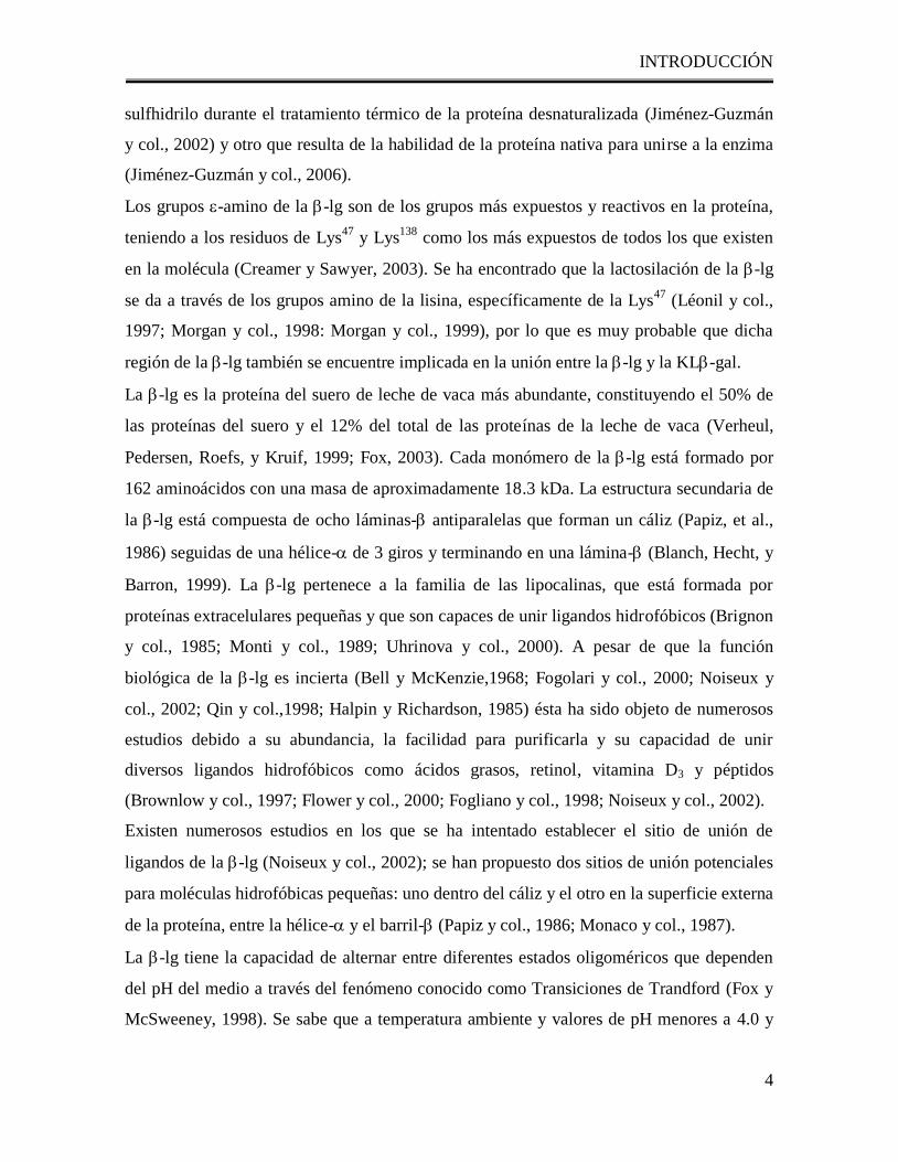
Los grupos -amino de la -lg son de los grupos más expuestos y reactivos en la proteína,
teniendo a los residuos de Lys47
y Lys138
como los más expuestos de todos los que existen
en la molécula (Creamer y Sawyer, 2003). Se ha encontrado que la lactosilación de la -lg
se da a través de los grupos amino de la lisina, específicamente de la Lys47
(Léonil y col.,
1997; Morgan y col., 1998: Morgan y col., 1999), por lo que es muy probable que dicha
región de la -lg también se encuentre implicada en la unión entre la -lg y la KL-gal.
La -lg es la proteína del suero de leche de vaca más abundante, constituyendo el 50% de
las proteínas del suero y el 12% del total de las proteínas de la leche de vaca (Verheul,
Pedersen, Roefs, y Kruif, 1999; Fox, 2003). Cada monómero de la -lg está formado por
162 aminoácidos con una masa de aproximadamente 18.3 kDa. La estructura secundaria de
la -lg está compuesta de ocho láminas- antiparalelas que forman un cáliz (Papiz, et al.,
1986) seguidas de una hélice- de 3 giros y terminando en una lámina- (Blanch, Hecht, y
Barron, 1999). La -lg pertenece a la familia de las lipocalinas, que está formada por
proteínas extracelulares pequeñas y que son capaces de unir ligandos hidrofóbicos (Brignon
y col., 1985; Monti y col., 1989; Uhrinova y col., 2000). A pesar de que la función
biológica de la -lg es incierta (Bell y McKenzie,1968; Fogolari y col., 2000; Noiseux y
col., 2002; Qin y col.,1998; Halpin y Richardson, 1985) ésta ha sido objeto de numerosos
estudios debido a su abundancia, la facilidad para purificarla y su capacidad de unir
diversos ligandos hidrofóbicos como ácidos grasos, retinol, vitamina D3 y péptidos
(Brownlow y col., 1997; Flower y col., 2000; Fogliano y col., 1998; Noiseux y col., 2002).
Existen numerosos estudios en los que se ha intentado establecer el sitio de unión de
ligandos de la -lg (Noiseux y col., 2002); se han propuesto dos sitios de unión potenciales
para moléculas hidrofóbicas pequeñas: uno dentro del cáliz y el otro en la superficie externa
de la proteína, entre la hélice- y el barril- (Papiz y col., 1986; Monaco y col., 1987).
La -lg tiene la capacidad de alternar entre diferentes estados oligoméricos que dependen
del pH del medio a través del fenómeno conocido como Transiciones de Trandford (Fox y
McSweeney, 1998). Se sabe que a temperatura ambiente y valores de pH menores a 4.0 y
INTRODUCCIÓN
5
mayores a 5.2, la proteína está formada principalmente por monómeros y dímeros.
Alrededor de pH 4.7, se forman estructuras oligoméricas más grandes tales como octámeros
(Verheul y col., 1999). En valores cercanos a un pH de 6.8 (el cual es el pH de la leche de
vaca) la -lg existe como un dímero (Fox y col., 1998). No existe información acerca de los
posibles sitios y mecanismos de interacción entre la KL-gal y el dímero de la -lg, que es
el estado oligomérico real de la -lg en las condiciones bajo las cuales se encuentran
reportados la mayoría de los estudios existentes. Aunque existen muchos estudios que
explican las interacciones de la -lg con moléculas hidrofóbicas a través del cáliz interno de
la proteína (Yang y col., 2008a; Kontopidis y col., 2002; Ragona y col., 2000; Wu y col.,
1999) este mecanismo no puede explicar la interacción con una proteína grande como lo es
la KL-gal y que debería ocurrir a través de una región muy expuesta de la proteína. En
2005, Tello y col., además de contribuir con estudios sobre la estructura secundaria de la
KL-gal, demostraron que existe una relación entre la estructura y la actividad de la KL-
gal dependiente del pH, misma que podría verse afectada con el fenómeno de interacción
con la -lg y por consiguiente relacionarse con la capacidad activadora. El propósito de este
trabajo fue describir el mecanismo de interacción entre la -galactosidasa de
Kluyveromyces lactis y la -lg para así contribuir con la explicación del fenómeno de
activación de la -galactosidasa de Kluyveromyces lactis por la -lg. Para cumplir dicho
objetivo se estudió tanto el papel de los grupos -amino de la -lg en el efecto activador y
en las interacciones entre la KL-gal y la -lg así como la importancia del dímero de la -
lactoglobulina en dicho fenómeno.
ANTECEDENTES
6
B. ANTECEDENTES
B. 1. Composición general de la leche
Dentro de la amplia gama de alimentos que el hombre ha seleccionado como parte de su
alimentación, la leche es el único que ha sido diseñado por la naturaleza para este
propósito; tiene una composición compleja de biomoléculas y es conocida y aceptada a
nivel mundial por su alta calidad nutrimental y por ser propia de cada especie (García-
Garibay y Gómez-Ruiz, 1996).
En la leche se encuentran disueltos una gran variedad de compuestos como son: lactosa,
grasa, proteínas, sales y otro tipo de compuestos en pequeñas cantidades. La composición
exacta de la leche no se ha podido definir en forma general, ya que ésta varía en función de
diversos factores como la raza, periodo de lactación y alimentación del mamífero del cual
provenga (Alais, 1991).
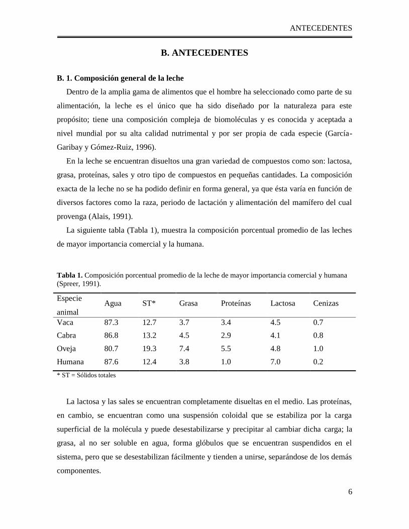
La siguiente tabla (Tabla 1), muestra la composición porcentual promedio de las leches
de mayor importancia comercial y la humana.
Tabla 1. Composición porcentual promedio de la leche de mayor importancia comercial y humana
(Spreer, 1991).
* ST = Sólidos totales
La lactosa y las sales se encuentran completamente disueltas en el medio. Las proteínas,
en cambio, se encuentran como una suspensión coloidal que se estabiliza por la carga
superficial de la molécula y puede desestabilizarse y precipitar al cambiar dicha carga; la
grasa, al no ser soluble en agua, forma glóbulos que se encuentran suspendidos en el
sistema, pero que se desestabilizan fácilmente y tienden a unirse, separándose de los demás
componentes.
Especie
animal Agua ST* Grasa Proteínas Lactosa Cenizas
Vaca 87.3 12.7 3.7 3.4 4.5 0.7
Cabra 86.8 13.2 4.5 2.9 4.1 0.8
Oveja 80.7 19.3 7.4 5.5 4.8 1.0
Humana 87.6 12.4 3.8 1.0 7.0 0.2
ANTECEDENTES
7
Para entender los cambios que se llevan a cabo en la leche como consecuencia de los
procesos a los que se le somete, es necesario conocer el comportamiento químico de sus
componentes que a continuación se describe brevemente.
El agua es el componente más abundante de la leche; su función esencial es la de actuar
como disolvente de los demás componentes. Sin embargo, en algunos derivados lácteos
puede estar como agua ligada químicamente o como agua libre. La presencia del agua
repercute directamente en la estabilidad de la leche, ya que el crecimiento bacteriano, así
como las reacciones no enzimáticas, son dependientes de la actividad de agua (aw). Por otro
lado, la actividad de agua (aw) tiene una gran influencia sobre el proceso de secado; pues
establece la cantidad de energía necesaria para llevar a cabo dicho proceso (Spreer, 1991).
De todos los componentes de la leche, la fracción formada por las grasas es la que más
varía y se encuentra en la leche en forma de glóbulos esféricos suspendidos en la fase
acuosa (Amiot, 1991). Entre los componentes grasos predominan los triglicéridos, que
constituyen el 98% de la grasa láctea, además de encontrarse pequeñas cantidades de di- y
monoglicéridos así como ácidos grasos libres. También se encuentran fosfolípidos,
colesterol, ésteres de colesterol y cerebrósidos.
Otros componentes se encuentran en cantidades muy pequeñas, pero pueden ser
importantes en las propiedades organolépticas o desde el punto de vista nutricio. Entre ellos
se pueden citar: las vitaminas liposolubles, principalmente A, D y E, junto con pequeñas
cantidades de vitamina K; los compuestos responsables del aroma y sabor como aldehídos,
cetonas y lactonas y los pigmentos carotenoides (Walstra y Jennes, 1984).
B.1. 1. Proteínas de la leche
Normalmente se distingue entre las caseínas, que precipitan a pH 4.6, y las proteínas del
suero que no precipitan con las caseínas a menos que previamente hayan sido
desnaturalizadas por el calor u otros tratamientos. Las proteínas del suero incluyen a la -
lactoalbúmina, -lactoglobulina, seroalbúmina e inmunoglobulinas (Amiot, 1991; Walstra
y Jennes, 1984).
ANTECEDENTES
8
B. 1. 1. 1. Caseínas
Las caseínas constituyen más del 80% de las proteínas totales de la leche (Walstra y
Jennes, 1984). Inicialmente se pensaba que la caseína era una sola proteína; actualmente se
sabe que en realidad el término caseína comprende a un grupo de proteínas que contienen
fosfato y que son propias de la leche. Son cuatro las de mayor importancia y se les
denomina caseínas primarias (Tabla 2) (Fox y McSweeney, 1998; Amiot, 1991; Walstra y
Jennes, 1984).
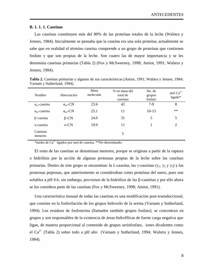
Tabla 2. Caseínas primarias y algunas de sus características (Amiot, 1991; Walstra y Jennes, 1984;
Varnam y Sutherland, 1994).
Nombre Abreviación
Masa
molecular
kDa
% en masa del
total de
caseínas
No. de
grupos
fosfato
mol Ca2+
ligado*
αs1-caseína αs1-CN 23.6 42 7-9 8
αs2-caseína αs2-CN 25.1 11 10-13 **
-caseína -CN 24.0 31 5 5
κ-caseína κ-CN 19.0 11 1 2
Caseínas
menores 5
*moles de Ca2+
ligados por mol de caseína, **No determinado.
El resto de las caseínas se denominan menores, porque se originan a partir de la ruptura
o hidrólisis por la acción de algunas proteasas propias de la leche sobre las caseínas
primarias. Dentro de éste grupo se encuentran: la λ-caseína, las γ-caseínas (γ1, γ2 y γ3) y las
proteosas peptonas, que anteriormente se consideraban como proteínas del suero, pues son
solubles a pH 4.6, sin embargo, provienen de la hidrólisis de las β-caseínas y por ello ahora
se les considera parte de las caseínas (Fox y McSweeney, 1998; Amiot, 1991).
Una característica inusual de todas las caseínas es una modificación post-transduccional,
que consiste en la fosforilación de los grupos hidroxilo de la serina (Varnam y Sutherland,
1994). Los residuos de fosfoserina (llamados también grupos fosfato), se concentran en
grupos y son responsables de la existencia de áreas hidrofílicas de fuerte carga negativa que
ligan, de manera proporcional al contenido de grupos serínfosfato, iones divalentes como
el Ca2+
(Tabla 2) sobre todo a pH alto (Varnam y Sutherland, 1994; Walstra y Jennes,
1984).
ANTECEDENTES
9
El carácter anfifílico de las caseínas y su fosforilación facilita las interacciones entre
ellas para formar complejos esféricos altamente hidratados conocidos como micelas y que
se forman bajo condiciones específicas de temperatura y fuerza iónica (Walstra y col.,
2001, Amiot, 1991). La estabilidad coloidal de las micelas de caseína se debe
principalmente a la κ-caseína y al fosfato de calcio coloidal (Walstra y Jennes, 1984).
B. 1. 1. 2. Proteínas del suero
Las proteínas del suero comprenden dos tipos de proteínas: las sintetizadas en la
glándula mamaria: β-lactoglobulina y α-lactoalbúmina; y las de origen sanguíneo: la
seroalbúmina e inmunoglobulinas. Tienen una estructura típica de proteínas globulares
compactas con una secuencia en la que los grupos no polares, polares y cargados tienen una
distribución relativamente uniforme; sufren un plegamiento intramolecular formándose
puentes disulfuro que las estabilizan ante los cambios de pH (Varnam y Sutherland, 1994).
Estas proteínas permanecen solubles en el suero, tanto si la leche se ha coagulado por
acidificación a pH 4.6, como si se ha hecho por vía enzimática (por ejemplo, en la
elaboración de queso por la acción de la quimosina). Por el contrario, el calentamiento de la
leche las desnaturaliza, es decir, provoca el desenrollamiento de la estructura globular de la
proteína y la consecuente precipitación de la misma; sin embargo, esta insolubilización
depende mucho del grado de calentamiento y las condiciones técnicas tales como la
acidificación, tratamientos previos o la presencia de otras proteínas.
Estas proteínas pueden ser separadas del suero por ultrafiltración. La fracción retenida
está enriquecida en estas proteínas y también pueden separarse por cromatografía de
intercambio iónico (Louquet y col., 1991).
La α-lactoalbúmina representa el 19.2% de las proteínas del suero. Su masa molecular es
de 14.4 kDa; es muy soluble en agua y su punto isoeléctrico es de 4.8 (Walstra y Jennes,
1984).
La albúmina sérica representa el 6.2 % de las proteínas de suero y es exactamente igual
que la albúmina del suero sanguíneo. Su masa molecular es de 66.2 kDa y tiene un punto
isoeléctrico de 4.7; es especialmente rica en lisina y cisteina y es muy soluble en agua
(Amiot, 1991; Walstra y Jennes, 1984).
ANTECEDENTES
10
Las inmunoglobulinas de la leche representan el 10.9% de las proteínas del suero. Se
caracterizan por tener una masa molecular elevada (entre 150 y 900 kDa) y porque están
glicosiladas (Walstra y Jennes, 1984). Se les llama inmunoglobulinas por sus importantes
propiedades inmunológicas, pues su presencia en gran proporción en el calostro es esencial
para transmitir al animal joven los anticuerpos necesarios para la lucha contra las
infecciones; además, se cree que contribuyen al sistema antibiótico de la leche cruda
(Amiot, 1991).
Existen otras proteínas que se encuentran en la leche en pequeñas cantidades: las que
están en la superficie de los glóbulos grasos de la leche, constituidas por una euglobulina, la
fosfatasa alcalina y la xantín-oxidasa. Además se han aislado en la leche una mucoproteína,
una lipoproteína y algunas ferroproteínas (lactoferrina y transferrina) (Amiot, 1991).
B. 1. 1. 2. 1. - Lactoglobulina
La β-lactoglobulina bovina (-lg) es la más importante de las proteínas del suero pues
constituye el 50.8% de las proteínas del suero y el 9.8 % del total de las proteínas de la
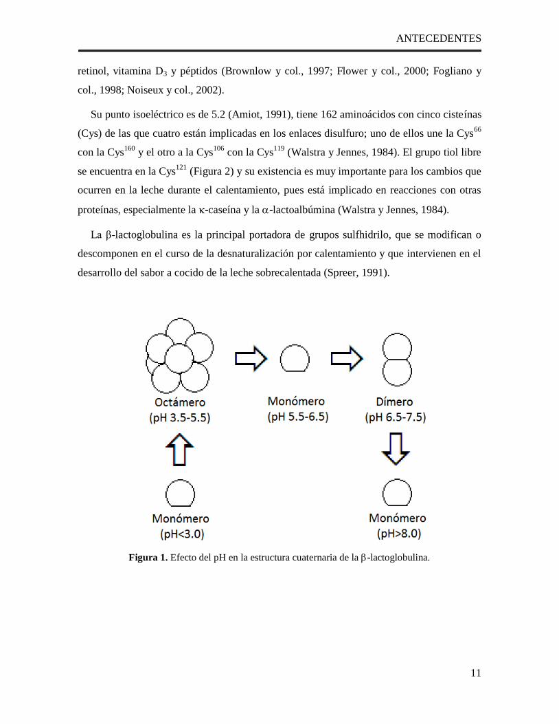
leche (Walstra y Jennes, 1984). Su masa molecular es de 18.3 kDa, pero en la literatura se
da a veces la de 36.6 kDa debido a que se presenta en la naturaleza como un dímero de dos
subunidades monoméricas entrecruzadas por dos puentes disulfuro (aproximadamente a pH
de 6.5) e incluso polímeros de más cadenas polipeptídicas dependiendo del pH (Figura 1)
(Ortiz, 2004; Walstra y Jennes, 1984).
La estructura secundaria de la -lg está compuesta de ocho láminas- antiparalelas que
forman un cáliz (Papiz, y col., 1986) seguidas de una hélice- de 3 giros y terminando en
una lámina- (Blanch y col., 1999) (Figura 2). La -lg pertenece a la familia de las
lipocalinas, que está formada por proteínas extracelulares pequeñas y que son capaces de
unir ligandos hidrofóbicos (Brignon y col., 1985; Monti y col., 1989; Uhrinova y col.,
2000), a pesar de que la función biológica de la -lg es incierta (Bell y McKenzie,1968;
Fogolari y col., 2000; Noiseux y col., 2002; Qin y col., 1998; Halpin y Richardson, 1985),
ésta ha sido objeto de numerosos estudios debido a su abundancia, la facilidad para
purificarla y su capacidad de unir diversos ligandos hidrofóbicos como ácidos grasos,
ANTECEDENTES
11
retinol, vitamina D3 y péptidos (Brownlow y col., 1997; Flower y col., 2000; Fogliano y
col., 1998; Noiseux y col., 2002).
Su punto isoeléctrico es de 5.2 (Amiot, 1991), tiene 162 aminoácidos con cinco cisteínas
(Cys) de las que cuatro están implicadas en los enlaces disulfuro; uno de ellos une la Cys66
con la Cys160
y el otro a la Cys106
con la Cys119
(Walstra y Jennes, 1984). El grupo tiol libre
se encuentra en la Cys121
(Figura 2) y su existencia es muy importante para los cambios que
ocurren en la leche durante el calentamiento, pues está implicado en reacciones con otras
proteínas, especialmente la -caseína y la -lactoalbúmina (Walstra y Jennes, 1984).
La β-lactoglobulina es la principal portadora de grupos sulfhidrilo, que se modifican o
descomponen en el curso de la desnaturalización por calentamiento y que intervienen en el
desarrollo del sabor a cocido de la leche sobrecalentada (Spreer, 1991).
Figura 1. Efecto del pH en la estructura cuaternaria de la -lactoglobulina.
ANTECEDENTES
12
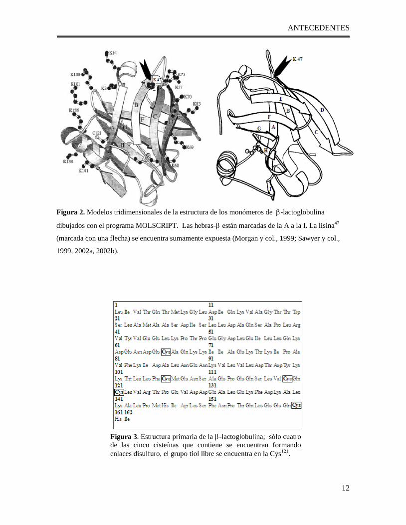
Figura 2. Modelos tridimensionales de la estructura de los monómeros de -lactoglobulina
dibujados con el programa MOLSCRIPT. Las hebras- están marcadas de la A a la I. La lisina47
(marcada con una flecha) se encuentra sumamente expuesta (Morgan y col., 1999; Sawyer y col.,
1999, 2002a, 2002b).
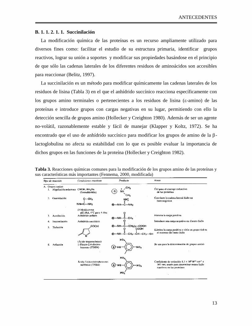
Figura 3. Estructura primaria de la -lactoglobulina; sólo cuatro
de las cinco cisteínas que contiene se encuentran formando
enlaces disulfuro, el grupo tiol libre se encuentra en la Cys121
.
ANTECEDENTES
13
B. 1. 1. 2. 1. 1. Succinilación
La modificación química de las proteínas es un recurso ampliamente utilizado para
diversos fines como: facilitar el estudio de su estructura primaria, identificar grupos
reactivos, lograr su unión a soportes y modificar sus propiedades basándose en el principio
de que sólo las cadenas laterales de los diferentes residuos de aminoácidos son accesibles
para reaccionar (Belitz, 1997).
La succinilación es un método para modificar químicamente las cadenas laterales de los
residuos de lisina (Tabla 3) en el que el anhídrido succínico reacciona específicamente con
los grupos amino terminales o pertenecientes a los residuos de lisina (-amino) de las
proteínas e introduce grupos con cargas negativas en su lugar, permitiendo con ello la
detección sencilla de grupos amino (Hollecker y Creighton 1980). Además de ser un agente
no-volátil, razonablemente estable y fácil de manejar (Klapper y Koltz, 1972). Se ha
encontrado que el uso de anhídrido succínico para modificar los grupos de amino de la -
lactoglobulina no afecta su estabilidad con lo que es posible evaluar la importancia de
dichos grupos en las funciones de la proteína (Hollecker y Creighton 1982).
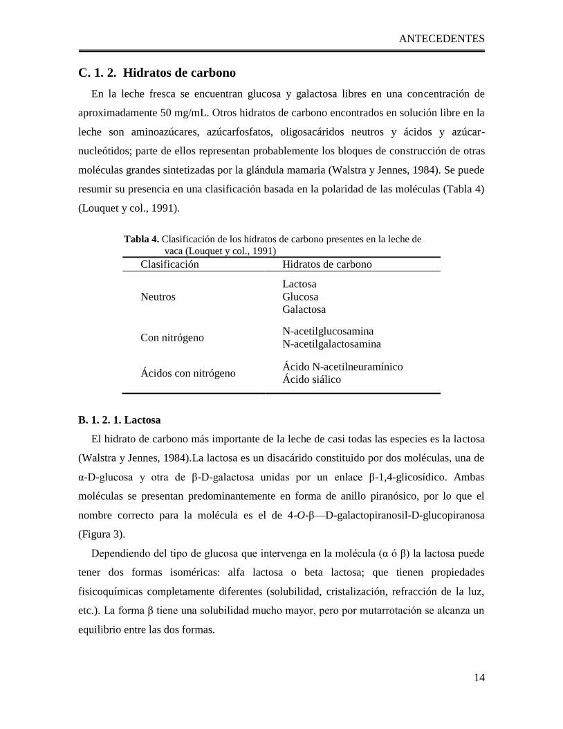
Tabla 3. Reacciones químicas comunes para la modificación de los grupos amino de las proteínas y
sus características más importantes (Fennema, 2000, modificada)
ANTECEDENTES
14
C. 1. 2. Hidratos de carbono
En la leche fresca se encuentran glucosa y galactosa libres en una concentración de
aproximadamente 50 mg/mL. Otros hidratos de carbono encontrados en solución libre en la
leche son aminoazúcares, azúcarfosfatos, oligosacáridos neutros y ácidos y azúcar-
nucleótidos; parte de ellos representan probablemente los bloques de construcción de otras
moléculas grandes sintetizadas por la glándula mamaria (Walstra y Jennes, 1984). Se puede
resumir su presencia en una clasificación basada en la polaridad de las moléculas (Tabla 4)
(Louquet y col., 1991).
Tabla 4. Clasificación de los hidratos de carbono presentes en la leche de
vaca (Louquet y col., 1991)
Clasificación Hidratos de carbono
Neutros
Lactosa
Glucosa
Galactosa
Con nitrógeno N-acetilglucosamina
N-acetilgalactosamina
Ácidos con nitrógeno Ácido N-acetilneuramínico
Ácido siálico
B. 1. 2. 1. Lactosa
El hidrato de carbono más importante de la leche de casi todas las especies es la lactosa
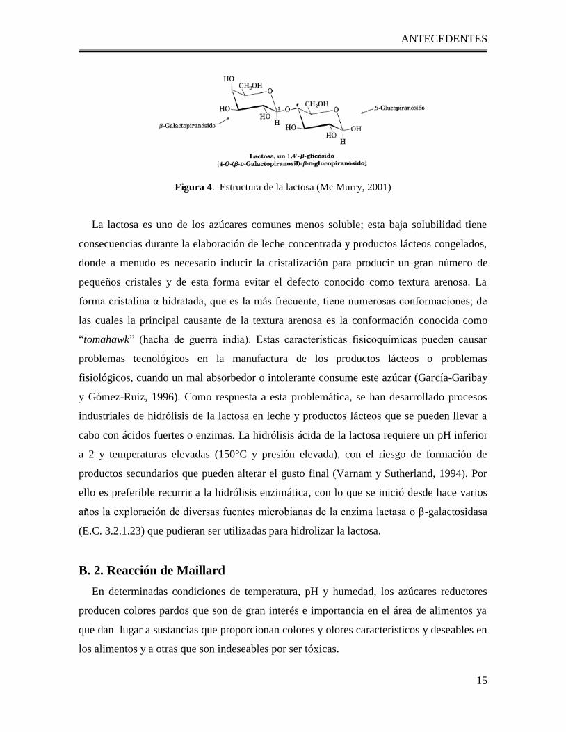
(Walstra y Jennes, 1984).La lactosa es un disacárido constituido por dos moléculas, una de
α-D-glucosa y otra de β-D-galactosa unidas por un enlace β-1,4-glicosídico. Ambas
moléculas se presentan predominantemente en forma de anillo piranósico, por lo que el
nombre correcto para la molécula es el de 4-O-β—D-galactopiranosil-D-glucopiranosa
(Figura 3).
Dependiendo del tipo de glucosa que intervenga en la molécula (α ó β) la lactosa puede
tener dos formas isoméricas: alfa lactosa o beta lactosa; que tienen propiedades
fisicoquímicas completamente diferentes (solubilidad, cristalización, refracción de la luz,
etc.). La forma β tiene una solubilidad mucho mayor, pero por mutarrotación se alcanza un
equilibrio entre las dos formas.
ANTECEDENTES
15
Figura 4. Estructura de la lactosa (Mc Murry, 2001)
La lactosa es uno de los azúcares comunes menos soluble; esta baja solubilidad tiene
consecuencias durante la elaboración de leche concentrada y productos lácteos congelados,
donde a menudo es necesario inducir la cristalización para producir un gran número de
pequeños cristales y de esta forma evitar el defecto conocido como textura arenosa. La
forma cristalina α hidratada, que es la más frecuente, tiene numerosas conformaciones; de
las cuales la principal causante de la textura arenosa es la conformación conocida como
―tomahawk‖ (hacha de guerra india). Estas características fisicoquímicas pueden causar
problemas tecnológicos en la manufactura de los productos lácteos o problemas
fisiológicos, cuando un mal absorbedor o intolerante consume este azúcar (García-Garibay
y Gómez-Ruiz, 1996). Como respuesta a esta problemática, se han desarrollado procesos
industriales de hidrólisis de la lactosa en leche y productos lácteos que se pueden llevar a
cabo con ácidos fuertes o enzimas. La hidrólisis ácida de la lactosa requiere un pH inferior
a 2 y temperaturas elevadas (150°C y presión elevada), con el riesgo de formación de
productos secundarios que pueden alterar el gusto final (Varnam y Sutherland, 1994). Por
ello es preferible recurrir a la hidrólisis enzimática, con lo que se inició desde hace varios
años la exploración de diversas fuentes microbianas de la enzima lactasa o β-galactosidasa
(E.C. 3.2.1.23) que pudieran ser utilizadas para hidrolizar la lactosa.
B. 2. Reacción de Maillard
En determinadas condiciones de temperatura, pH y humedad, los azúcares reductores
producen colores pardos que son de gran interés e importancia en el área de alimentos ya
que dan lugar a sustancias que proporcionan colores y olores característicos y deseables en
los alimentos y a otras que son indeseables por ser tóxicas.
ANTECEDENTES
16
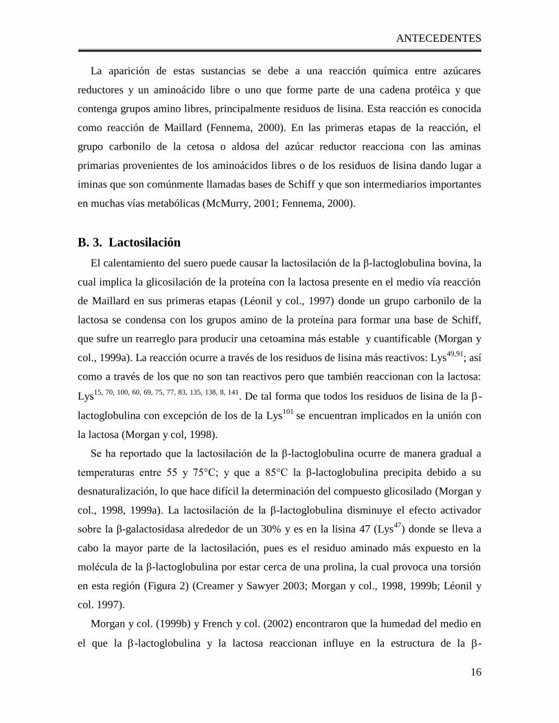
La aparición de estas sustancias se debe a una reacción química entre azúcares
reductores y un aminoácido libre o uno que forme parte de una cadena protéica y que
contenga grupos amino libres, principalmente residuos de lisina. Esta reacción es conocida
como reacción de Maillard (Fennema, 2000). En las primeras etapas de la reacción, el
grupo carbonilo de la cetosa o aldosa del azúcar reductor reacciona con las aminas
primarias provenientes de los aminoácidos libres o de los residuos de lisina dando lugar a
iminas que son comúnmente llamadas bases de Schiff y que son intermediarios importantes
en muchas vías metabólicas (McMurry, 2001; Fennema, 2000).
B. 3. Lactosilación
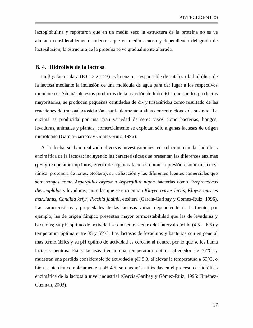
El calentamiento del suero puede causar la lactosilación de la β-lactoglobulina bovina, la
cual implica la glicosilación de la proteína con la lactosa presente en el medio vía reacción
de Maillard en sus primeras etapas (Léonil y col., 1997) donde un grupo carbonilo de la
lactosa se condensa con los grupos amino de la proteína para formar una base de Schiff,
que sufre un rearreglo para producir una cetoamina más estable y cuantificable (Morgan y
col., 1999a). La reacción ocurre a través de los residuos de lisina más reactivos: Lys49,91
; así
como a través de los que no son tan reactivos pero que también reaccionan con la lactosa:
Lys15, 70, 100, 60, 69, 75, 77, 83, 135, 138, 8, 141
. De tal forma que todos los residuos de lisina de la -
lactoglobulina con excepción de los de la Lys101
se encuentran implicados en la unión con
la lactosa (Morgan y col, 1998).
Se ha reportado que la lactosilación de la β-lactoglobulina ocurre de manera gradual a
temperaturas entre 55 y 75°C; y que a 85°C la β-lactoglobulina precipita debido a su
desnaturalización, lo que hace difícil la determinación del compuesto glicosilado (Morgan y
col., 1998, 1999a). La lactosilación de la β-lactoglobulina disminuye el efecto activador
sobre la β-galactosidasa alrededor de un 30% y es en la lisina 47 (Lys47
) donde se lleva a
cabo la mayor parte de la lactosilación, pues es el residuo aminado más expuesto en la
molécula de la β-lactoglobulina por estar cerca de una prolina, la cual provoca una torsión
en esta región (Figura 2) (Creamer y Sawyer 2003; Morgan y col., 1998, 1999b; Léonil y
col. 1997).
Morgan y col. (1999b) y French y col. (2002) encontraron que la humedad del medio en
el que la -lactoglobulina y la lactosa reaccionan influye en la estructura de la -
ANTECEDENTES
17
lactoglobulina y reportaron que en un medio seco la estructura de la proteína no se ve
alterada considerablemente, mientras que en medio acuoso y dependiendo del grado de
lactosilación, la estructura de la proteína se ve gradualmente alterada.
B. 4. Hidrólisis de la lactosa
La β-galactosidasa (E.C. 3.2.1.23) es la enzima responsable de catalizar la hidrólisis de
la lactosa mediante la inclusión de una molécula de agua para dar lugar a los respectivos
monómeros. Además de estos productos de la reacción de hidrólisis, que son los productos
mayoritarios, se producen pequeñas cantidades de di- y trisacáridos como resultado de las
reacciones de transgalactosidación, particularmente a altas concentraciones de sustrato. La
enzima es producida por una gran variedad de seres vivos como bacterias, hongos,
levaduras, animales y plantas; comercialmente se explotan sólo algunas lactasas de origen
microbiano (García-Garibay y Gómez-Ruiz, 1996).
A la fecha se han realizado diversas investigaciones en relación con la hidrólisis
enzimática de la lactosa; incluyendo las características que presentan las diferentes enzimas
(pH y temperatura óptimos, efecto de algunos factores como la presión osmótica, fuerza
iónica, presencia de iones, etcétera), su utilización y las diferentes fuentes comerciales que
son: hongos como Aspergillus oryzae o Aspergillus niger; bacterias como Streptococcus
thermophilus y levaduras, entre las que se encuentran Kluyveromyes lactis, Kluyveromyces
marxianus, Candida kefyr, Picchia jadinii, etcétera (García-Garibay y Gómez-Ruiz, 1996).
Las características y propiedades de las lactasas varían dependiendo de la fuente; por
ejemplo, las de origen fúngico presentan mayor termoestabilidad que las de levaduras y
bacterias; su pH óptimo de actividad se encuentra dentro del intervalo ácido (4.5 – 6.5) y
temperatura óptima entre 35 y 65°C. Las lactasas de levaduras y bacterias son en general
más termolábiles y su pH óptimo de actividad es cercano al neutro, por lo que se les llama
lactasas neutras. Estas lactasas tienen una temperatura óptima alrededor de 37°C y
muestran una pérdida considerable de actividad a pH 5.3, al elevar la temperatura a 55°C, o
bien la pierden completamente a pH 4.5; son las más utilizadas en el proceso de hidrólisis
enzimática de la lactosa a nivel industrial (García-Garibay y Gómez-Ruiz, 1996; Jiménez-
Guzmán, 2003).
ANTECEDENTES
18
B. 5. -Galactosidasa de Kluyveromyces lactis.
La -galactosidasa de Kluyveromyces lactis (EC 3.2.1.23) (KL-gal) es la enzima más
ampliamente utilizada en la industria láctea para resolver tanto el problema de intolerancia
a la lactosa como problemas técnicos relacionados con la baja solubilidad de la lactosa y su
tendencia a la cristalización, que generan problemas técnicos de precipitación; formación
de grumos y arenosidades indeseables en productos lácteos con un alto contenido de sólidos
como son: helados, leches condensadas y azucaradas, cajetas y flanes (Mahoney, 1997;
García-Garibay, 1993; García-Garibay, 1992; Gekas y López-Leyva, 1985). Tiene una
masa molar aproximada de 117.62 KDa y se ha reportado que existen diversas formas
oligoméricas de la enzima, con actividad en las formas dimérica y tetramérica (Becerra y
col., 1998; Tello-Solis y col., 2005). A pesar de ser la lactasa de mayor uso comercial, la
estructura de la KL-lg no ha sido muy estudiada. Tello-Solis y col. (2005), determinaron
mediante estudios con dicroismo circular que la KL-gal es básicamente una proteína-,
formada por un 22% de giros, 14% de láminas- paralelas, 25% de láminas- antiparalelas,
34% de estructura desordenada y tan solo un 5% de hélice-.
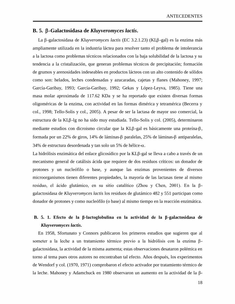
La hidrólisis enzimática del enlace glicosídico por la KLβ-gal se lleva a cabo a través de un
mecanismo general de catálisis ácida que requiere de dos residuos críticos: un donador de
protones y un nucleófilo o base, y aunque las enzimas provenientes de diversos
microorganismos tienen diferentes propiedades, la mayoría de las lactasas tiene al mismo
residuo, el ácido glutámico, en su sitio catalítico (Zhou y Chen, 2001). En la β-
galactosidasa de Kluyveromyces lactis los residuos de glutámico 482 y 551 participan como
donador de protones y como nucleófilo (o base) al mismo tiempo en la reacción enzimática.
B. 5. 1. Efecto de la β-lactoglobulina en la actividad de la β-galactosidasa de
Kluyveromyces lactis.
En 1958, Sfortunato y Connors publicaron los primeros estudios que sugieren que al
someter a la leche a un tratamiento térmico previo a la hidrólisis con la enzima β-
galactosidasa, la actividad de la misma aumenta; estas observaciones desataron polémica en
torno al tema pues otros autores no encontraban tal efecto. Años después, los experimentos
de Wendorf y col. (1970, 1971) comprobaron el efecto activador por tratamiento térmico de
la leche. Mahoney y Adamchuck en 1980 observaron un aumento en la actividad de la β-
ANTECEDENTES
19
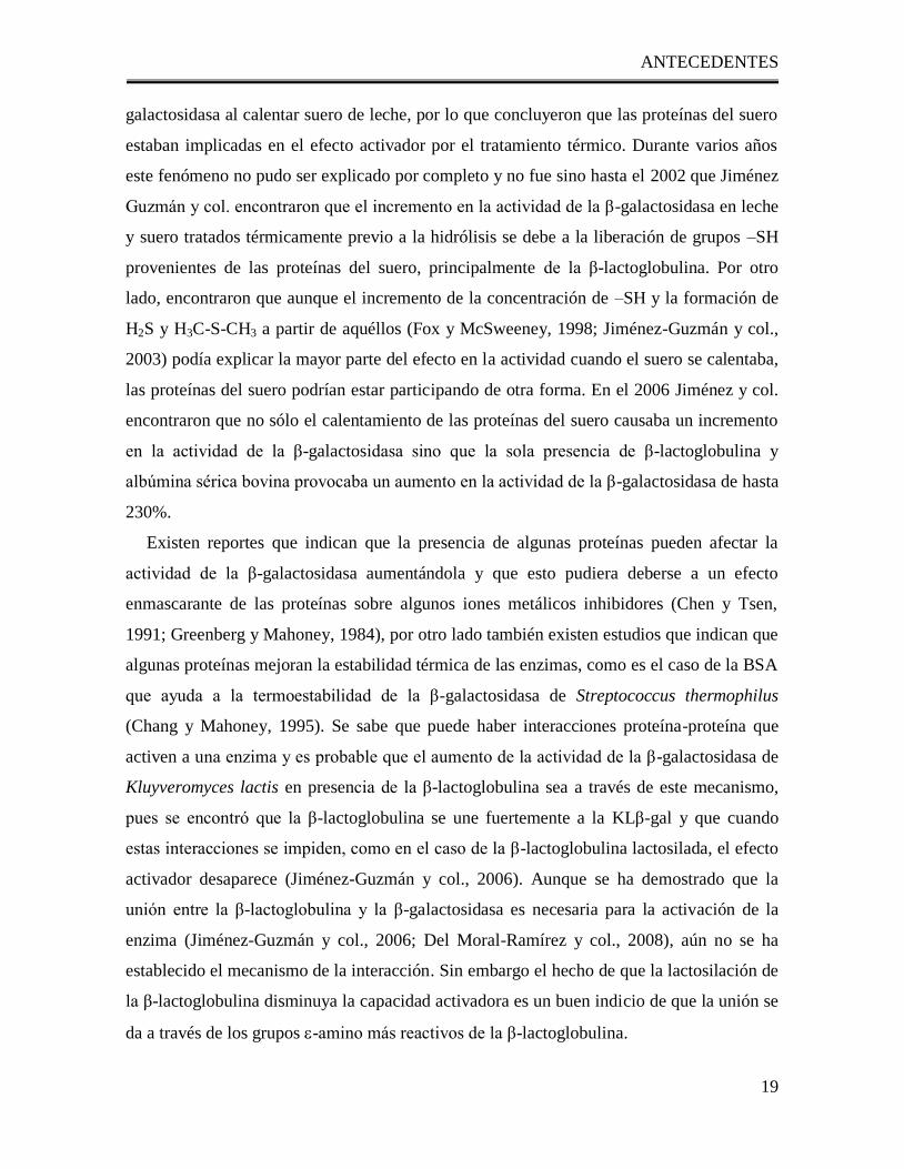
galactosidasa al calentar suero de leche, por lo que concluyeron que las proteínas del suero
estaban implicadas en el efecto activador por el tratamiento térmico. Durante varios años
este fenómeno no pudo ser explicado por completo y no fue sino hasta el 2002 que Jiménez
Guzmán y col. encontraron que el incremento en la actividad de la β-galactosidasa en leche
y suero tratados térmicamente previo a la hidrólisis se debe a la liberación de grupos –SH
provenientes de las proteínas del suero, principalmente de la β-lactoglobulina. Por otro
lado, encontraron que aunque el incremento de la concentración de –SH y la formación de
H2S y H3C-S-CH3 a partir de aquéllos (Fox y McSweeney, 1998; Jiménez-Guzmán y col.,
2003) podía explicar la mayor parte del efecto en la actividad cuando el suero se calentaba,
las proteínas del suero podrían estar participando de otra forma. En el 2006 Jiménez y col.
encontraron que no sólo el calentamiento de las proteínas del suero causaba un incremento
en la actividad de la β-galactosidasa sino que la sola presencia de β-lactoglobulina y
albúmina sérica bovina provocaba un aumento en la actividad de la β-galactosidasa de hasta
230%.
Existen reportes que indican que la presencia de algunas proteínas pueden afectar la
actividad de la β-galactosidasa aumentándola y que esto pudiera deberse a un efecto
enmascarante de las proteínas sobre algunos iones metálicos inhibidores (Chen y Tsen,
1991; Greenberg y Mahoney, 1984), por otro lado también existen estudios que indican que
algunas proteínas mejoran la estabilidad térmica de las enzimas, como es el caso de la BSA
que ayuda a la termoestabilidad de la β-galactosidasa de Streptococcus thermophilus
(Chang y Mahoney, 1995). Se sabe que puede haber interacciones proteína-proteína que
activen a una enzima y es probable que el aumento de la actividad de la β-galactosidasa de
Kluyveromyces lactis en presencia de la β-lactoglobulina sea a través de este mecanismo,
pues se encontró que la β-lactoglobulina se une fuertemente a la KLβ-gal y que cuando
estas interacciones se impiden, como en el caso de la β-lactoglobulina lactosilada, el efecto
activador desaparece (Jiménez-Guzmán y col., 2006). Aunque se ha demostrado que la
unión entre la β-lactoglobulina y la β-galactosidasa es necesaria para la activación de la
enzima (Jiménez-Guzmán y col., 2006; Del Moral-Ramírez y col., 2008), aún no se ha
establecido el mecanismo de la interacción. Sin embargo el hecho de que la lactosilación de
la β-lactoglobulina disminuya la capacidad activadora es un buen indicio de que la unión se
da a través de los grupos -amino más reactivos de la β-lactoglobulina.
ANTECEDENTES
20
B. 6. Esterificación de proteínas.
Desde inicios del siglo XX, se han estudiado diversos métodos para esterificar los
grupos carboxilo de las proteínas (Blackburn, Carter, & Phillips, 1941; Blackburn &
Phillips, 1944; Fraenkel-Conrat & Olcott, 1945). El uso de ácido clorhídrico como
catalizador para esterificar proteínas y polipéptidos fue sugerido por primera vez en 1932
(Felix & Reindl, 1932) y actualmente se sabe que concentraciones bajas de iones hidrógeno
(entre 0.02 y 0.1M) son suficientes para catalizar la esterificación completa de muchos
ácidos carboxílicos con alcohol metílico a temperatura ambiente en 24 horas. Tales
condiciones son considerablemente más suaves que las que se emplean normalmente, por lo
que pueden ser utilizadas para modificar químicamente sustancias lábiles como son las
proteínas.
B. 7. Uso de programas de cómputo para la construcción de un modelo
molecular y docking de una proteína El acelerado crecimiento de la información sobre las macromoléculas se traduce en una
labor de investigación sumamente importante e imprescindible, sin embargo la misma
demanda de información ha obligado a los investigadores a construir herramientas que les
permitan cumplir con su tarea en un corto tiempo para atender a la demanda de
información. Para ello se ha recurrido a la informática y se han creado programas de
cómputo que, por su disponibilidad y difusión en el medio científico, han podido ser
mejorados continuamente brindando con ello una alta confiabilidad en los resultados
obtenidos.
Anteriormente, para poder obtener un modelo tridimensional de una proteína era
necesario un arduo trabajo experimental, sin embargo actualmente basta contar con un
modelo cristalográfico con alta homologia a la proteína cuya estructura es desconocida para
poder construir un modelo tridimensional con una precisión comparable a la que se tendría
usando los métodos experimentales de mediana resolución (Krieger y col., 2003). El
modelaje por homología se ha convertido en un método ampliamente usado para poder
conocer la estructura tridimensional de una proteína y con ello obtener gran cantidad de
información sobre ella. Además, el modelaje por homología tiene una amplia gama de
aplicaciones como: diseño de mutantes, predicción de una función, identificación de sitios
ANTECEDENTES
21
de unión, planeación de experimentos, etc. (Fisher y col., 2003) es por esto que es necesario
saber cómo se construye y cómo se puede validar el modelo obtenido.
Por otra parte, una vez obtenido el modelo, es necesario optimizarlo y para ello se han
creado programas de cómputo para analizar los cambios energéticos que sufrirían los
átomos de una molécula si se les sometiera a un gran número de condiciones que, si se
intentara hacer experimentalmente, el tiempo sería demandante y quizá nunca suficiente
como para llevarlo a cabo. Por tal razón, la dinámica molecular que se hace con los
programas de cómputo también se ha convertido en una herramienta indispensable en el
modelaje molecular. Finalmente, el saber dónde se unen los diferentes tipos de ligandos a
una proteína es una tarea a la que muchos grupos de trabajo dedican gran parte de su
tiempo. Actualmente se cuenta con programas que ayudan a predecir las interacciones tanto
de ligandos de tamaño pequeño como de proteínas completas incluso sin tener una idea de
cuál o cuáles son los sitios de interacción (docking ciego) (Hetényi y col., 2002). Lo
anterior permite dirigir la atención del investigador hacia determinados puntos de unión con
una certeza mayor a la que tendría si no conociera dónde sería probable la interacción, con
lo que es posible reducir el tiempo de experimentación y obtener un gran número de
ventajas tanto científicas como económicas.
OBJETIVOS
22
C. OBJETIVOS
C. 1. Objetivo General
Caracterizar el mecanismo de interacción entre la -galactosidasa de Kluyveromyces
lactis y la -lactoglobulina, así como su relación con el efecto activador de la β-lg en la
actividad de la KLβ-gal
C. 2. Objetivos Particulares
Obtener un modelo de la estructura tridimensional del monómero y el dímero de la β-
lactoglobulina.
Determinar, mediante el estudio de los modelos tridimensionales y docking ciego, cuáles
son los grupos amino más expuestos y reactivos de la -lactoglobulina.
Evaluar el efecto del pH en las interacciones entre la -galactosidasa de Kluyveromyces
lactis y en el efecto activador.
Bloquear los grupos amino de la -lactoglobulina por medio de una reacción de
succinilación.
Evaluar por medio de cromatografía de afinidad el efecto de la succinilación de la β-lg
en su interacción con la -galactosidasa de Kluyveromyces lactis y en su capacidad
activadora.
Construir un modelo tridimensional del monómero y del dímero de la -galactosidasa de
Kluyveromyces lactis.
Determinar, mediante el estudio de los modelos tridimensionales y docking ciego, cuáles
son los grupos carboxilo más expuestos y reactivos de la KL-gal.
Bloquear los grupos carboxilo de la -galactosidasa de Kluyveromyces lactis por medio
de una reacción de esterificación con metanol.
Evaluar por medio de cromatografía de afinidad el efecto de la esterificación de la KL-
gal en la interacción con la -lactoglobulina y el efecto activador.
RESULTADOS
23
D. 1. Role of Lysine -Amino Groups of -Lactoglobulin on Its Activating
Effect of Kluyveromyces lactis Galactosidase.
Del Moral-Ramírez, E., Domínguez-Ramírez, L., Cruz-Guerrero, A. E., Rodríguez-
Serrano, G. M., García-Garibay, M., Gómez-Ruiz, L., & Jiménez-Guzmán, J.
(2008). Role of Lysine -Amino Groups of -Lactoglobulin on Its Activating Effect
of Kluyveromyces lactis -Galactosidase.
Journal of Agricultural and Food Chemistry, 56, 5859-5863.
RESULTADOS
29
D. 2. Determination of the probable interaction site of bovine -
lactoglobulin dimer with electrophilic molecules.
Del Moral-Ramírez, E., Domínguez-Ramírez, L., Cruz-Guerrero, A. E., Gómez-Ruiz, L.,
Rodríguez-Serrano, G. M., García-Garibay, M., & Jiménez-Guzmán, J. (2012).
Determination of the probable interaction site of bovine β-lactoglobulin dimer with
electrophilic molecules.
Enviado a: Food Chemistry
RESULTADOS
30
Short Communication 1
Determination of the probable interaction site of bovine -lactoglobulin 2
dimer with electrophilic molecules 3
4
Running Title: Probable interaction site of β-lg dimer with electrophilic molecules 5
6
Elizabeth Del Moral-Ramírez1, Lenin Domínguez-Ramírez
2, Alma E. Cruz-Guerrero
1, 7
Lorena Gómez-Ruiz1, Gabriela M. Rodríguez-Serrano
1, Mariano García-Garibay
1,3, Judith 8
Jiménez-Guzmán1*
9
10
1Departamento de Biotecnología, Universidad Autónoma Metropolitana, Iztapalapa, 11
Mexico City, Mexico 12
2Molecular and Cellular Biology, College of Biological Sciences, University of California 13
at Davis, Davis Ca. USA 14
3División de Ciencias Biológicas y de la Salud, Universidad Autónoma Metropolitana, 15
Lerma, Lerma de Villada, México 16
17
*Corresponding author: Departamento de Biotecnología, Universidad Autónoma 18
Metropolitana, Iztapalapa, AP 55-535, Mexico City, 09340, Mexico. E-mail: 19
[email protected], phone:+(52)(55)5804-4720, Fax: +(52)(55)5804- 4712 20
21
22
RESULTADOS
31
ABSTRACT 23
24
The probable interaction site of bovine -lactoglobulin dimer with electrophile molecules 25
was studied. Blind dockings of -lactoglobulin dimer with lactose and succinic anhydride 26
used as model ligands showed that Lys138
of one monomer and Lys141
of the other are the 27
most probable to interact with both lactose and succinic anhydride (Final Docking 28
Energies -3.30 and -3.11 kcal mol-1
respectively). Moreover, the dimer formation of -29
lactoglobulin is necessary to strengthen the interactions due to the participation of Lysine138
30
of one of -lg monomers and Lys141
of the other. Besides binding to Lys138
and 31
Lys141
electrophiles form hydrogen bonds with other nearby exposed amino acids of both 32
monomers; this may strengthen the interaction by approaching and stabilizing the ligands in 33
a claw-like structure so that the nucleophilic attack may take place. 34
35
KEYWORDS: -lactoglobulin dimer, blind docking, electrophile, lysine -amino groups 36
37
1. Introduction 38
Bovine -lactoglobulin (-lg) is the major whey protein conforming up to 50% of whey 39
proteins and 12% of whole cow milk proteins (Verheul, Pedersen, Roefs, & Kruif, 1999; 40
Fox, 2003). Each -lg monomer consists of 162 aminoacids with a mass of approximately 41
18.3 kDa. Secondary structure of -lg is composed of eight up and down antiparallel -42
strands which form a calyx (Papiz, et al., 1986) followed by a three turn -helix and ending 43
with a -strand (Blanch, Hecht, & Barron, 1999). -Lg belongs to the lipocalin family 44
which constitutes small, extracellular proteins that are capable of binding hydrophobic 45
ligands (Brignon, Chtorou, & Ribadeau-Dumas, 1985; Monti, Mermoud, & Jolles, 1989; 46
Uhrinova, Smith, Jameson, Uhrin, Sawyer, & Barlow, 2000) and although its biological 47
function is uncertain (Bell & McKenzie,1968; Fogolari, Ragona, Licciardi, Romagnoli, 48
Michelutti, Ugolini, & Molinari, 2000; Noiseux, Gauthier, & Turgeon, 2002; Qin, Bewley, 49
Creamer, Baker, Baker, & Jameson, 1998; Halpin & Richardson, 1985) it has been subject 50
of numerous studies because of its abundance, ease of purification and its ability to bind 51
RESULTADOS
32
small hydrophobic ligands such as fatty acids, retinol and peptides (Brownlow et al., 1997; 52
Flower, North, & Sansom, 2000; Fogliano et al., 1998; Noiseux et al., 2002). 53
Many studies have attempted to establish the ligand-binding site of -lg (Noiseux et al., 54
2002) and despite of remaining uncertain, two potential binding sites have been postulated 55
for hydrophobic and small molecules: one inside the calyx and the other at the outer surface 56
of the protein between the -helix and the -barrel (Papiz et al., 1986; Monaco, Zanottti, 57
Spadon, Bolognesi, Sawyer, & Eliopoulos, 1987). Recent studies showed that besides 58
binding small, polar and non polar ligands, -lactoglobulin can also bind to Kluyveromyces 59
lactis -galactosidase (KL-gal) increasing its activity (Jiménez-Guzmán et al., 2006, Del 60
Moral-Ramírez, et al., 2008). It has been recently reported that the interaction between 61
these proteins is very likely to occur through lysine -amino groups of -lg and studies 62
using molecular docking of the monomer and succinic anhydride used as a model ligand for 63
the protein showed that the interactions between -lg and KL-gal may specifically occur 64
through Lys
(Del Moral-Ramírez et al., 2008). 65
Oligomeric association and dissociation behavior of -lg has been widely studied by 66
different experimental techniques. Bovine -lg oligomeric states change as a function of 67
pH, phenomenon known as Tandford Transitions (Fox &McSweeney, 1998) and it has been 68
found that at room temperature and pH values below 4.0 and above 5.2 the protein consists 69
predominantly of monomers and dimers. Around pH 4.7, larger oligomeric structures as 70
octamers are formed (Verheul et al., 1999). At around pH 6.8, which is cow’s milk pH, -lg 71
exists as a dimer (Fox et al., 1998). 72
Many studies explain the interactions of -lg with hydrophobic molecules through the 73
internal calix of the protein (Yang et al., 2008a; Kontopidis, Holt, & Sawyer, 2002; Ragona 74
et al., 2000; Wu, Pérez, Puyol, & Sawyer, 1999); however this mechanism cannot explain 75
the interaction with a large protein such as KL-gal, suggesting that the interactions should 76
occur through a very exposed region of the protein. As far as we know, there are only a few 77
studies reporting this kind of interactions, and there is very limited information about the 78
possible interaction sites or mechanisms. Since at most experimental conditions bovine -lg 79
exists as a dimer, this work aimed to compare the probable interaction sites of the dimer 80
with those previously reported for the monomer (Del Moral-Ramírez et al, 2008). 81
RESULTADOS
33
Molecular docking of -lactoglobulin dimer with lactose and succinic anhydride was 82
carried out to determine the probable binding site of -lactoglobulin dimer with 83
Kluyveromyces lactis -galactosidase. 84
85
2. Materials and methods 86
2.1 Three-Dimensional Modeling 87
88
Bovine -lactoglobulin dimer three-dimensional (3D) model was built with PyMOL 89
software (DeLanoScientific, 2009) using the X-Ray diffraction coordinates of bovine -90
lactoglobulin triclinic crystal from growth at pH 6.5 and at 1.8 Å resolution found in the 91
Protein Data Bank (PDB) database (PDB ID: 1BEB) (Brownlow et al., 1997) 92
93
2.2 Molecular Docking 94
Blind docking was performed to estimate the binding energies between all -lg dimer 95
aminoacid residues and lactose or succinic anhydride using AutoDock 3.05 software (The 96
Scripps Research Institute, 2006). The runs were submitted at 25ºC and were clustered at a 97
range of RMSD of 5 Å. The 3D models for lactose and succinic anhydride were drawn at 98
The Dundee PRODRG2 Server site (http://davapc1.bioch.dundee.ac.uk/programs/prodrg/). 99
100
3. Results and discussion 101
102
3.1 Bovine -lactoglobulin dimer model and molecular docking 103
A 3D model of -lg dimer was built in order to study the exposition and the possible steric 104
hindrance that the different lysines in the molecule would show when reacting with lactose, 105
succinic anhydride and/or another protein (Figure 1). Potential -lg binding sites for small 106
hydrophobic molecules have been largely studied and have shown to be located in highly 107
hydrophobic areas of the monomer(Yang et al., 2008a; Yang et al., 2008b;Kontopidis, Holt, 108
& Sawyer, 2002; Ragona et al., 2000; Wu, Pérez, Puyol, & Sawyer, 1999; Ragona, 109
Pusterla, Zetta, Monaco, & Molinari, 1997); the binding site may vary as the dimer is 110
formed,but it is always located in the same region of the protein as shown in figure 111
RESULTADOS
34
1,whereas the binding site of small charged molecules seems to be nonspecific (Noiseux et 112
al., 2002; Wenbing et al, 2011). Lysine -amino groups are some of the most exposed and 113
reactive ones in -lg, with Lys47
and Lys138
being the most exposed (Creamer & Sawyer, 114
2003);it has also been reported that lactolation of -lg occurs through the amino groups of 115
lysine, specifically Lys47
(Léonil, Mollé, Fauquant, Maubois, Pearce, & Bouhallab, 1997; 116
Collin, D’Alfonso, & Baldini, 2000). 117
Despite the function of -lg has not been well established, its probable function as a 118
transporter protein has led to many studies focused on determining the binding sites of -lg 119
for different ligands. Most of ligand-protein interaction studies have been performed in 120
conditions involving the dimeric state of -lg; however,the techniques for determination of 121
ligand binding sites require digestion of -lg and therefore the loss of the dimer. As 122
interactions in many studies take place under conditions where the dimer is formed, and 123
given the difficulty for determining binding sites without hydrolising the protein, blind 124
docking of -lg dimer with lactose and succinic anhydride was performed to compare the 125
probable binding site of the dimer with those reported for the monomer and hydrolysates. 126
Docking results for lactose and -lg dimer (Figure 2) showed that Lys138
and Lys141
were 127
the aminoacids with the most favorable interaction energy at pH 7.0 (FDE=-3.30 kcal mol-
128
1) and thus the most probable to interact with lactose. Since a great variation in binding 129
sites has been found, it was recently proposed that ligand exchange among different sites of 130
-lg might occur in solution and that Lys138
and Lys141
are involved in such phenomenon 131
(Hu, et al., 2010; Ragona et al., 2000);our results agree with this finding, but it is 132
remarkable that the molecular docking performed showed that among all aminoacids 133
involved in the interactions, Lys
and Lys141
had the same docking energy but they 134
correspond to different monomers (Figure 2). This suggests that the dimeric state of -lg is 135
important for the interaction since both residues are required for it. This may also explain 136
the variation in binding sites found by other authors when hydrolysates are used, since upon 137
hydrolysis the ligand may remain in one or the other monomer. 138
Léonil et al. (1997) reported for the first time a binding site for the lactolation of -lg. The 139
fact that the binding occurs only through lysines suggests the importance of -lg-amino 140
groups in such reaction in which a nucleophilic attack must take place between an amino 141
RESULTADOS
35
group and an electrophile such as lactose. As shown in figure 2, besides binding to Lys138
142
and Lys141
lactose forms hydrogen bonds with other nearby exposed amino acids of both 143
monomers; this may strengthen the interaction by approaching and stabilizing lactose in a 144
claw-like structure so that the nucleophilic attack may take place. The former can explain 145
experimental evidence of strong and stable interactions found between -lg and larger 146
proteins such as KL-gal, in which -lg-amino groups of very exposed regions of the 147
proteins are involved (Del Moral-Ramírez et al., 2008; Jiménez-Guzmán et al., 2006). 148
In order to determine where the nucleophilic attack would take place in -lg dimer, succinic 149
anhydride (SA) was used as a stronger electrophile for docking studies. As with lactose, 150
Lys138
and Lys141
were the aminoacids with the most favorable interaction energy at pH 7.0 151
(FDE=-3.11 kcal mol-1
) and thus the most probable to interact with SA (Figure 3) via the 152
same mechanism proposed for lactose in which the ligand is stabilized by a claw-like 153
structure forming hydrogen bonds with some nearby aminoacids.Our results suggest that 154
besides being a highly electophilic region, Lys
of one -lg monomer and Lys141
of the 155
other may be a specific binding site for electrophilic molecules. 156
157
4. Conclusions 158
Our results show that the interaction sites found for -lg dimer and succinic anhydride or 159
lactose are consistent with those found for the monomer (Del Moral-Ramírez et al., 2008); 160
however, despite several studies pointing Lysine47
as the main amino acid involved in the 161
reaction with lactose (Léonil et al., 1997), this study suggests that in the case of the dimer 162
the interaction with the highly electrophilic molecules lactose and succinic anhidryde is 163
more likely to occur through Lysine138
. Moreover, the dimer formation of -lactoglobulin is 164
necessary to strengthen the interactions due to the participation of Lysine138
of one of -lg 165
monomers and Lys141
of the other: as the dimer associates, the regions around Lys138
and 166
Lys141
of both monomers form a claw-like structure which may establish strong interactions 167
through several electrophilic and hydrogen bonds (Figures 2 and 3) which could also 168
explain the interactions with larger molecules such as KLβ-lg which according to Jimenez-169
Guzman et al. (2006) results in a very stable binding. 170
171
RESULTADOS
36
5. Abbreviations used 172
RMSD: Root-mean-square deviation; FDE: Final Docking Energy 173
174
6. References 175
Bell, K., & McKenzie, H. A. (1964).-lactoglobulins.Nature, 204, 1275-1279. 176
Blanch, E. W., Hecht, L., & Barron, L. D. (1999). New insight into the pH- dependent 177
conformational changes in bovine -lactogloulin from Raman optical activity. Protein 178
Science, 8,1362-1367. 179
Brignon, G., Chtorou, A., & Ribadeau-Dumas, B. (1985). Does -lactoglobulin occur in 180
human milk? Journal of Dairy Research, 52, 249-254. 181
Brownlow, S., Cabral, J. H. M., Cooper, R., Flower, D. R., Yewdall, S. J., Polikarpov, I., 182
North, A. C. T., & Sawyer, L. (1997). Bovine -lactoglobulin at 1.8 Ǻ resolution– still an 183
enigmatic lipocalin. Structure, 5, 481-495. 184
Collin, M., D’Alfonso, L., &Baldini, G. (2000). New insight on -lactoglobulinbindig sites 185
by 1-anilinonaphtalene-8-sulfonate fluorescence decay. Protein Science, 9, 1968-1974. 186
Creamer, L. K., & Sawyer, L. (2003).Beta lactoglobulin.Ecyclopedia of Dairy Sciences 187
Del Moral-Ramírez, E., Domínguez-Ramírez, L., Cruz-Guerrero, A. E., Rodríguez-Serrano, 188
G. M., García-Garibay, M., Gómez-Ruiz, L., & Jiménez-Guzmán, J. (2008). Role of Lysine 189
-Amino Groups of -Lactoglobulin on Its Activating Effect of Kluyveromyces lactis -190
Galactosidase.Journal of Agricultural and Food Chemistry, 56, 5859-5863. 191
Flower, D. R., North, A. C. T., & Sansom, C. E. (2000). The lipocalin protein family: 192
structural and sequence overview. Biochimica et Biophysica Acta, 1482, 9-24. 193
Fogliano, V., Monti, M. S., Visconti, A., Randazzo, G., Facchiano, M. A., Colonna, G., & 194
Ritienti, A. (1998). Identification of a beta-lactoglobulin lactosylation site. Biochimica et 195
Biophysica Acta, 1338, 295-304. 196
Fogolari, F., Ragona, L., Licciardi, S., Romagnoli, S., Michelutti, R., Ugolini, R., & 197
Molinari, H. (2000). Electrostatic properties of bovine -lactoglobulin. Proteins: Structure 198
Function and Bioinformatics, 39, 4, 317-330. 199
RESULTADOS
37
Fox, P. F. (2003). Milk proteins: general and historical aspects. In P. F. Fox, & P. L. H. 200
McSweeney (Eds.), Advanced Dairy Chemistry Volume 1 (pp 1-48). New York: Kluwer 201
Academic/Plenum Publishers. 202
Fox, P. F., & McSweeney, P. L. H. (1998). Dairy Chemistry and Biochemistry. New York: 203
Kluwer Academic/Plenum Publishers. 204
Halpin, M. I., & Richardson, T. (1985).Selected Functionality Changes of -Lactoglobulin 205
upon Esterification of Side-Chain Carboxyl Groups.Journal of DairyScience, 68, 3189-206
3198. 207
Jiménez-Guzmán, J., Sarabia-Leos, C., Cruz-Guerrero, A., Rodríguez-Serrano, G., López-208
Munguía, A., Gómez-Ruiz, L., & García-Garibay, M. (2006). Interaction between β-209
lactoglobulin and lactase and its effect on enzymatic activity. International Dairy Journal, 210
16, 1169-1173. 211
Léonil, J., Mollé, D., Fauquant, J., Maubois, J. L., Pearce, R. J., &Bouhallab, S. (1997). 212
Characterization by Ionization Mass Spectrometry of Lactosylβ-Lactoglobulin Conjugates 213
Formed During Heat Treatment of Milk and Whey and Identification of One Lactose-214
Binding Site.Journal of Dairy Science, 80, 2270-2281. 215
Monaco, H. L, Zanotti, G., Spadon, P., Bolognesi, M., Sawyer, L., & Eliopoulos, E. E. 216
(1987). Crystal structure of the trigonal form of bovine beta-lactoglobulin and of its 217
complex with retinol at 2.5 Å resolution. Journal of Molecular Biology. 197, 695-706. 218
Monti, J. C., Mermoud, A. F., Jolles, P. (1989). Antibovine-lactoglobulin antibodies react 219
with a human lactoferrin fragment and bovine -lactoglobulin present in human milk. 220
Experientia, 45, 178-180. 221
Noiseux, I., Gauthier, S. L., & Turgeon, S. (2002). Interactions between Bovine -222
lactoglobulin and Peptides under Different Physicochemical Conditions.Journal of 223
Agricultural and Food Chemistry, 50, 1587-1592. 224
Papiz, M. Z., Sawyer, L., Eliopoulos, E. E., North, A. C. T., Findlay, J. B. C., 225
Sivaprasadarao, R., Jones, T. A., Newcomer, M. E., & Kraulis, P. J. (1986). The structure 226
of -lactoglobulin and its similarity to plasma retinol-binding protein. Nature, 324, 383-227
385. 228
RESULTADOS
38
Qin, B. Y., Bewley, M. C., Creamer, L. K., Baker, H. M., Baker, E. N., & Jameson, G. B. 229
(1998). Structural Basis of the Tanford Transition of Bovine -Lactoglobulin. 230
Biochemistry, 37, 14014-14023. 231
Ragona, L., Fogolari, F., Zetta., L., Perez, D. M., Puyol, P., De Kruif, K., Lohr, F., 232
Ruterjans, H., & Molinari, H. (2000). Bovine beta-lactoglobulin: interaction studies with 233
palmitic acid. Protein Science, 9, 1347-1356. 234
Uhrinova, S., Smith, M. H., Jameson, G. B., Uhrin, D., Sawyer, L., & Barlow, P. N. 235
(2000).Structural changes accompanying pH-induced dissociation of the -lactoglobulin 236
dimer. Biochemistry, 39, 3565-3574. 237
Ragona, L., Pusterla, F., Zetta, L., Monaco, H. L., & Molinari, H. (1997). Identification of a 238
conserved hydrophobic cluster in partially folded bovine beta-lactoglobulin at pH 2. 239
Folding and Design, 2, 281-290. 240
Verheul, M., Pedersen, J. S., Roefs, S. P. F. M., & Kruif, K. G. (1999). Association 241
behavior of native -lactoglobulin. Biopolymers, 49, 11-20. 242
Wenbing, H., Jianan, L., Qun, L., Yumiao, H., Kui, W., Shuang, L., Shaoxiang, X., & Fuyi, 243
W. (2011). Elucidation of the binding sites of sodium dodecil sulfate to -lactoglobulin 244
using hydrogen/deuterium exchange mass spectrometry combined with docking simulation. 245
Mass Spectrometry, 25, 1429-1436. 246
Yang, M. C., Guan, H. H., Liu, M.Y., Lin, Y. H., Yang, J. M., Chen, W. L., Chen, C. J., & 247
Mao, S. J. T. (2008a). Crystal Structure of a secondary vitamin D3 binding site of milk 248
beta-lactoglobulin. Proteins, 71, 3, 1197-1210. 249
Yang, M. C., Guan, H. H., Yang, J. M., Ko, C. N., Liu, M.Y., Lin, Y. H., Huang, Y. C., 250
Chen, C. J., & Mao, S. J. T. (2008b). Rational Design for Crystallization of -251
Lactoglobulin and Vitamin D3 Complex: revealing a Secondary Binding Site. Crystal 252
Growth and Design, 8, 12, 4268-4276. 253
254
255
256
257
258
RESULTADOS
39
Figure Captions 259
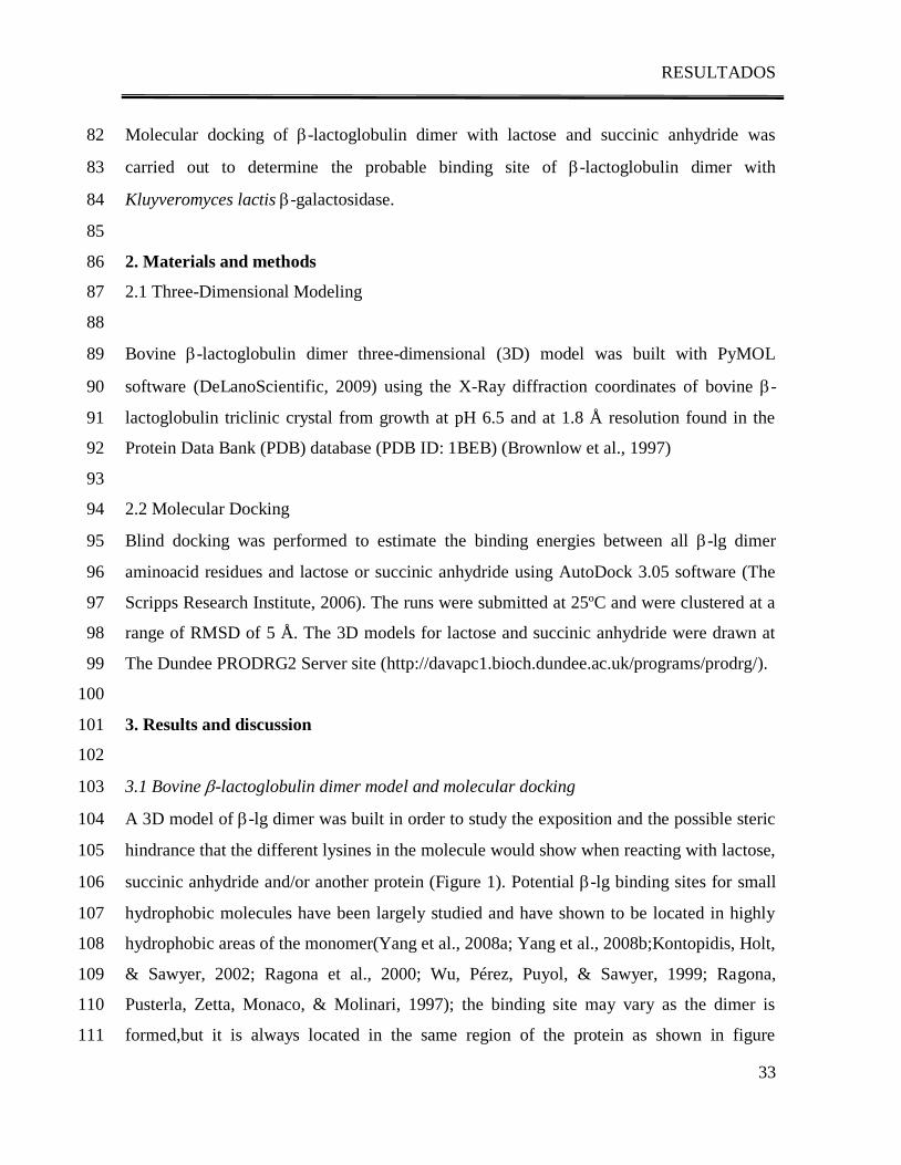
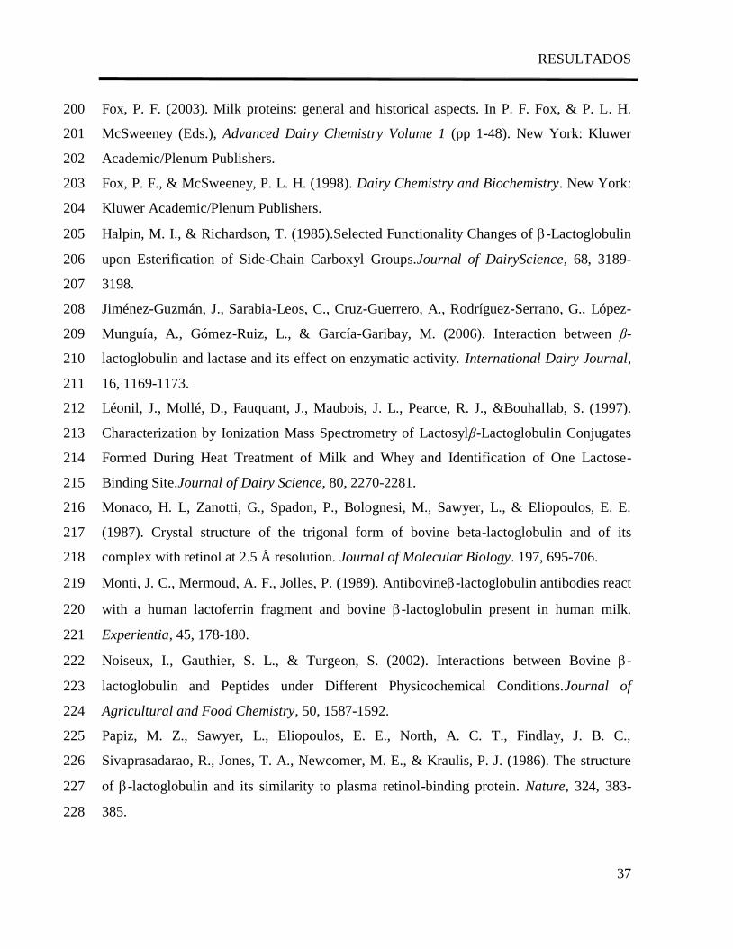
Figure 1. Bovine -lactoblobulin dimer showing hydrophobic potential binding sites (calyx 260
and outer surface) and reactive lysines. 261
262
Figure 2. Molecular docking of lactose (gray and red) and -lactoglobulin´s dimer showing 263
lactose interaction with Lys141
of one -lactoglobulin monomer (green) and with Lys138
of 264
the other -lactoglobulin´s monomer (cyan). 265
266
Figure 3. Molecular docking of succinic anhydride (SA) (gray and red) and -lactoglobulin 267
dimer showing lactose interaction with Lys141
of one -lactoglobulin´s monomer (green) 268
and with Lys138
of the other -lactoglobulin monomer (cyan). 269
270
Figures 271
Figure 1 272
273
Figure 1. Bovine -lactoblobulin dimer showing hydrophobic potential binding sites (calyx 274
and outer surface) and reactive lysines. 275
276
277
RESULTADOS
40
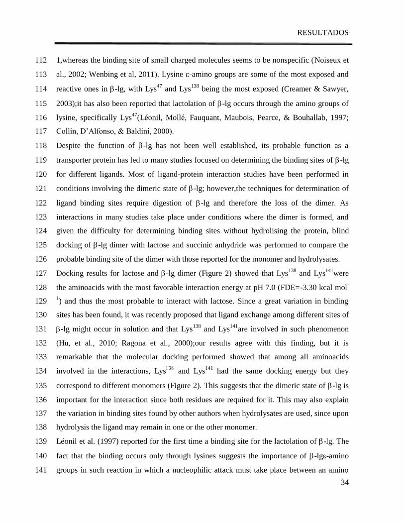
Figure 2 278
279
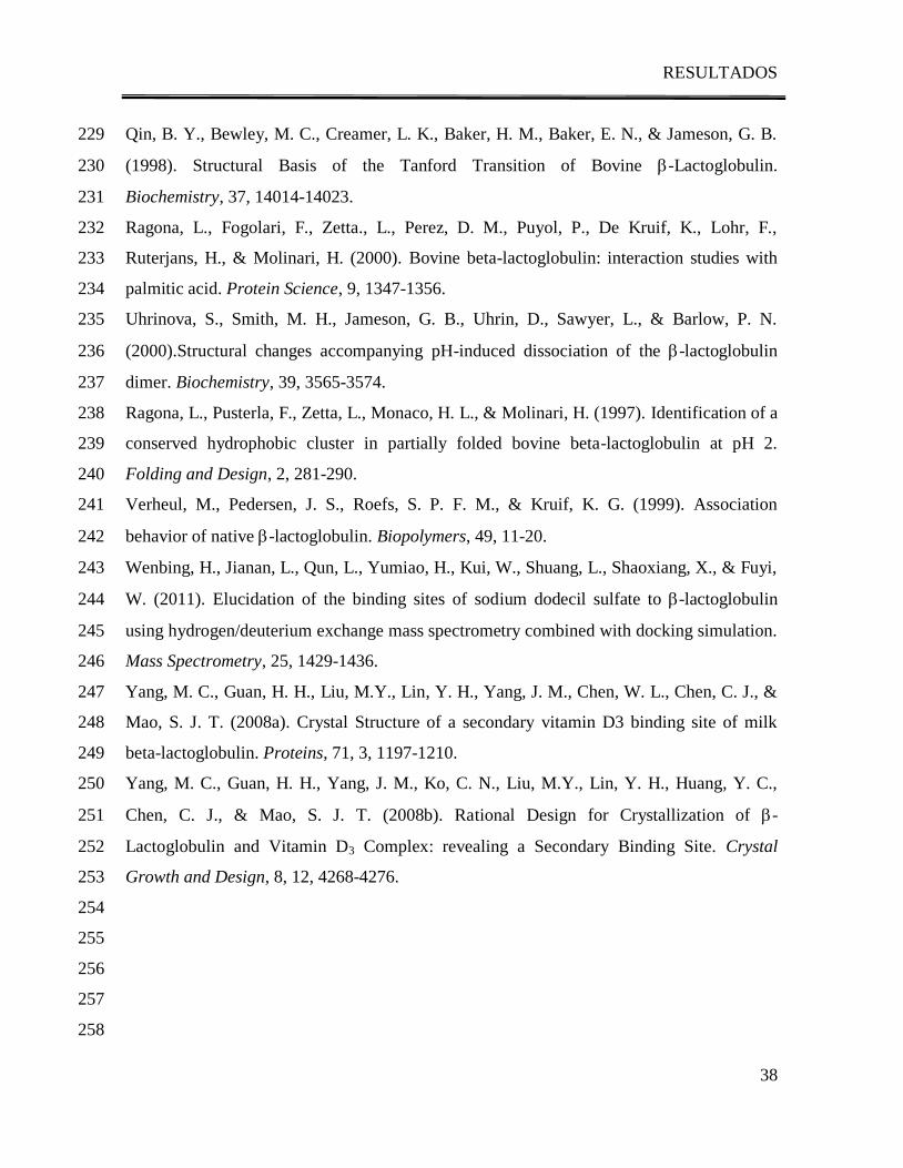
Figure 2. Molecular docking of lactose (gray and red) and -lactoglobulin dimer showing 280
lactose interaction with Lys141
of one -lactoglobulin monomer (green) and with Lys138
of 281
the other -lactoglobulin monomer (cyan). 282
283
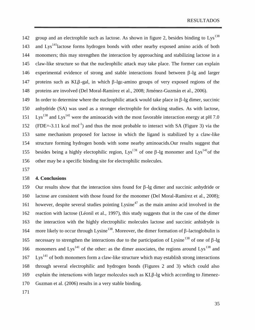
Figure 3 284
285
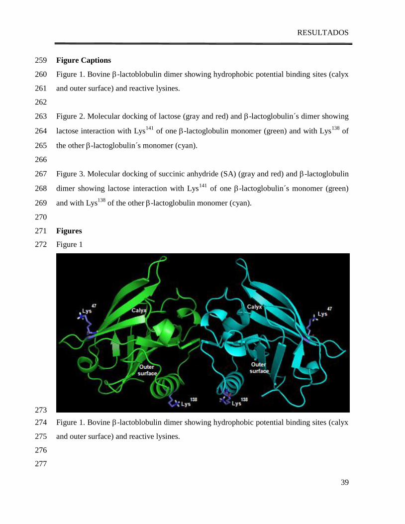
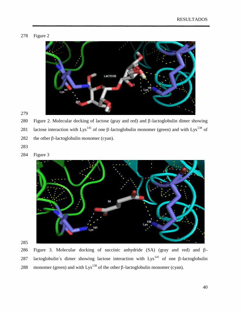
Figure 3. Molecular docking of succinic anhydride (SA) (gray and red) and -286
lactoglobulin´s dimer showing lactose interaction with Lys141
of one -lactoglobulin 287
monomer (green) and with Lys138
of the other -lactoglobulin monomer (cyan). 288
RESULTADOS
41
D. 3. Effect of pH on the interaction of -lactoglobulin with
Kluyveromyces lactis -galactosidase and its effect on enzymatic activity.
Del Moral-Ramírez, E., Domínguez-Ramírez, L., Pérez-Rangel, M. C., Cruz-
Guerrero, A. E., Gómez-Ruiz, L., Rodríguez-Serrano, G. M., García-Garibay, M., &
Jiménez-Guzmán, J. (2012). Effect of pH on the interaction of β-lactoglobulin with
Kluyveromyces lactis β-galactosidase and its effect on enzymatic activity.
Enviado a: Biochimica et Biophysica Acta
RESULTADOS
42
Effect of pH on the interaction of -lactoglobulin with Kluyveromyces 1
lactis -galactosidase and its effect on enzymatic activity. 2
3
Running Title: Effect of pH on activation and interaction of -lg with KL-gal. 4
5
Elizabeth Del Moral-Ramírez1, Lenin Domínguez-Ramírez
2, María del Carmen Pérez-6
Rangel1, Alma E. Cruz-Guerrero
1, Lorena Gómez-Ruiz
1, Gabriela M. Rodríguez-Serrano
1, 7
Mariano García-Garibay1,3
, Judith Jiménez-Guzmán1*
8
9
1Departamento de Biotecnología, Universidad Autónoma Metropolitana, Iztapalapa, 10
Mexico City, Mexico 11
2Molecular and Cellular Biology, College of Biological Sciences, University of California 12
at Davis, Davis Ca, USA 13
3División de Ciencias Biológicas y de la Salud, Universidad Autónoma Metropolitana, 14
Lerma, Lerma de Villada, México 15
16
*Corresponding author: Departamento de Biotecnología, Universidad Autónoma 17
Metropolitana, Iztapalapa, AP 55-535, Mexico City, 09340, Mexico. E-mail: 18
[email protected], phone:+(52)(55)5804-4720, Fax: +(52)(55)5804- 4712 19
20
21
RESULTADOS
43
ABSTRACT 22
Some reports have established the activating effect of Bovine -lactoglobulin (-lg) on 23
Kluyveromyces lactis -galactosidase (KL-gal) activity, suggesting that the interaction 24
between -lg and the enzyme could be responsible for this effect. Since both structure-25
activity of KL-gal and dimer formation of -lg are pH dependent, the present work studies 26
the effect of pH on the interaction of -lactoglobulin with Kluyveromyces lactis -27
galactosidase and its effect on enzymatic activity. The presence of β-lg has an activating 28
effect on KL-gal, which is stronger at pH 7.0 and absent at pHs 6.0 and 8.5. Comparison 29
of the changes on the activation by pH with the changes on the interactions between β-lg 30
and KL-gal showed a direct correlation: as the interaction between β-lg and KL-gal 31
increases the activity increases. Furthermore, it was observed that the dimeric state of β-lg 32
is essential for both the interaction and the activation of the enzyme. The activating effect 33
of β-lg yielded a higher activity than the one found at the optimum pH in buffer solution 34
(142% activation) suggesting that the interaction between both proteins could help to reach 35
an even more active conformation of KL-gal. It also shifts the optimum pH from 7.5 in 36
buffer solution to 7.0 in the presence of the β-lg suggesting that at this pH -lg promotes 37
the KL-gal conformational state in which the active site is more accessible to the 38
substrate. 39
40
41
Key words: -lactoglobulin, Kluyveromyces lactis -galactosidase, protein-protein 42
interaction 43
44
RESULTADOS
44
INTRODUCTION 45
Kluyveromyces lactis β-galactosidase (KL-gal) is by far the most important commercial 46
lactase used in the dairy industry. Several reports have established that the presence of 47
some proteins in the reaction medium may affect the activity of β-gal (Greenberg & 48
Mahoney, 1984; Chen & Tsen, 1991). Jiménez-Guzmán et al. (2006) studied the effect of 49
whey proteins in β-gal activity and demonstrated that the activity of Kluyveromyces lactis 50
β-gal increased when it was measured in the presence of either β-lactoglobulin (β-lg) or 51
bovine serum albumin; this finding is particularly interesting since these proteins are 52
available in milk and whey, which are the natural reaction media for this enzyme in dairy 53
processing. 54
Recent studies have demonstrated that -lactoglobulin (-lg), the major whey protein in 55
cow milk, enhaces lactase activity up to 230% by binding to KL-gal through lysine -56
amino groups probably involving Lysine138
of -lg (Jiménez-Guzmán et al., 2006, Del 57
Moral-Ramírez, et al., 2008). It is well known that -lg monomers can associate to form 58
different oligomeric structures and then dissociate into the monomers as a function of pH 59
by the phenomenon known as Trandford Transitions (Fox & McSweeney, 1998). It has 60
been found by different experimental techniques that at room temperature and pH values 61
below 4.0 and above 5.2 the protein consists predominantly of monomers and dimers. 62
Larger oligomeric structures as octamers are formed at around pH 4.7 (Verheul et al., 63
1999). At pH 6.8, which is cow’s milk pH, -lg exists as a dimer (Fox et al., 1998). It has 64
been recently reported that -lg dimer can interact with electrophilic molecules such as 65
lactose and succinic anhydride and that the dimer formation of -lactoglobulin is necessary 66
to strengthen these interactions due to the participation of Lysine138
of one of -lg 67
monomers and Lys141
of the other. Since -lg interactions with KL-gal occur through -68
amino groups, it is very likely that the region of -lg dimer involved in the interaction with 69
electrophilic molecules may be the same involved in the interaction with KL-gal. 70
Although there is scarce information on KL-gal structure, in 2005 Tello-Solis et al. found 71
by circular dichroism that KL-gal is mainly a -type protein consisting of 22% turns, 14% 72
parallel -sheet, 25% antiparallel -sheet, 34% unordered structure and only 5% -helix, 73
they also showed that there is a structure-activity relationship as a function of pH. 74
RESULTADOS
45
This work aimed to study the effect of pH on the interaction of -lactoglobulin with 75
Kluyveromyces lactis -galactosidase and its effect on enzymatic activity since both, 76
structure-activity of KL-gal and dimer formation of -lg are pH dependent. 77
78
MATERIALS AND METHODS 79
Enzyme Activity Measurement 80
The source of KL-gal was a commercial enzyme preparation, Maxilact LX-5000, (Gist 81
Brocades, Delft, The Netherlands). All reactions were carried out at 37°C by dilution of 82
1:400 of Maxilact LX-5000 into 0.05 M potassium phosphate buffer at pH values of 6.0, 83
6.5, 7.0, 7.5, 8.0 and 8.5. A solution of 0.034 M ortho-nitro-phenyl--D-galactoside 84
(ONPG) (Sigma Chemical Co. St. Louis MO, USA) was used as substrate and enzyme 85
activity was measured spectrophotometrically at 410 nm on the basis of ortho-nitro-phenol 86
(ONP) release after mixing 0.2 mL of ONPG solution with 0.1 mL of enzyme solution in 87
2.7 mL of phosphate buffer at each pH value. The hydrolysis rate (vo) for each pH value 88
was calculated from the linear portion of data of ONP production versus time. One enzyme 89
unit (U) was defined as the amount of enzyme that hydrolyses 1 mol of substrate (ONPG) 90
in 1 min at 37°C and the corresponding pH (6.0, 6.5, 7.0, 7.5, 8.0 or 8.5). Specific activity 91
was calculated dividing the enzyme units by the protein concentration of the sample 92
determined according to Lowry, Rosegrough, Farr, & Randall (1951). 93
KL-Gal activity of Maxilact LX-5000 was also measured in the presence of 0.3 mg/mL of 94
native -lg, at different pH (6.0, 6.5, 7.0, 7.5, 8.0 and 8.5). 95
Protein-Enzyme Interaction 96
Affinity chromatography was used to determine -lg interaction with KL-gal using a 97
Eupergit (Röhm GmbH & Co. Darmstadt, Germany) support with immobilized -lg at pH 98
6.0, 6.5, 7.0, 7.5, 8.0 and 8.5. Immobilization was performed following the instructions of 99
the producer. Controls without ligand were prepared at each pH by blocking the active 100
oxyrane groups of the support with glycine. After immobilization, the amount of 101
immobilized protein was calculated through the difference between the protein in solution 102
before and after interacting with the support the efficiency of immobilization was 0.640, 103
RESULTADOS
46
0.634, 0.687, 0.712 0.699 and 0.705 mol/gsupport at pH 6.0, 6.5, 7.0, 7.5, 8.0 and 8.5, 104
respectively. 105
Maxilact LX-5000, determined by electrophoresis to contain a mixture of eight proteins, 106
was used as the KL-gal sample. Because of the differences in the amount of protein 107
immobilized at each pH, -gal samples were prepared to have a concentration according to 108
a molar ratio of 0.05 mol-gal/molimmobilized -lg for each pH. Immobilized ligands and the 109
controls were allowed to interact with -gal by directly adding 0.1 g of support with 110
immobilized ligands to 3 mL of KL-gal sample and shaking for 1 h, then the supernatant 111
was separated from the support by centrifugation (3220g, 15 min) using a Beckman J2-MI 112
centrifuge (Beckman Instruments, Palo Alto CA, USA), and its specific activity was 113
measured using ONPG as a substrate. 114
Statistical Analyses. 115
Each experiment was performed three times. Data were analyzed by means of a variance 116
test (ANOVA); in some cases, a Tukey test was performed. All statistical analyses were 117
carried out using the statistical analysis software Statistica 5.0 (Stat Soft, Tulsa OK, USA), 118
with p < 0.05 used as a threshold of statistical significance. 119
120
RESULTS AND DISCUSSION 121
Figure 1 shows the variation of ONPG hydrolysis rate (vo) by KL-gal as a function of pH. 122
It was observed that lactase activity is low at around pH 6.0 and 8.5 consistent with that 123
reported by Tello et al. (2005) and Kim, Lim, & Kim (1997). The maximum vo was found 124
at pH 7.5, and activity variation found at different pH values from 6.0 to 8.5 is consistent 125
with that reported by Tello et al. (2005) who found an important increase in lactase activity 126
when pH changed from 6.5 to 7.0 and suggested that pH changes cause small modifications 127
in KL-gal secondary structure and that the increase in enzyme activity is mostly caused by 128
small local changes in structure such as the charges in some residues. Furthermore, when 129
pH increases from 7.0 to 7.5 (Figure 1) hydrolysis rate reaches its maximum, suggesting 130
that this change in pH may cause an important modification on KL-gal secondary 131
structure and thus a considerable increase in lactase activity. 132
RESULTADOS
47
When lactase activity was measured in the presence of 0.3 mg/mL -lg at different pH 133
values (Figure 1) an increase in vo was observed at pH between 6.5 – 8.0 (α < 0.0002), 134
confirming that the presence of -lg enhances lactase activity as reported by Jiménez-135
Guzmán et al., 2006 and Del Moral et al., 2008. At pHs of 6.0 and 8.5 -lg did not activate 136
KL-gal (α > 0.05) showing that the activation effect is dependent on pH. Optimum pH, at 137
which the maximum activity was reached, shifted from 7.5 to 7.0 in the presence of -lg. 138
Some reports have already established the activating effect of some whey proteins such as 139
-lg or bovine serum albumin on KL-gal activity (Jiménez-Guzmán et al, 2002; Jiménez-140
Guzmán et al, 2006). Jiménez-Guzman et al (2006) suggested that this effect could be 141
related to the ability of the protein to bind to the enzyme. It is very likely that as pH 142
changes, charges in the residues of -lg are modified, changing the protein’s ability to bind 143
to the enzyme. In order to study the effect of pH on the interaction of -lg with KL-gal, 144
affinity chromatography was performed at different pH values immobilizing β-lg on 145
Eupergit, and allowing the immobilized protein to interact with a solution of Maxilact 146
LX5000 at different pHs. A control for each pH was used in which no protein was 147
immobilized, but the reactive groups of the resin were blocked with glycine. 148
The specific activity of the control was not significantly different from that of the initial 149
lactase solution at any of the pHs tested (α > 0.05 for all cases) meaning that lactase was 150
not specifically bound to the support without protein (Figure 2). When the same solution of 151
lactase (Maxilact LX5000), containing eight proteins as determined by electrophoresis, was 152
mixed with the support containing β-lg as ligand it was observed that at pH 6.0 and 8.5 the 153
specific activity of the supernatant remained the same as that of the control (α= 0.9794 and 154
0.5430 for pH 6.0 and 8.5 respectively) (Table 1) demonstrating that at these pHs, there is 155
no specific interaction between β-lg and KL-gal. At pH between 6.5 and 8.0 the specific 156
activity of the enzyme solution that interacted with immobilized β-lg changed significantly 157
with respect to the control (support without ligand, α<0.00033 in all cases), reaching a 158
maximum diminution of 70% at pH 7.0 showing a specific interaction between β-lg and 159
KL-gal, which decreased as pH increased further more (Table 1). This suggests that KL-160
gal bound specifically to β-lg and that the interaction was favored at pH 7.0. At pHs below 161
or above 7.0, the interaction gradually decreased until it was not observable at pH 6.0 or 162
RESULTADOS
48
8.5. When comparing the changes on the activation caused by pH with the changes on the 163
interactions between β-lg and KL-gal (Figure 3), a direct correlation was observed: as the 164
interaction between β-lg and KL-gal is stronger the activation increases. 165
Tello et al (2005) established that small changes in KL-gal structure might cause 166
significant changes in enzyme’s activity. Mbuyi-Kalala, Schnek, & Léonis (1988) reported 167
that -galactosidase activity is influenced by some ionizable groups, prototropic groups and 168
by conformational changes. Jurado, Camacho, & Luzón (2003) reported that the effect of 169
pH on lactase activity is usually described by a kinetic model which involves the 170
dissociation of one or two protons in -gal. Although KL-gal has its most stable 171
conformation at pH 7.0 (Tello et al., 2005) this is not the conformational state at which it 172
reaches the highest activity which is at pH 7.5 (Figure 1) and in which conformational 173
changes caused by pH variation may leave the active site more accessible to the substrate, 174
increasing enzyme activity. The interaction between KL-gal and -lg could cause a 175
structural change on the enzyme leading to the exposure of the active site and thus a more 176
active conformation. 177
Many studies have attempted to establish the ligand-binding site of -lg (Noiseux et al., 178
2002); two potential binding sites have been postulated for hydrophobic and small 179
molecules: one inside the calyx and the other at the outer surface of the protein between the 180
-helix and the -barrel (Papiz et al., 1986; Monaco, Zanottti, Spadon, Bolognesi, Sawyer, 181
& Eliopoulos, 1987). Jiménez-Guzmán et al., 2006 and Del Moral-Ramírez, et al., 2008 182
showed that besides binding small, polar and non-polar ligands, -lactoglobulin can also 183
bind to Kluyveromyces lactis -galactosidase increasing its activity and that the interaction 184
between these proteins is very likely to occur through lysine -amino groups of -lg, 185
specifically Lys138
. 186
Sawyer, Kontopidis, & Wu (1999) reported that lysine, tyrosine and tryptophan residues 187
show a variable reactivity that is dependent on the media, and that cysteine121
(Cys121
) has a 188
free thiol group with a reactivity that is dependent on pH and structural changes that take 189
place at pH values from 6 to 8. 190
It has also been reported that the quaternary structure of -lg undergoes structural changes 191
when pH is modified (Verheul et al, 1999). At pH 6.0 and 8.5 at which no activation or 192
RESULTADOS
49
interaction with KL-gal was observed, β-lg exists as a monomer, while between 6.5 and 193
8.0 where both interaction and activation were observed, it exists as a dimer, showing that 194
the dimeric state of β-lg is essential for both the interaction and the activation of the 195
enzyme. 196
In buffer solution, as pH approximates 7.5 the most active conformation of KL-gal is 197
reached (Figure 1); however, in the presence of the -lg dimer the interaction between both 198
proteins could help to reach an even more active conformation of KL-gal, shifting the 199
optimum pH from 7.5 in the absence of -lg to 7.0 in the presence of the protein and 200
achieving a rate of hydrolysis significantly higher (α = 0.00018) than the one obtained in 201
the optimum pH in the absence of -lg (142% activation with respect to optimum pH in 202
buffer solution), suggesting that at this pH -lg promotes the KL-gal conformational state 203
in which the active site is more accessible to the substrate. 204
205
CONCLUSIONS 206
The presence of β-lg has an activating effect on KL-gal activity, which is stronger at pH 207
7.0 and absent at pHs 6.0 and 8.5 showing that the activation effect is dependent on pH. 208
When comparing the changes on the activation caused by pH with the changes on the 209
interactions between β-lg and KL-gal a direct correlation was observed: as the interaction 210
between β-lg and KL-gal is stronger the activation increases. Furthermore, when the 211
oligomeric structure of β-lg was compared to the pHs at which both interaction and 212
activation were found, it was observed that the dimeric state of β-lg is essential for both the 213
interaction and the activation of the enzyme. The fact that in the presence of β-lg the 214
activity observed was significantly higher than the one found at the optimum pH in buffer 215
solution suggests that the interaction between both proteins could help to reach an even 216
more active conformation of KL-gal, shifting the optimum pH from 7.5 in buffer solution 217
to 7.0 in the presence of the protein suggesting that at this pH -lg promotes the KL-gal 218
conformational state in which the active site is more accessible to the substrate. 219
220
RESULTADOS
50
REFERENCES 221
1. Brownlow, S., Cabral, J. H. M., Cooper, R., Flower, D. R., Yewdall, S. J., Polikarpov, 222
I., North, A. C. T., & Sawyer, L. (1997). Bovine -lactoglobulin at 1.8 Ǻ resolution– 223
still an enigmatic lipocalin. Structure, 5, 481-495. 224
2. Chen, J. Y. & Tsen, H. Y. (1991). Effect of milk and milk constituents on heath 225
stability of lactase from Saccharomyces lactis, J. Chinese Agric. Chem. Soc., 29, 4, 226
456-464. 227
3. Del Moral-Ramírez, E., Domínguez-Ramírez, L., Cruz-Guerrero, A. E., Rodríguez-228
Serrano, G. M., García-Garibay, M., Gómez-Ruiz, L., & Jiménez-Guzmán, J. (2008). 229
Role of Lysine -Amino Groups of -Lactoglobulin on Its Activating Effect of 230
Kluyveromyces lactis -Galactosidase. Journal of Agricultural and Food Chemistry, 231
56, 5859-5863. 232
4. Fox, P. F., & McSweeney, P. L. H. (1998). Milk proteins. In Dairy Chemistry and 233
Biochemistry. Blackie Academic & Professional: United Kingdom, 187-192. 234
5. García-Garibay, M. (1992). Recuperación de enzimas intracelulares de interés 235
industrial. -Galactosidasa de levadura. Ciencia (Mex.), 43, 23-33. 236
6. Gekas, V., López-Leyva, M. (1985). Hydrolysis of lactose: a literature review. Process 237
Biochem. 20, 2-11. 238
7. Greenberg, N. A., & Mahoney, R. R. (1994). The activity of lactase Streptococcus 239
thermophilus in milk and whey. Food Chem., 15, 4, 307-313. 240
8. Jiménez-Guzmán, J., Cruz-Guerrero, A.E., Rodríguez-Serrano, G., López-Munguía, 241
A., Gómez-Ruiz, L., & García-Garibay, M. (2002). Enhancement of lactase activity in 242
milk by reactive sulfhydryl groups induced by heat treatment. Journal of Dairy 243
Science, 85, 2497-2502. 244
9. Jiménez-Guzmán, J., Sarabia-Leos, C., Cruz-Guerrero, A., Rodríguez-Serrano, G., 245
López-Munguía, A., Gómez-Ruiz, L., & García-Garibay, M. (2006). Interaction 246
between β-lactoglobulin and lactase and its effect on enzymatic activity. International 247
Dairy Journal, 16, 1169-1173. 248
10. Léonil, J., Mollé, D., Fauquant, J., Maubois, J. L., Pearce, R. J., & Bouhallab, S. 249
(1997). Characterization by Ionization Mass Spectrometry of Lactosyl β-Lactoglobulin 250
RESULTADOS
51
Conjugates Formed During Heat Treatment of Milk and Whey and Identification of 251
One Lactose-Binding Site. Journal of Dairy Science, 80, 2270-2281. 252
11. Lowry, O., Rosebrough, N., Farr, A., & Randall, R. (1951). Protein measurement with 253
the Folin phenol reagent, Biol. Chem., 193, 265. 254
12. Mahoney, R. R. (1997). Lactose: Enzymatic modification. In Advanced Dairy 255
Chemistry, 2nd
ed., Fox, P. F., Ed.: Chapman & Hall: London, Vol. 3, p 95. 256
13. Tello-Solis, S., Jiménez-Guzmán, J., Sarabia-Leos, C., Gómez Ruiz, L., Cruz-257
Guerrero, A. E., Rodríguez-Serrano, G., & García-Garibay, M. (2005). Determination 258
of the Secondary Structure of Kluyveromyces lactis -Galactosidase and its Structure-259
Activity Relationship as a Function of the pH. 260
14. Verheul, M., Pedersen, J. S., Roefs, S. P. F. M., & Kruif, K. G. (1999). Association 261
behavior of native -lactoglobulin. Biopolymers, 49, 11-20. 262
263
264
FIGURE CAPTIONS 265
266
Figure 1. Effect of pH on ONPG hydrolysis rate (vo) by KL-gal and over the effect of -lg 267
on KL-gal activity. 268
269
Figure 2. Effect of pH on the interaction between β-lg and KL-gal determined by affinity 270
chromatography. 271
272
Figure 3. Relation between the activating effect of β-lg and its interaction with KL-gal. 273
274
Table 1. Effect of pH on the activation of KL-gal by β-lg. 275
276
Table 2. Interaction between β-lg and KL-gal as determined by affinity chromatography. 277
278
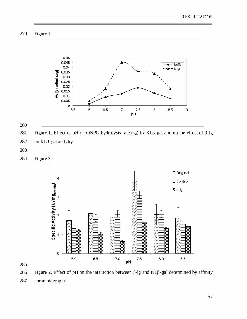
RESULTADOS
52
Figure 1 279
280
Figure 1. Effect of pH on ONPG hydrolysis rate (vo) by KL-gal and on the effect of -lg 281
on KL-gal activity. 282
283
Figure 2 284
285
Figure 2. Effect of pH on the interaction between β-lg and KL-gal determined by affinity 286
chromatography. 287
0
0.005
0.01
0.015
0.02
0.025
0.03
0.035
0.04
0.045
0.05
5.5 6 6.5 7 7.5 8 8.5 9
Vo
[
mo
l/m
l seg
]
pH
buffer
b-lg
0
1
2
3
4
6.0 6.5 7.0 7.5 8.0 8.5
Spec
ific
Act
ivit
y (U
/mg p
rote
in)
pH
Original
Control
b-lg
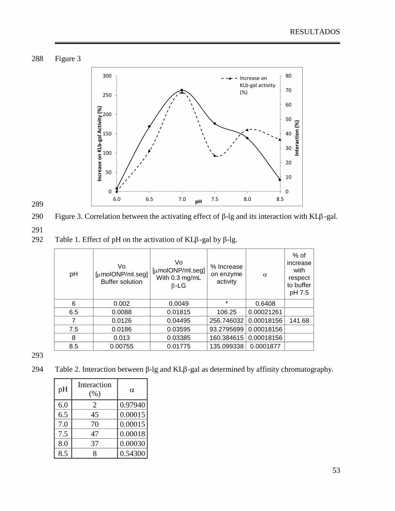
RESULTADOS
53
Figure 3 288
289
Figure 3. Correlation between the activating effect of β-lg and its interaction with KL-gal. 290
291
Table 1. Effect of pH on the activation of KL-gal by β-lg. 292
pH
Vo
[molONP/ml.seg] Buffer solution
Vo
[molONP/ml.seg] With 0.3 mg/mL
-LG
% Increase on enzyme
activity
% of increase
with respect to buffer pH 7.5
6 0.002 0.0049 * 0.6408
6.5 0.0088 0.01815 106.25 0.00021261
7 0.0126 0.04495 256.746032 0.00018156 141.68
7.5 0.0186 0.03595 93.2795699 0.00018156
8 0.013 0.03385 160.384615 0.00018156
8.5 0.00755 0.01775 135.099338 0.0001877
293
Table 2. Interaction between β-lg and KL-gal as determined by affinity chromatography. 294
pH Interaction
(%)
6.0 2 0.97940
6.5 45 0.00015
7.0 70 0.00015
7.5 47 0.00018
8.0 37 0.00030
8.5 8 0.54300
0
10
20
30
40
50
60
70
80
0
50
100
150
200
250
300
6.0 6.5 7.0 7.5 8.0 8.5
Inte
ract
ion
(%
)
Incr
eas
e o
n K
Lb-g
al A
ctiv
ity
(%)
pH
Increase onKLb-gal activity(%)
RESULTADOS
54
D. 4. Role of carboxyl groups of Kluyveromyces lactis -galactosidase in
the interaction with -lactoglobulin and its activating effect.
Del Moral-Ramírez, E., Domínguez-Ramírez, L., Cruz-Guerrero, A. E., Gómez-Ruiz, L.,
Rodríguez-Serrano, G. M., García-Garibay, M., & Jiménez-Guzmán, J. (2012). Role of
carboxyl groups of Kluyveromyces lactis β-galactosidase in the interaction with β-
lactoglobulin and its activating effect.
Enviado a: Enzyme and Microbial Technology
RESULTADOS
55
Role of carboxyl groups of Kluyveromyces lactis -galactosidase in the 1
interaction with -lactoglobulin and its activating effect. 2
3
Running Title: Study of the interaction between KL-gal and -lg. 4
5
Elizabeth Del Moral-Ramírez1, Lenin Domínguez-Ramírez
1,2, Alma E. Cruz-Guerrero
3, 6
Lorena Gómez-Ruiz3, Gabriela M. Rodríguez-Serrano
3, Mariano García-Garibay
1,3, Judith 7
Jiménez-Guzmán1*
8
9
1División de Ciencias Biológicas y de la Salud, Universidad Autónoma Metropolitana, 10
Lerma, Lerma de Villada, México
11
2Molecular and Cellular Biology, College of Biological Sciences, University of California 12
at Davis, Davis Ca. USA
13
3Departamento de Biotecnología, Universidad Autónoma Metropolitana, Iztapalapa, 14
Mexico City, Mexico
15
16
*Corresponding author: Departamento de Biotecnología, Universidad Autónoma 17
Metropolitana, Iztapalapa, AP 55-535, Mexico City, 09340, Mexico. E-mail: 18
[email protected], phone:+(52)(55)5804-4720, Fax: +(52)(55)5804- 4712
19
RESULTADOS
56
ABSTRACT 20
Reports have established the activating effect of -lactoglobulin (-lg) on Kluyveromyces 21
lactis -galactosidase (KL-gal), suggesting that the interaction between -lg and KLβ-gal 22
is directly responsible for this effect. Since lysine ɛ-amino groups of β-lg have been found 23
essential for both the interaction and activation of KLβ-gal, the interaction with KL-gal is 24
expected to occur via electrophilic groups such as side-carboxyl groups of the enzyme. The 25
aim of this work was to determine the role of side-carboxyl groups of KLβ-gal on both the 26
interaction with β-lg and its activating effect. A 3D model was constructed for KLβ-gal. 27
After excluding carboxyl amino acids that are involved in the formation of the dimer, it was 28
observed that only a few are exposed and likely to interact with β-lg. Blind docking of 29
KLβ-gal with lysine showed that the amino acid that could be involved in the interaction is 30
Glu592
(FDE= -4.55 kcal/mol). Esterification of the most exposed and reactive carboxyl 31
groups of KLβ-gal, among which Glu592
is found, resulted in the loss of the enzyme’s 32
ability to interact with β-lg. When lactase activity of the esterified KLβ-gal derivatives was 33
determined in the presence of β-lg it was observed that as long as there are carboxyl groups 34
available β-lg has an activating effect on the enzyme. When those groups were blocked by 35
esterification no activating effect was observed, suggesting that carboxyl groups of KLβ-gal 36
are essential for both, the ability of the enzyme to interact with β-lg and its capacity to be 37
activated by it. 38
39
40
KEYWORDS: blind docking, carboxyl groups, esterification, electrophile. 41
RESULTADOS
57
INTRODUCTION 42
Among several microbial -galactosidases, Kluyveromyces lactis β-galactosidase (KLβ-gal) 43
is by far the most important commercial lactase used in the dairy industry (Gekas & Lopez-44
Leiva, 1985; German, 1997; Nijipels, 1981). KL-Gal is mainly a -type protein consisting 45
of 22% turns, 14% parallel -sheet, 25% antiparallel -sheet, 34% unordered structure and 46
only 5% -helix (Tello-Solis et al., 2005). Enzymatic hydrolysis of the glycosidic bond by 47
-galactosidase takes place via general acid catalysis that requires two critical residues: a 48
proton donor and a nucleophile/base, and although the enzymes derived from various 49
microbial origins have different properties, such as molecular weight, protein chain length, 50
and the position of the active site, most -galactosidases have the same residue, a glutamic 51
acid, at their catalytic site (Zhou & Chen, 2001). Kluyveromyces lactis β-galactosidase has 52
glutamic acid482
(Glu482
) and glutamic acid551
(Glu551
) as the proton donor and the 53
nucleophile/base, respectively, in the enzymatic reaction (Zhou et al., 2001). 54
Several reports have established that the presence of some proteins in the reaction medium 55
may affect the activity of KLβ-gal; this raises the possibility that the proteins prevent the 56
inhibitory effect of ions in the reaction medium (Greenberg & Mahoney, 1984; Chen & 57
Tsen, 1991). Jiménez-Guzmán et al. (2006) studied the effect of whey proteins in KLβ-gal 58
activity and demonstrated that the activity of Kluyveromyces lactis β-gal increased when it 59
was measured in the presence of either β-lactoglobulin (β-lg) or bovine serum albumin; this 60
finding is particularly interesting since these proteins are available in milk and whey, which 61
are the natural reaction media for this enzyme in dairy processing. Affinity chromatography 62
assays demonstrated that KLβ-gal bound specifically to -lg, resulting in enzyme 63
activation. Moreover, heating pure β-lg further increased β-gal activity, but heating it in the 64
presence of lactose diminished its activating effect. Other reports have established that 65
heating β-lg in the presence of lactose resulted in a reaction between the protein and the 66
sugar (lactosylation) (Léonil, Mollé, Fauquant, Maubois, Pierce, & Bouhallab, 1997) which 67
diminished the binding capacity of the protein to the enzyme and therefore its activating 68
effect (Jiménez-Guzmán et al., 2006). It has been recently reported that -lg dimmer can 69
interact with electrophilic molecules such as lactose and succinic anhydride and that the 70
dimer formation of -lactoglobulin is necessary to strengthen these interactions due to the 71
RESULTADOS
58
participation of Lysine138
of one of -lg monomers and Lys141
of the other. Since -lg 72
interactions with KL-gal occur through -amino groups, it is very likely that the region of 73
-lg dimmer involved in the interaction with electrophilic molecules may be the same 74
involved in the interaction with KL-gal via electrophilic groups such as the side-carboxyl 75
groups of the enzyme. 76
The aim of this work was to determine the role of the side-carboxyl groups of 77
Kluyveromyces lactis β-galactosidase on both the interaction with β-lg and the activating 78
effect. 79
80
MATERIALS AND METHODS 81
Kluyveromyces lactis -galactosidase three-Dimensional Modeling 82
Kluyveromyces lactis -galactosidase three-dimensional (3D) model was built by homology 83
modeling with Modeller v9.2 software (Eswar et al., 2006; Marti-Renom et al., 2000; Sali, 84
1993; Fiser, Do, & Sali, 2000). The template corresponded to X-ray diffraction coordinates 85
of E.coli -galactosidase A chains (PDB ID: 1JZ7 and 1JZ8) (Juers et al., 2001) at 1.5 Å 86
(E=9x10-125
and E=3x10-124
respectively) with a 48% of homology. The best model had a 87
DOPE of -103795.781 kJ/mol. 88
Molecular Docking 89
Blind docking was performed to estimate the binding energies between all KL-gal amino 90
acid residues and lactose or lysine using AutoDock 3.05 software (The Scripps Research 91
Institute, 2006). The runs were submitted at 25ºC and were clustered at a range of RMSD 92
of 2 Å. The 3D models for lactose and lysine were drawn at The Dundee PRODRG2 Server 93
site (http://davapc1.bioch.dundee.ac.uk/programs/prodrg). 94
Kluyveromyces lactis -galactosidase Esterification 95
Methyl alcohol gradually and specifically blocks the side-chain carboxyl groups of KL-96
gal. Maxilact LX 5000 containing 461.92 mgprotein/mL was used as source of KL-gal. A 97
solution of methyl alcohol (Sigma Chemical Co., St. Louis, MO, USA) containing 1% of 98
protein and 0.05 N of hydrochloric acid (Sigma Chemical Co., St. Louis, MO, USA) was 99
prepared. The mixture was left under vigorous agitation for two hours at room temperature 100
according to Fraenkel-Conrat & Olcott (1945). Samples with KL-gal ester derivatives 101
RESULTADOS
59
were taken at the beginning of reaction (E0), after 1 hour (E1) and after two hours (E2) of 102
reaction; they were centrifuged at 2057 x g for 5 minutes and diluted 1:1 (vol/vol) with 103
deionized, distilled water and dialyzed in the cold against 10 volumes of 0.001 M HCl for 4 104
hours. In order to determine the degree of esterification, a general method to quantify 105
carboxyl groups of proteins employing the formation of a colored hydroxamate-ferric ion 106
complex was used according to Halpin & Richardson (1985): 500 L of 1.0 N 107
hydroxylamine hydrochloride (Aldrich Cemical Co., Milwahkee, MI, USA) were added 108
slowly while vortexing to 500 L of E0, E1, E2 or diluted Maxilact LX 5000, followed by 109
the addition of 200 L of 6.0 N potassium hydroxide (JT Baker, Xalostoc, Mexico). The 110
samples were heated at 74°C for 55 min and then cooled in ice. Thereafter, 1.6 mL of 1.0 N 111
HCl and 75 mL of 5.0% (wt/vol) ferric chloride hexahydrate (Sigma Chemical Co., St. 112
Louis, MO, USA), were added to each sample, with vortexing. Absorbance of the ferric 113
ion-hydroxamate complex was read at 475 nm within 30 min. An equimolar concentration 114
of KL-gal served as blank. 115
L-Glutamic acid -monoethyl ester (Sigma Chemical Co., St. Louis, MO, USA) was used 116
to prepare a standard curve for molar absorption of the ferric ion-hydroxamate complex, 117
using the foregoing procedure. The blank contained an equimolar amount of L-glutamic 118
acid (Sigma Chemical Co., St. Louis, MO, USA) treated in the same manner as the 119
corresponding ester. 120
Enzyme Activity Measurement 121
Maxilact LX-5000 was used as source of KL-gal (Gist Brocades, Delft, The Netherlands). 122
All reactions were carried out at 37°C by dilution of 1:10000 of Maxilact LX-5000 or 123
esther derivatives (E0, E1 and E2) into 0.05 M potassium phosphate buffer at pH 7.0. A 124
solution of 0.034 M ortho-nitro-phenyl--D-galactoside (ONPG) (Sigma Chemical Co. St. 125
Louis MO, USA) was used as substrate, and enzyme activity was measured 126
spectrophotometrically at 410 nm on the basis of ortho-nitro-phenol (ONP) release. 127
Reactions were carried out by mixing 67 L of ONPG solution with 33 L of enzyme 128
solution in 900 L of phosphate buffer. The hydrolysis rate (vo) was calculated from the 129
linear portion of data of ONP production versus time. One enzyme unit (U) was defined as 130
the amount of enzyme that hydrolyses 1 mol of substrate (ONPG) in 1 min at 37°C and 131
RESULTADOS
60
pH 7.0. Specific activity was calculated dividing the enzyme units by the protein 132
concentration of the sample determined according to Bradford (1976). 133
In order to evaluate the activating effect of lg in some experiments activity was 134
determined in the presence of 0.3 mg/mL lg. 135
Protein-Enzyme Interaction 136
Affinity chromatography was used to determine -lg interaction with KL-gal using a 137
Eupergit (Röhm GmbH & Co. Darmstadt, Germany) support with immobilized -lg at pH 138
7.0. Immobilization was performed following the instructions of the producer. Controls for 139
each derivative without ligand were prepared by blocking the active oxyrane groups of the 140
support with glycine 0.05 M. After immobilization, the amount of immobilized protein was 141
calculated through the difference between the protein in solution before and after 142
interacting with the support, the efficiency of immobilization was 1.6568 mol-lg/gsupport at 143
pH 7.0. 144
Derivatives E0 and E1 were used as the KL-gal samples. Samples were prepared to have a 145
concentration according to a molar ratio of 0.05 mol-gal/molimmobilized-lg. Immobilized 146
ligand and the controls were allowed to interact with -gal by directly adding 0.1 g of 147
support with immobilized ligands to 1.5 mL of KL-gal samples and shaking for 30 min, 148
then the supernatant was separated from the support by centrifugation (3220 x g, 15 min) 149
with an Eppendorf Centrifuge 5810 R (Eppendorf AG, Hamburg, Germany), and its 150
specific activity was measured using ONPG as a substrate. 151
Statistical Analyses. 152
Each experiment was performed three times. Data were analyzed by means of a variance 153
test (ANOVA); in some cases, a Tukey test was performed. All statistical analyses were 154
carried out using the statistical analysis software Statistica 5.0 (Stat Soft, Tulsa OK, USA), 155
with p < 0.05 used as a threshold of statistical significance. 156
157
RESULTS AND DISCUSSION 158
Recent studies have demonstrated that -lactoglobulin (-lg), the major whey protein in 159
cow milk, enhances lactase activity up to 230% by binding to KL-gal through lysine -160
amino groups probably involving Lys138
of -lg (Jiménez-Guzmán et al., 2006, Del Moral-161
RESULTADOS
61
Ramírez, et al., 2008) and suggesting that the interaction mechanism might be via the 162
nucleophilic attack of Lys138
of -lg to an electrophile group in KL-gal. 163
In order to study how KL-gal would interact with nucleophiles such as lactose, lysine 164
and/or other protein, a 3D model of KL-gal was built using the X-ray diffraction 165
coordinates of E.coli -galactosidase A chains (PDB ID: 1JZ7 and 1JZ8) as template (Juers 166
et al., 2001). 167
Blind docking of KL-gal with lactose showed that the amino acids with the most favorable 168
interaction energy at pH 7.0 were Glu482
and Glu551
(FDE= -4.82 kcal/mol) which are 169
consistent with the catalytic sites reported for KL-gal (Zhou et al., 2001). 170
Since Lys138
of β-lg has been suggested to be the amino acid most likely responsible for the 171
interaction between KL-gal and β-lg, blind docking of KL-gal with lysine was 172
performed, and 250 possible binding sites were displayed. Is has been reported that at pH 173
7.0, which is the pH at which our studies were performed, KL-gal exists as a dimer (Tello-174
Solis et al., 2005) because of this, a 3D model of the KL-gal dimer was built to select the 175
possible amino acids interacting with lysine (Figure 1). The selection criteria for the 176
possible interaction site of KL-gal with lysine was based on the most favorable binding 177
energy as well as on the exclusion of those amino acids located at the dimer interface and 178
those of the catalytic site, which due to steric hindrance, or the loss of enzyme activity 179
would clearly not be available to interact with another protein. By selecting with these 180
criteria, Glu592
was found to be the most probable interaction site with lysine (FDE= -4.55 181
kcal/mol) (Figure 1). 182
It is well known that carboxyl groups of proteins can be chemically modified through 183
gradual esterification with alcohols of low molecular weight by adding low concentrations 184
of hydrogen ions (0.02 to 0.1 M) to catalyze the reaction in mild conditions (Feeney, 1977; 185
Halpin et al., 1985). Kluyveromyces lactis -galactosidase has 157 esterificable groups per 186
monomer (156 combined glutamic acid and aspartic acid residues and 1 carboxyl end 187
group). In order to determine if side-carboxyl groups of KL-gal actively participate in the 188
interaction with β-lg, KL-gal carboxyl groups were esterified for 1(E1) or 2 (E2) hours. A 189
sample was taken immediately after mixing all reagents (E0) in order to discard the effect 190
of the reagents in lactase activity. 191
RESULTADOS
62
Esterification was monitored to determine the degree of side-carboxyl groups blocked in 192
the different esterified derivatives. After 1 and 2 hours of reaction, 3.9% and 16.5% of 193
carboxyl groups of KL-gal respectively were esterified. It is very likely that after 1 hour, 194
the first side-chain carboxyl groups to be esterified were the most reactive and exposed 195
groups, thus the most probable to interact with -lg. According to the results of the blind 196
docking, such exposed and thus reactive side-carboxyl group in KL-gal, could be Glu592
197
(Figure 1). 198
Activity of KL-gal ester derivatives obtained at 1 and 2 hours of esterification (E1 and E2) 199
was measured to ensure that the enzyme was still active after esterification process. It was 200
observed that activity significantly decreased after 1hour of reaction (α=0.007) and 201
completely disappeared after 2 hours (Figure 2); because of this, E2 was excluded of all 202
further experiments. As reported by Zhou et al. (2001), Kluyveromyces lactis β-203
galactosidase has Glu482
and Glu551
at its catalytic site, it is very likely that after 2 hours of 204
esterification these residues were esterified and thus activity was lost. 205
Jiménez-Guzmán et al. (2006) demonstrated that the activating effect of β-lg could be due 206
to the interaction of β-lg with KLβ-gal. In order to study the effect of KL-gal side-207
carboxyl groups in the interaction with β-lg, the capacity of KL-gal ester derivative (E1) 208
to bind to immobilized β-lg was compared with that of unesterified KL-gal (E0) through 209
affinity chromatography. 210
Interaction tests were performed immobilizing β-lg on Eupergit, and allowing the esterified 211
derivatives (E0 and E1) to interact with the immobilized ligand. In order to discard 212
unspecific interactions, a control for each derivative (CE0 or CE1) was used in which no 213
protein was immobilized, but the reactive oxyran groups of the resin were blocked with 214
glycine. 215
Specific activity relates the activity ratio to the amount of protein present in a given sample, 216
and therefore helps to establish the proportion of enzyme among other contaminating 217
proteins; when the interaction with the ligand is unspecific any of the proteins may be 218
bound to the support but their proportions in the eluting mixture would not change; 219
however, if there were specific interactions between one protein (in this case KLβ-gal) and 220
the ligand, its proportion in the eluting mixture would decrease. Results showed that the 221
RESULTADOS
63
specific activity of the control was not significantly different from that of the initial KLβ-222
gal solution (α = 0.1483 and 0.2841 for CE0 and CE1 respectively) meaning that KLβ-gal 223
was not specifically bound to the support without protein (Figure 3). When E0 was allowed 224
to interact with the immobilized β-lg, lactase activity of the supernatant significantly 225
decreased (α = 0.03) with respect to the control, suggesting that while the carboxyl groups 226
of KLβ-gal are not esterified, there is still a specific interaction between KLβ-gal and β-lg. 227
On the other hand, when E1 (containing the esterified KLβ-gal) was allowed to interact 228
with immobilized β-lg specific activity of the supernatant remained the same as in the 229
control (Figure 3, α = 0.061), suggesting that when the carboxyl groups of KLβ-gal were 230
blocked by the esterification, its ability to bind to β-lg was lost. 231
Blind docking of KLβ-gal and lysine showed that the most exposed and reactive carboxyl 232
groups of the enzyme would be the most likely to participate in an interaction with β-lg 233
(Figure 1). On the other hand, after 1 hour of esterification, only the most reactive and 234
exposed groups would be esterified; the fact that the interaction between β-lg and KLβ-gal 235
was lost as the side-carboxyl groups of KLβ-gal were esterified up to 3.9%, suggests that 236
these groups could be essential for the interaction between the enzyme and the protein. 237
The interaction of KLβ-gal with β-lg could induce some structural changes of the enzyme, 238
and according to Tello-Solis et al. (2005), those changes could induce an important 239
activation of Kluyveromyces lactis β-galactosidase. In order to evaluate the effect of KLβ-240
gal esterification in its activation by β-lg, lactase activity of the esterified derivatives (E0 241
and E1) was determined in the presence of 0.3 mg/mL β-lg (Figure 2). Results showed that 242
when KLβ-gal is not esterified (E0), the presence of β-lg in the reaction media induces a 243
significant activation of the enzyme (18.75%, α=0.0254). Jiménez-Guzmán et al (2006) and 244
Del Moral-Ramírez et al (2008) had reported an important activation of the enzyme by β-lg 245
of up to 235%, even though our results showed a significant activation of the enzyme, it 246
was lower than the one reported before. This could be explained by the fact that in order to 247
discard the effects of the esterification reagents in lactase activity, enzyme activity was 248
determined in the presence of hydrochloric acid. Hydrochloric acid is known to diminish 249
enzyme activity. On the other hand, when KLβ-gal was esterified (E1), the presence of β-lg 250
did not induce any significant activation of the enzyme (α=0.4808), suggesting that the 251
availability of carboxyl groups of KLβ-gal is essential for the activation. 252
RESULTADOS
64
When comparing the changes on the activation caused by esterification of KLβ-gal with the 253
changes on the interactions between β-lg and KL-gal a direct correlation was observed: as 254
long as β-lg may interact with KL-gal its activating effect is observable. Tello-Solis et al. 255
(2005) established that small changes in KL-gal structure may cause significant changes 256
in enzyme’s activity; these changes may be induced by different factors, such as pH or an 257
interaction with another protein. Our results suggest that the interaction with -lg promotes 258
a conformational change of KL-gal leading to a state in which the active site is more 259
accessible to the substrate and thus, activity is enhanced. 260
261
CONCLUSIONS 262
A 3D model was constructed for KLβ-gal, and after excluding the carboxyl amino acids 263
that are involved in the formation of the dimer, it was observed that only a few carboxylic 264
amino acids are exposed and likely to interact with β-lg. Blind docking of KLβ-gal with 265
lysine showed that the most exposed and reactive amino acid that could be involved in the 266
interaction with β-lg is glu592
(FDE= -4.55 kcal/mol). Esterification of the most exposed 267
and reactive carboxyl groups of KLβ-gal among which glu592
is found, resulted in the loss 268
of the enzyme’s ability to interact with β-lg. When lactase activity of the esterified KLβ-gal 269
was determined in the presence of β-lg no activating effect was observed. 270
When comparing the changes on the activation caused by esterification of KLβ-gal with the 271
changes on the interactions between β-lg and KL-gal a direct correlation was observed: as 272
long as there are carboxyl groups available for the interaction with β-lg, its activating effect 273
is observable; this suggests that carboxyl groups of KLβ-gal are essential for both, the 274
ability of the enzyme to interact with β-lg and its capacity to be activated by it. The 275
interaction with -lg could induce a conformational change of KL-gal leading to a state in 276
which the active site is more accessible to the substrate and thus, activity is enhanced. 277
278
ABBREVIATIONS USED 279
RMSD: Root-mean-square deviation; FDE: Final Docking Energy; DOPE: Discrete 280
Optimized Protein Energy 281
REFERENCES 282
RESULTADOS
65
● Bradford, M. M. (1976). A rapid and sensitive method for the quantification of 283
microgram quantities of protein utilizing the principle of protein-dye binding. 284
Analyt. Biochem., 72, 248-254. 285
● Chen, J. Y. & Tsen, H. Y. (1991). Effect of milk and milk constituents on heat stability 286
of lactase from Saccharomyces lactis. Journal of the Chinese Agricultural 287
Chemical Society, 29, 456-464. 288
● Del Moral-Ramírez, E., Domínguez-Ramírez, L., Cruz-Guerrero, A. E., Rodríguez-289
Serrano, G. M., García-Garibay, M., Gómez-Ruiz, L., & Jiménez-Guzmán, J. 290
(2008). Role of Lysine -Amino Groups of -Lactoglobulin on Its Activating Effect 291
of Kluyveromyces lactis -Galactosidase. Journal of Agricultural and Food 292
Chemistry, 56, 5859-5863. 293
● Eswar, N., Marti-Renom, M. A., Webb B., Madhusudhan, M. S., Eramian, D., Shen, 294
M., Pieper, U., & Sali, A. (2006). Comparative Protein Structure Modeling With 295
MODELLER. Current Protocols in Bioinformatics, John Wiley & Sons, Inc., 296
Supplement 15, 5.6.1-5.6.30. 297
● Feeney, R. E. (1997). Chemical modifications of food proteins. In Feeney, R. E. & 298
Whitaker, J. R. (Eds.), Food proteins: Improvements through chemical end 299
enzymatic modification. Advances through chemical and enzymatic modification 300
(pp 3-3b). Washington, DC: American Chemical Society. 301
● Fiser, A., Do, R.K., & Sali, A. (2000). Modeling of loops in protein structures, Protein 302
Science, 9, 1753-1773. 303
● Fraenkel-Conrat, H. L. & Olcott, H. S. (1945). Esterification of proteins with alcohols 304
of low molecular weight. The Journal of Biological Chemistry, 161, 259-268. 305
● Gekas, V. & Lopez-Leiva, M. (1985). Hydrolysis of lactose: a literature review. 306
Process Biochemistry, 20, 2-12. 307
● German, J. H. (1997). Applied enzymology of lactose hydrolysis. Milk Powders for the 308
Future, 81-87. 309
● Greenberg, N. A. & Mahoney, R. R. (1984). The activity of lactase (Streptococcus 310
thermophilus) in milk and sweet whey. Food Chemistry, 15, 307-313. 311
RESULTADOS
66
● Halpin, M. I., & Richardson, T. (1985). Selected Functionality Changes of -312
Lactoglobulin upon Esterification of Side-Chain Carboxyl Groups. Journal of Dairy 313
Science, 68, 3189-3198. 314
● Jiménez-Guzmán, J., Sarabia-Leos, C., Cruz-Guerrero, A., Rodríguez-Serrano, G., 315
López-Munguía, A., Gómez-Ruiz, L., & García-Garibay, M. (2006). Interaction 316
between β-lactoglobulin and lactase and its effect on enzymatic activity. 317
International Dairy Journal, 16, 1169-1173. 318
● Juers, D. H.,Heightman, T. D.,Vasella, A.,McCarter, J. D.,Mackenzie, L.,Withers, S. 319
G.,& Matthews, B. W. (2001). A structural view of the action of Escherichia coli 320
(lacZ) beta-galactosidase. Biochemistry, 40, 14781-14794 321
● Léonil, J., Mollé, D., Fauquant, J., Maubois, J. L., Pearce, R. J., & Bouhallab, S. 322
(1997). Characterization by Ionization Mass Spectrometry of Lactosyl β-323
Lactoglobulin Conjugates Formed During Heat Treatment of Milk and Whey and 324
Identification of One Lactose-Binding Site. Journal of Dairy Science, 80, 2270-325
2281. 326
● Marti-Renom, M.A., Stuart, A., Fiser, A., Sánchez, R., Melo, F., & Sali, A. (2000). 327
Comparative protein structure modeling of genes and genomes. Annual Review of 328
Biophysics and Biomolecular Structure, 29, 291-325. 329
● Nijipels, H. H. (1991). Lactases and their applications. In Enzyme and Food 330
Processing. Applied Science Publishers: London, 89-104. 331
● Sali, A. & Blundell, T.L. (1993). Comparative protein modelling by satisfaction of 332
spatial restraints. Journal of Molecular Biology, 234, 779-815. 333
● Tello-Solis, S., Jiménez-Guzmán, J., Sarabia-Leos, C., Gómez Ruiz, L., Cruz-334
Guerrero, A. E., Rodríguez-Serrano, G., & García-Garibay, M. (2005). 335
Determination of the Secondary Structure of Kluyveromyces lactis -Galactosidase 336
and its Structure-Activity Relationship as a Function of the pH. Journal of 337
Agricultural and Food Chemistry, 53, 10200-10204. 338
● Zhou, Q. Z. K. & Chen, X. D. (2001). Effects of temperature and pH on the catalytic 339
activity of the immobilized -galactosidase from Kluyveromyces lactis. Biochemical 340
Engineering Journal, 9, 33-40. 341
RESULTADOS
67
FIGURE CAPTIONS 342
343
Figure 1. Probable interaction site of lysine with KL-gal dimer. Glu592
(yellow spheres) is 344
far from the catalytic site (red spheres) and is highly exposed. 345
346
Figure 2. Effect of β-lg on the activity of esterified derivatives of KL-gal. 347
348
Figure 3. Specific activity of KL-gal in esterified derivatives (E0 and E1) after affinity 349
chromatography with immobilized -lg. Initial refers to the specific activity in the solution 350
before affinity chromatography. Control refers to the specific activity obtained when no 351
ligand was immobilized in the support. Final refers to the specific activity obtained after 352
affinity chromatography. 353
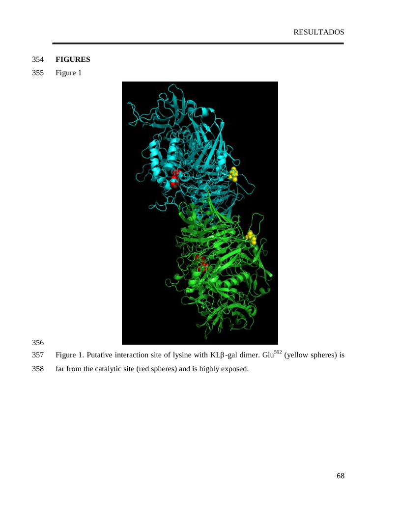
RESULTADOS
68
FIGURES 354
Figure 1 355
356
Figure 1. Putative interaction site of lysine with KL-gal dimer. Glu592
(yellow spheres) is 357
far from the catalytic site (red spheres) and is highly exposed. 358
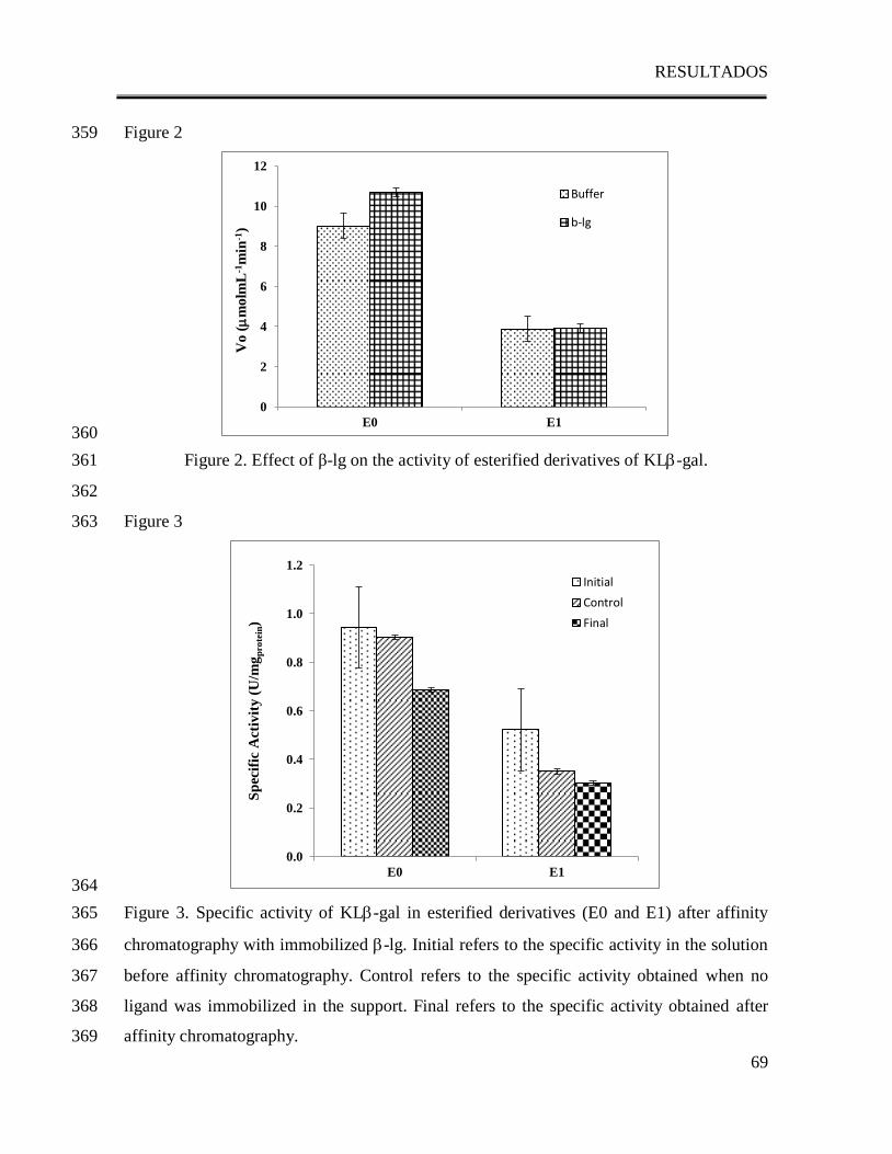
RESULTADOS
69
Figure 2 359
360
Figure 2. Effect of β-lg on the activity of esterified derivatives of KL-gal. 361
362
Figure 3 363
364
Figure 3. Specific activity of KL-gal in esterified derivatives (E0 and E1) after affinity 365
chromatography with immobilized -lg. Initial refers to the specific activity in the solution 366
before affinity chromatography. Control refers to the specific activity obtained when no 367
ligand was immobilized in the support. Final refers to the specific activity obtained after 368
affinity chromatography. 369
0
2
4
6
8
10
12
E0 E1
Vo (
molm
L-1
min
-1)
Buffer
b-lg
0.0
0.2
0.4
0.6
0.8
1.0
1.2
E0 E1
Sp
ecif
ic A
ctiv
ity (
U/m
gp
ro
tein
)
Initial
Control
Final
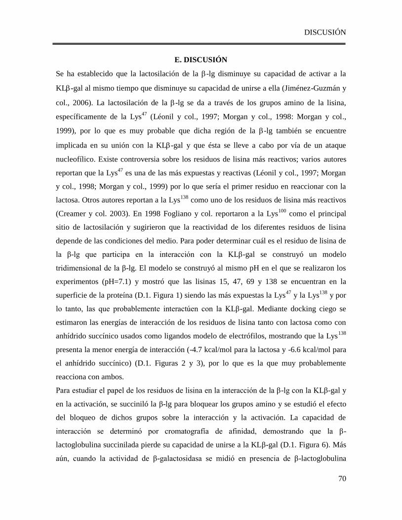
DISCUSIÓN
70
E. DISCUSIÓN
Se ha establecido que la lactosilación de la -lg disminuye su capacidad de activar a la
KL-gal al mismo tiempo que disminuye su capacidad de unirse a ella (Jiménez-Guzmán y
col., 2006). La lactosilación de la -lg se da a través de los grupos amino de la lisina,
específicamente de la Lys47
(Léonil y col., 1997; Morgan y col., 1998: Morgan y col.,
1999), por lo que es muy probable que dicha región de la -lg también se encuentre
implicada en su unión con la KL-gal y que ésta se lleve a cabo por vía de un ataque
nucleofílico. Existe controversia sobre los residuos de lisina más reactivos; varios autores
reportan que la Lys47
es una de las más expuestas y reactivas (Léonil y col., 1997; Morgan
y col., 1998; Morgan y col., 1999) por lo que sería el primer residuo en reaccionar con la
lactosa. Otros autores reportan a la Lys138
como uno de los residuos de lisina más reactivos
(Creamer y col. 2003). En 1998 Fogliano y col. reportaron a la Lys100
como el principal
sitio de lactosilación y sugirieron que la reactividad de los diferentes residuos de lisina
depende de las condiciones del medio. Para poder determinar cuál es el residuo de lisina de
la β-lg que participa en la interacción con la KLβ-gal se construyó un modelo
tridimensional de la β-lg. El modelo se construyó al mismo pH en el que se realizaron los
experimentos (pH=7.1) y mostró que las lisinas 15, 47, 69 y 138 se encuentran en la
superficie de la proteína (D.1. Figura 1) siendo las más expuestas la Lys47
y la Lys138
y por
lo tanto, las que probablemente interactúen con la KLβ-gal. Mediante docking ciego se
estimaron las energías de interacción de los residuos de lisina tanto con lactosa como con
anhídrido succínico usados como ligandos modelo de electrófilos, mostrando que la Lys138
presenta la menor energía de interacción (-4.7 kcal/mol para la lactosa y -6.6 kcal/mol para
el anhídrido succínico) (D.1. Figuras 2 y 3), por lo que es la que muy probablemente
reacciona con ambos.
Para estudiar el papel de los residuos de lisina en la interacción de la β-lg con la KLβ-gal y
en la activación, se succiniló la β-lg para bloquear los grupos amino y se estudió el efecto
del bloqueo de dichos grupos sobre la interacción y la activación. La capacidad de
interacción se determinó por cromatografía de afinidad, demostrando que la β-
lactoglobulina succinilada pierde su capacidad de unirse a la KLβ-gal (D.1. Figura 6). Más
aún, cuando la actividad de β-galactosidasa se midió en presencia de β-lactoglobulina
DISCUSIÓN
71
succinilada, se perdió el efecto activador (D.1. Figura 5) y la actividad permaneció igual a
la del control sin β-lactoglobulina. Debido a que la succinilación bloquea los grupos ε-
amino, lo anterior demuestra claramente que el bloqueo de los grupos ε-amino de la β-lg
disminuye las interacciones entre la β-lg y la KLβ-gal, provocando la pérdida del efecto
activador demostrando que los residuos de lisina son esenciales tanto para la interacción
como para la activación.
Los monómeros de la β-lg se pueden asociar para formar diferentes estructuras
oligoméricas y luego disociarse en monómeros en función del pH (Verheul et al., 1999; Fox
& McSweeney, 1998; Fox et al., 1998); el pH en el que realizaron los experimentos en este
trabajo promueve la formación del dímero de la β-lg. Los sitios potenciales de unión de la
β-lg con moléculas hidrofóbicas pequeñas se han estudiado ampliamente, y se ha
encontrado que estos se localizan en una región altamente hidrofóbica del monómero (Yang
y col. 2008a; Yang y col., 2008b, Kontopidis y col., 2002; Ragona y col. 2000; Wu y col.,
1999; Ragona y col., 1997). El sitio de unión para las moléculas hidrofóbicas puede variar
conforme el dímero se forma pero siempre se localiza en la región del cáliz de la β-lg,
mientras que el sitio de unión para moléculas hidrofílicas parece no ser tan específico
(Noiseux y col., 2002; Wenbing y col., 2011).
Se estudió el probable sitio de interacción del dímero de la β-lg con moléculas
electrofílicas. Para ello, se construyó un modelo del dímero con las condiciones en las que
se llevaron a cabo los experimentos (D.2. Figura 1) y se estudió por docking molecular
cuáles serían los aminoácidos con mejor probabilidad de participar en la interacción. Se
hicieron dockings ciegos del dímero de la β-lg con lactosa y anhídrido succínico utilizados
como ligandos modelo de electrófilos (D.2. Figura 2), la Lys138
de uno de los monómeros y
la Lys141
del otro, son los que muestran una mayor probabilidad de interactuar tanto con la
lactosa como con el anhídrido succínico (energías finales de docking, o EDF, de -3.30 y -
3.11 kcal/mol, respectivamente). Es importante señalar que tanto en el monómero como en
el dímero de la β-lg, los residuos con las mejores probabilidades de interactuar con los
ligandos son residuos de lisina y que en ambos casos la Lys138
participa en la interacción.
Lo anterior sugiere que los residuos de lisina son sumamente importantes en la interacción,
en especial la Lys138
. Más aún, los resultados obtenidos en el docking del dímero con los
ligandos sugieren que la formación del dímero de la β-lg es necesaria para fortalecer las
DISCUSIÓN
72
interacciones debido a la participación de la Lys138
de uno de los monómeros y la Lys141
del
otro. En esta región del dímero se observó que además de unirse a la Lys138
y a la Lys141
,
los electrófilos forman puentes de hidrógeno con otros aminoácidos cercanos y expuestos
de ambos monómeros (D.2. Figuras 2 y 3); esto podría fortalecer la interacción mediante el
acercamiento y la estabilización de los ligandos en una estructura tipo ―pinza‖, de tal
manera que el ataque nucleofílico pueda llevarse a cabo.
Debido a que tanto la estructura y actividad de la KLβ-gal, así como la formación del
dímero de la β-lg son dependientes del pH, se estudió también el efecto del pH en la
interacción de la β-lg con la KLβ-gal y en la actividad enzimática. Para ello se determinó el
% de activación de la KLβ-gal por la β-lg a diferentes valores de pH (D.3. Tabla 1); se
observó que la β-lg tiene un efecto activador sobre la KLβ-gal, el cual es más fuerte a pH
7.0 y completamente ausente a valores de pH de 6.0 y 8.5. Al estudiar el efecto del pH en la
interacción entre la β-lg y la KLβ-gal se observó un comportamiento similar (D.3. Figura
2): la interacción es mucho más fuerte a pH de 7.0 (70%), y la capacidad de interacción
disminuye gradualmente hasta desaparecer por completo a valores de pH de 6.0 y 8.5.
Cuando se comparan los cambios en la activación provocados por el pH con los cambios en
las interacciones entre la β-lg y la KLβ-gal se observa una correlación (D.3. Figura 3):
conforme la interacción entre la β-lg y la KLβ-gal es más fuerte, la activación aumenta. Se
sabe que a temperatura ambiente y valores de pH menores a 4.0 y mayores a 5.2, la β-lg
está formada principalmente por monómeros y dímeros. Alrededor de pH 4.7, se forman
estructuras oligoméricas más grandes tales como octámeros (Verheul et al., 1999). En
valores cercanos a un pH de 7.0 la -lg existe como un dímero (Fox et al., 1998). Al
comparar la estructura cuaternaria de la β-lg a diferentes valores de pH con su capacidad de
interactuar y activar a la KLβ-gal, se observó que el estado dimérico de la β-lg es esencial
tanto para la interacción como para la activación de la enzima. Lo anterior refuerza los
resultados obtenidos con el docking, pues explican la importancia del dímero al formar una
estructura tipo ―pinza‖ que estabilice la interacción con los ligandos. Por otro lado, al
caracterizar el efecto del pH en la capacidad de activación de la enzima por la β-lg, se
observó que a pH 7.0, en el que la interacción es más fuerte, el efecto activador de la β-lg
dio lugar a una actividad más alta aún que la encontrada en el pH óptimo en solución
amortiguadora (142%). Este resultado sugiere que la interacción entre ambas proteínas
DISCUSIÓN
73
podría ayudar a alcanzar una conformación de la KLβ-lg aún más activa, desplazando el pH
óptimo de 7.5 (solo solución amortiguadora), a 7.0 (en presencia de β-lg); a pH 7.0 la fuerte
interacción con la β-lg promueve un estado conformacional de la KLβ-gal en el que el sitio
activo es más accesible para el sustrato.
Dado que la interacción entre ambas proteínas es esencial para la activación, es importante
conocer qué región de la KLβ-gal podría ser la responsable de la interacción con la β-lg. En
vista de que se ha encontrado que los grupos ε-amino de la β-lg son esenciales tanto para la
interacción como para la activación de la KLβ-gal, puede asumirse que la interacción
tendría que suceder a través de grupos electrofílicos tales como los grupos carboxilo de la
enzima. Para comprobarlo, se estudió el papel de los grupos carboxilo de la KLβ-gal tanto
en la interacción con la β-lg como en el efecto activador. Para esto se construyó un modelo
tridimensional de la KLβ-gal y se hizo un docking molecular con lisina (la contraparte por
el lado de la β-lg); este docking mostró más de 250 posibles sitios de interacción. Para
reducir el número de posibilidades se empleó la siguiente estrategia: dado que al pH de
estudio la KLβ-gal es un dímero, se construyó un modelo del dímero de KLβ-gal y se
localizaron los aminoácidos que el docking del monómero había resaltado. Después de
excluir a los aminoácidos carboxílicos que están involucrados en la formación del dímero y
a los que forman parte del sitio activo de la enzima, se observó que sólo unos cuantos se
encuentran expuestos y por lo tanto podrían interactuar con la β-lg. El docking ciego de la
KLβ-gal con lisina mostró que el aminoácido con la menor energía de interacción es el
Glu592
(EFD=-4.5kcal/mol) (D.4. Figura 1). Los grupos carboxilo de las proteínas pueden
ser modificados químicamente por medio de su esterificación gradual con alcoholes de bajo
peso molecular adicionando concentraciones bajas de iones hidrógeno para catalizar la
reacción en condiciones suaves (Feeney, 1977). Para estudiar el papel de los aminoácidos
carboxílicos más expuestos de la KLβ-gal se bloquearon estos grupos por medio de una
reacción de esterificación. Al esterificar los grupos carboxilo más expuestos y reactivos de
la KLβ-gal, entre los cuales se encuentra el Glu592
, se observó la pérdida de la capacidad de
la enzima para unirse a la β-lg (D.4. Figura 3). Además, al determinar la actividad de la
enzima esterificada en presencia de β-lg la actividad no mostró diferencia significativa con
el control (D.4. Figura 2), sugiriendo que los grupos carboxilo de la KLβ-gal son esenciales
DISCUSIÓN
74
tanto para la capacidad de la enzima para interactuar con la β-lg como para poder ser
activada por ésta.
CONCLUSIONES
75
F. CONCLUSIONES
El hecho de que no se detectara interacción ni activación al bloquearse los grupos ε-
amino de la β-lg, sugiere que estos grupos podrían estar involucrados en la habilidad de la
proteína para unirse a la enzima y por lo tanto activarla. Los resultados obtenidos en este
trabajo mostraron que los sitios de interacción encontrados para el dímero de la β-lg y el
anhídrido succínico o lactosa, son consistentes con aquellos encontrados para el monómero.
Sin embargo, a pesar de que diversos estudios señalan a la Lys47
, como el principal residuo
involucrado en la reacción con lactosa, este estudio sugiere que en el caso del dímero, la
interacción con moléculas altamente electrofílicas como la lactosa y el anhídrido succínico
sucede más probablemente a través de la Lys138
. Además, se demostró que la formación del
dímero de la β-lg es necesaria para fortalecer las interacciones debido a la participación de
la Lys138
de uno de los monómeros y la Lys141
del otro: conforme el dímero se forma las
regiones cercanas a la Lys138
y a la Lys141
, podrían formar una estructura tipo ―pinza‖ que
podría establecer interacciones fuertes a través de varios enlaces de hidrógeno y
electrofílicos que también podrían explicar las interacciones con moléculas más grandes
como la KLβ-gal.
Por otro lado, también se demostró que la presencia de la β-lg tiene un efecto
activador sobre la KLβ-gal que es más fuerte a pH 7.0 y que desaparece a valores de pH de
6.0 y 8.5, mostrando que el efecto activador es dependiente del pH. Cuando se compararon
los cambios en la activación ocasionados por el pH con los cambios en las interacciones
entre la β-lg y la KLβ-gal, se observó una correlación directa: conforme la interacción entre
β-lg y la KLβ-gal es más fuerte, la activación aumenta. Es más, cuando la estructura
oligomérica de la β-lg se comparó con los valores de pH en los que se encontró tanto
interacción como activación, se observó que el estado dimérico de la β-lg es esencial tanto
para la interacción como para la activación de la enzima. El hecho de que, a pH 7.0 en
presencia de β-lg la actividad observada fue significativamente mayor que la encontrada en
el pH óptimo en solución amortiguadora, sugiere que la interacción entre ambas proteínas
podría ayudar a alcanzar una conformación de la KLβ-gal aún más activa, desplazando el
pH óptimo de 7.5 en solución amortiguadora a 7.0 en presencia de la proteína, sugiriendo
que a este pH, la β-lg promueve un estado conformacional de la KLβ-gal en el cual, el sitio
activo es más accesible para el sustrato.
CONCLUSIONES
76
También se construyó un modelo tridimensional para la KLβ-gal, y después de
excluir los aminoácidos carboxílicos que están involucrados en la formación del dímero, se
observó que sólo unos cuantos aminoácidos están expuestos y que probablemente podrían
interactuar con la β-lg. El docking ciego de la KLβ-gal con lisina mostró que el aminoácido
más expuesto y reactivo que podría estar involucrado en la interacción con la β-lg es el
Glu592
(EFD=-4.5kcal/mol). La esterificación de los grupos carboxilo de la KLβ-gal más
expuestos y reactivos, entre los cuales se encuentra el Glu592
, dio lugar a la pérdida de la
capacidad de la enzima para unirse a la β-lg. Cuando se determinó la actividad de la KLβ-
gal esterificada en presencia de la β-lg ésta no mostró diferencia significativa con el
control. Al comparar los cambios en la actividad ocasionados por la esterificación de la
KLβ-gal con los cambios en las interacciones entre la β-lg y la KLβ-gal se observó una
correlación directa: mientras existen grupos carboxilo disponibles para la interacción con la
β-lg, no hay diferencia significativa de la actividad con respecto al control; esto sugiere que
los grupos carboxilo de la KL β-gal son esenciales tanto para la capacidad de la enzima
para interactuar con la β-lg como para su capacidad de ser activada por ésta.
Todo lo anterior sugiere que la interacción con la β-lg podría inducir un cambio
conformacional de la KLβ-gal en el que el sitio activo sea más accesible para el sustrato y
por lo tanto la actividad aumente.
REFERENCIAS
77
H. REFERENCIAS
Amiot, J., Ciencia y Tecnología de la Leche Principios y aplicaciones, Acribia,
Zaragoza, España (1991), 18-37.
Becerra, M., Cerdán, E., & González-Siso, M. I. (1998). Micro-scale purification of
Kluyveromyces lactis reveals that dimeric and tetrameric forms are active.
Biotechnoogy Techniques, 12, 3, 253-256.
Bell, K., & McKenzie, H. A. (1964). -Lactoglobulins. Nature, 204, 1275-1279.
Blackburn, S., Carter, E. G. H., & Phillips, H. (1941). The methylation of wool with
methyl sulphate and methyl halides. Biochemical Journal, 35, 627-639.
Blackburn, S. & Phillips, H. (1944). Experiments on the methylation and acetylation of
wool, silk fibroin, collagen and gelatin. Biochemical Journal, 38, 171-178.
Blanch, E. W., Hecht, L., & Barron, L. D. (1999). New insight into the pH- dependent
conformational changes in bovine -lactogloulin from Raman optical activity. Protein
Science, 8,1362-1367.
Bradford, M. M. (1976). A rapid and sensitive method for the quantification of
microgram quantities of protein utilizing the principle of protein-dye binding.
Analytical Biochemistry, 72, 248-254.
Brignon, G., Chtorou, A., & Ribadeau-Dumas, B. (1985). Does -lactoglobulin occur
in human milk? Journal of Dairy Research, 52, 249-254.
Brownlow, S., Cabral, J. H. M., Cooper, R., Flower, D. R., Yewdall, S. J., Polikarpov,
I., North, A. C. T., & Sawyer, L. (1997). Bovine -lactoglobulin at 1.8 Ǻ resolution–
still an enigmatic lipocalin. Structure, 5, 481-495.
Chen, J. Y. & Tsen, H. Y. (1991). Effect of milk and milk constituents on heat stability
of lactase from Saccharomyces lactis. Journal of the Chinese Agricultural Chemical
Society, 29, 456-464.
Collin, M., D’Alfonso, L., &Baldini, G. (2000). New insight on -lactoglobulin bindig
sites by 1-anilinonaphtalene-8-sulfonate fluorescence decay. Protein Science, 9, 1968-
1974.
REFERENCIAS
78
Creamer, L.K.; Sawyer, L. Beta lactoglobulin. In: Encyclopedia of Dairy Sciences;
Roginski, H.; Fuquay, J.W.; Fox, P.F., Eds.; Academic Press: London UK, 2003; Vol
3, pp 1932-1938.
Creighton, T. E. (1989). Protein Structure A Practical Approach, IRL Press, Oxford,
Great Britain, 148-153.
Del Moral-Ramírez, E., Domínguez-Ramírez, L., Cruz-Guerrero, A. E., Rodríguez-
Serrano, G. M., García-Garibay, M., Gómez-Ruiz, L., & Jiménez-Guzmán, J. (2008).
Role of Lysine -Amino Groups of -Lactoglobulin on Its Activating Effect of
Kluyveromyces lactis -Galactosidase. Journal of Agricultural and Food Chemistry,
56, 5859-5863.
Eswar, N., Marti-Renom, M. A., Webb B., Madhusudhan, M. S., Eramian, D., Shen,
M., Pieper, U., & Sali, A. (2006). Comparative Protein Structure Modeling With
MODELLER. Current Protocols in Bioinformatics, John Wiley & Sons, Inc.,
Supplement 15, 5.6.1-5.6.30.
Feeney, R. E. (1997). Chemical modifications of food proteins. In Feeney, R. E. &
Whitaker, J. R. (Eds.), Food proteins: Improvements through chemical end enzymatic
modification. Advances through chemical and enzymatic modification (pp 3-3b).
Washington, DC: American Chemical Society.
Fennema, O. R., Química de los Alimentos, Acribia, Zaragoza, España (2000), 204-
206, 394-397.
Fiser, A., Do, R.K., & Sali, A. (2000). Modeling of loops in protein structures, Protein
Science, 9, 1753-1773.
Fisher, A. y Sali, A. (2003). Modeller: Generation and Refinement of Homology-Based
Protein Structure Models. Methods in Enzymology, 374, 461-491.
Flower, D. R., North, A. C. T., & Sansom, C. E. (2000). The lipocalin protein family:
structural and sequence overview. Biochimica et Biophysica Acta, 1482, 9-24.
Fogliano, V., Monti, M. S., Visconti, A., Randazzo, G., Facchiano, M. A., Colonna, G.,
& Ritienti, A. (1998). Identification of a beta-lactoglobulin lactosylation site.
Biochimica et Biophysica Acta, 1338, 295-304.
REFERENCIAS
79
Fogolari, F., Ragona, L., Licciardi, S., Romagnoli, S., Michelutti, R., Ugolini, R., &
Molinari, H. (2000). Electrostatic properties of bovine -lactoglobulin. Proteins:
Structure Function and Bioinformatics, 39, 4, 317-330.
Fox, P. F. (2003). Milk proteins: general and historical aspects. In P. F. Fox, & P. L. H.
McSweeney (Eds.), Advanced Dairy Chemistry Volume 1 (pp 1-48). New York:
Kluwer Academic/Plenum Publishers.
Fox, P. F., McSweeney, P. L. H., Cap. 9: Heat induced changes in milk, Dairy
Chemistry and Biochemistry, Blackie Academic and Professional Press, Reino Unido
(1998), 21-39, 146-150, 188-192.
Fraenkel-Conrat, H. L. & Olcott, H. S. (1945). Esterification of proteins with alcohols
of low molecular weight. The Journal of Biological Chemistry, 161, 259-268.
French, S. J., Harper, W. J., Kleinholz, N. M., Jones, R. B. y Green-Church, K. B.,
―Maillard Reaction Induced Lactose Attachment to Bovine -lactoglobulin:
Electrospray Ionization and Matrix-Assisted Laser Desorption/Ionization
Examination‖, J. Agric. Food Chem., 50 (2002), 820-823.
García Garibay, M. & Gómez Ruiz, L. (1996). Usos de β-galactosidasas microbianas
para reducir el contenido de lactosa en leche y productos lácteos. Revista de
Investigación Clínica, 48 (supl.), 51-61.
García-Garibay, M.. (1992). Recuperación de enzimas intracelulares de interés
industrial. -Galactosidasa de levadura. Ciencia (Mex.), 43, 23-33.
García-Garibay, M., Gómez-Ruíz, L., Revah Moissev, S. (1993). Biotecnología de
productos lácteos. In García-Garibay, M., Quintero Ramírez, R. y López- Munguía, A.,
Biotecnología Alimentaria. Limusa, México, Cap. 9, 153-161.
Gekas, V. & Lopez-Leiva, M. (1985). Hydrolysis of lactose: a literature review.
Process Biochemistry, 20, 2-12.
German, J. H. (1997). Applied enzymology of lactose hydrolysis. Milk Powders for the
Future, 81-87.
Greenberg, N. A. & Mahoney, R. R. (1984). The activity of lactase (Streptococcus
thermophilus) in milk and sweet whey. Food Chemistry, 15, 307-313.
REFERENCIAS
80
Halpin, M. I., & Richardson, T. (1985). Selected Functionality Changes of -
Lactoglobulin upon Esterification of Side-Chain Carboxyl Groups.Journal of Dairy
Science, 68, 3189-3198.
Hetényi, C. & van der Spoel, D. (2002). Efficient docking of peptides to prteins
without prior knowledge of the binding site. Protein Science, 11, 1729-1737.
Hollecker, M. & Creighton, T. E. (1982). Effect on Protein Stability of Reversing the
Charge on Amino Groups. Biochimica et Biophysica Acta, 701, 395-404.
Hollecker, M. & Creighton, T.E. (1982). Counting integral numbers of amino groups
per polypeptide chain. FEBS Letters, 119, 187-189.
Jiménez-Guzmán, J., Cruz-Guerrero, A.E., Rodríguez-Serrano, G., López-Munguía,
A., Gómez-Ruiz, L., & García-Garibay, M. (2002). Enhancement of lactase activity in
milk by reactive sulfhydryl groups induced by heat treatment. Journal of Dairy
Science, 85, 2497-2502.
Jiménez-Guzmán, J., Sarabia-Leos, C., Cruz-Guerrero, A., Rodríguez-Serrano, G.,
López-Munguía, A., Gómez-Ruiz, L., & García-Garibay, M. (2006). Interaction
between β-lactoglobulin and lactase and its effect on enzymatic activity. International
Dairy Journal, 16, 1169-1173.
Juers, D. H., Heightman, T. D.,Vasella, A.,McCarter, J. D.,Mackenzie, L.,Withers, S.
G.,& Matthews, B. W. (2001). A structural view of the action of Escherichia coli
(lacZ) beta-galactosidase. Biochemistry, 40, 14781-14794
Krieger, E., Nabours, S. B., & Vriend, G. (2003). In Bourne, P. E y Weissig, H., Cap.
25. Homology Modeling, Structural Bioinformatics. Wiley-Liss, Inc.
Kuwata, K., Hoshino, M., Forge, V., Era, S., Batt, C.A. & Goto, Y. (1999). Solution
structure and dynamics of bovine beta-lactoglobulin A. Protein Science, 8, 2541-2545.
Léonil, J., Mollé, D., Fauquant, J., Maubois, J. L., Pearce, R. J., & Bouhallab, S.
(1997). Characterization by Ionization Mass Spectrometry of Lactosyl β-Lactoglobulin
Conjugates Formed During Heat Treatment of Milk and Whey and Identification of
One Lactose-Binding Site. Journal of Dairy Science, 80, 2270-2281.
REFERENCIAS
81
López, P., Rosado, J. L., Palma, M., González, C. & Valencia, M. (1996). Mala
digestión de lactosa. Su definición, su prevalencia en México y sus implicaciones en el
consumo de la leche. Revista de Investigación Clínica, 48 (supl.), 15-22.
Louquet, M. F., Bonjean-Linczowsky, Y., Keilling, J. & Wilde, R. (1991). In Leche y
Productos lácteos Vaca-Oveja-Cabra. Ed.: Acribia: España, 11-25, 53-56.
Lowry, O., Rosebrough, N., Farr, A., & Randall, R. (1951). Protein measurement with
the Folin phenol reagent, Biol. Chem., 193, 265.
Mahoney, R. R. (1997). Lactose: Enzymatic modification. In Advanced Dairy
Chemistry, 2nd
ed., Fox, P. F., Ed.: Chapman & Hall: London, Vol. 3, p 95.
Mandrich, L., Caputo, E., Martin, B. M., Rossi, M., & Manco, G.. ―The Aes protein
and the monomeric b-galactosidase from Escherichia coli form a non-covalent
complex‖, J. Biol. Chem., 277 (2002), 48241–48247.
Marti-Renom, M.A., Stuart, A., Fiser, A., Sánchez, R., Melo, F., & Sali, A. (2000).
Comparative protein structure modeling of genes and genomes. Annual Review of
Biophysics and Biomolecular Structure, 29, 291-325.
Mc Murry, J. (2001). In Química Orgánica, Ed.: Thomson: México, pp1057.
Monaco, H. L, Zanotti, G., Spadon, P., Bolognesi, M., Sawyer, L., & Eliopoulos, E. E.
(1987). Crystal structure of the trigonal form of bovine beta-lactoglobulin and of its
complex with retinol at 2.5 Å resolution. Journal of Molecular Biology. 197, 695-706.
Monti, J. C., Mermoud, A. F., Jolles, P. (1989). Antibovine -lactoglobulin antibodies
react with a human lactoferrin fragment and bovine -lactoglobulin present in human
milk. Experientia, 45, 178-180.
Morgan, F., Bouhallab, S., Henry, G., Maubois, J. L. & Léonil, J. (1998). Lactolation
of -lactoglobulin Monitored by Electrospray Ionisation Mass Spectrometry.
International Dairy Journal, 8, 95-98.
Morgan, F., Gwénaële, H., Le Graët, Y., Mollé, D., Léonil, J. & Bouhallab, S. (1999a).
Resistance of -lactoglobulin-bound Lactose to the HydroLysis by -galactosidase.
International Dairy Journal, 9, 813-816.
Morgan, F., Mollé, D., Henry, G., Vénien, A., Léonil, J., Peltre, G., Levieux, D.,
Maubois, J. L. & Bouhallab, S. (1999b). Glycation of Bovine -lactoglobulin: effect on
REFERENCIAS
82
the Protein Structure. International Journal of Food Science & Technology., 34, 429,
435.
Morgan, F., Vénien, A., Bouhallab, S., Mollé, D., Léonil, J., Peltre, G. & Levieux, D.
(1999). Modification of Bovine -lactoglobulin by Glycation in a Powered State or in
an Aqueous Solution: Immunochemical Characterization. Journal of Agricultural and
Food Chemistry, 47, 4543, 4548.
Nijipels, H. H. (1991). Lactases and their applications. In Enzyme and Food
Processing. Applied Science Publishers: London, 89-104.
Noiseux, I., Gauthier, S. L., & Turgeon, S. (2002). Interactions between Bovine -
lactoglobulin and Peptides under Different Physicochemical Conditions.Journal of
Agricultural and Food Chemistry, 50, 1587-1592.
Ortiz-Chao, P. A., ―Estudio de la interacción de las proteínas del suero de leche con la
-galactosidasa de Kluyveromyces lactis y su efecto en la actividad‖ (Tesis de
licenciatura), Universidad Nacional Autónoma de México, México (2004), 48-80.
Papiz, M. Z., Sawyer, L., Eliopoulos, E. E., North, A. C. T., Findlay, J. B. C.,
Sivaprasadarao, R., Jones, T. A., Newcomer, M. E., & Kraulis, P. J. (1986). The
structure of -lactoglobulin and its similarity to plasma retinol-binding protein. Nature,
324, 383-385.
Qin, B. Y., Bewley, M. C., Creamer, L. K., Baker, H. M., Baker, E. N., & Jameson, G.
B. (1998). Structural Basis of the Tanford Transition of Bovine -Lactoglobulin.
Biochemistry, 37, 14014-14023.
Ragona, L., Fogolari, F., Zetta., L., Perez, D. M., Puyol, P., De Kruif, K., Lohr, F.,
Ruterjans, H., & Molinari, H. (2000). Bovine beta-lactoglobulin: interaction studies
with palmitic acid. Protein Science, 9, 1347-1356.
Ragona, L., Pusterla, F., Zetta, L., Monaco, H. L., & Molinari, H. (1997). Identification
of a conserved hydrophobic cluster in partially folded bovine beta-lactoglobulin at pH
2. Folding and Design, 2, 281-290.
Sali, A. & Blundell, T.L. (1993). Comparative protein modelling by satisfaction of
spatial restraints. Journal of Molecular Biology, 234, 779-815.
REFERENCIAS
83
Sawyer, L., Barlow, P. N., Boland, M. J., Creamer. L. K.,Helen D., Edwards, P. J.B.,
Holt, C., Jameson, G. B., Kontopidis, G., Norris, G, E., Uhrínová, S. & Wu, S.
(2002b). Erratum to: Milk protein structure- what can it tell the dairy industry?
International Dairy Journal, 12, 709.
Spreer, E., Lactología Industrial. Leche Preparación y elaboración. Máquinas,
Instalaciones y aparatos. Productos lácteos, traducción de Oscar Dignoes Torres-
Quevedo, Acribia, España (1991), 11-21.
Tello-Solis, S., Jiménez-Guzmán, J., Sarabia-Leos, C., Gómez Ruiz, L., Cruz-
Guerrero, A. E., Rodríguez-Serrano, G., & García-Garibay, M. (2005). Determination
of the Secondary Structure of Kluyveromyces lactis -Galactosidase and its Structure-
Activity Relationship as a Function of the pH. Journal of Agricultural and Food
Chemistry, 53, 10200-10204.
Uhrinova, S., Smith, M. H., Jameson, G. B., Uhrin, D., Sawyer, L., & Barlow, P. N.
(2000). Structural changes accompanying pH-induced dissociation of the -
lactoglobulin dimer. Biochemistry, 39, 3565-3574.
Varnam, A. H. & Sutherland, J. P. (1994). In Milk and milk Products Technology,
Chemistry and Microbiology. Chapman and Hall: Great Britain, 1-10
Verheul, M., Pedersen, J. S., Roefs, S. P. F. M., & Kruif, K. G. (1999). Association
behavior of native -lactoglobulin. Biopolymers, 49, 11-20.
Walstra, P., Geurts, T. J., Noomen, A., Jellema, A. & van Boekel, M. A. J. S. (2001).
In Ciencia de la leche y tecnología de los productos lácteos. Ed.: Acribia: España, pp
39-43, 91-94, 109-114.
Walstra, P., Jennes, J. (1984). In Química y Física Lactológica. Ed.: Acribia: España,
pp 84-105.
Wenbing, H., Jianan, L., Qun, L., Yumiao, H., Kui, W., Shuang, L., Shaoxiang, X., &
Fuyi, W. (2011). Elucidation of the binding sites of sodium dodecil sulfate to -
lactoglobulin using hydrogen/deuterium exchange mass spectrometry combined with
docking simulation. Mass Spectrometry, 25, 1429-1436.
REFERENCIAS
84
Wendorf, W. L., Amundsen, C. H., y Olson, N. F., & Garver, J. C. (1971). Use of yeast
-galactosidase in milk and milk products Journal of Milk and Food Technology, 34, 6,
294.
Wendorf, W. L., Amundson, C. C., y Olson, N. F. (1970). The effect of heath treatment
of milk upon the hydrolyzability of lactose by the enzyme lactase. Journal of Milk and
Food Technology, 22, 9, 377-379.
Yang, M. C., Guan, H. H., Liu, M.Y., Lin, Y. H., Yang, J. M., Chen, W. L., Chen, C.
J., & Mao, S. J. T. (2008a). Crystal Structure of a secondary vitamin D3 binding site of
milk beta-lactoglobulin. Proteins, 71, 3, 1197-1210.
Yang, M. C., Guan, H. H., Yang, J. M., Ko, C. N., Liu, M.Y., Lin, Y. H., Huang, Y. C.,
Chen, C. J., & Mao, S. J. T. (2008b). Rational Design for Crystallization of -
Lactoglobulin and Vitamin D3 Complex: revealing a Secondary Binding Site. Crystal
Growth and Design, 8, 12, 4268-4276.
Zhou, Q. Z. K. & Chen, X. D. (2001). Effects of temperature and pH on the catalytic
activity of the immobilized -galactosidase from Kluyveromyces lactis. Biochemical
Engineering Journal, 9, 33-40.
Recommended