Embed Size (px)
Citation preview
Encephalitozoonosis (Nosematosis)of the cornea
N. ASHTON AND P. A. WIRASINHAFrom the Department of Pathology, Institute of Ophthalmology, University of London, and the EyeHospital, Colombo, Ceylon
Infection of mammals by protozoa of the genus Nosema, of the class Microsporidea of thesubphylum Cnidospora (Fig. I) which are characterized by the possession of polarfilaments, was first described in the brain and kidneys of a rabbit by Wright and Craighead(1922), and named Encephalitozoon cunicudi by Levaditi, Nicolau, and Schoen (1923). Sincethat time the parasite has been found in every major group of animals, including other pro-tozoa, helminths, insects, fish, amphibians, reptiles, and possibly birds (cf. Shadduck andPakes, I97I), but only a single acceptable example of human infection has been reported(Matsubayashi, Koike, Mikata, Takei, and Hagiwara, I959). The patient was a 9-year-oldJapanese boy who had developed meningo-encephalitis (the eyes were said to be normal);oval bodies resembling Nosema cuniculi were found in the cerebrospinal fluid and urine andthese organisms were propagated in mice. While this diagnosis was thought to be beyondreasonable doubt by Petri (i969), it had been questioned by others on the grounds thatmice (which also harbour microsporidia) were used for inoculation (Innes, Zeman, Frankel,and Borners, I962).
CNIDOSPORA
Superclass
Class MYXOSPORIDEA MICROSPORIDEA FIG. I
Subclass
Order 3 MICROSPORIDIANosema
The present case, wherein the cornea was invaded by microsporidia which on morpho-logical grounds were classified as Nosema, is therefore of exceptional rarity and of interest inbeing the first report of ocular involvement in man.The literature up to recent times is fully covered by Petri (I969) and Shadduck and
Pakes (I971), who also discuss the classification of the parasite. Until the early I960sthe original name of Encephalitozoon cuniculi persisted, but Nosema cuniculi was then proposedas more appropriate (Lainson, Garnham, Killick-Kendrick, and Bird, I964). Shadduckand Pakes (197), however, found marked ultrastructural differences between the micro-sporidian parasite of the rabbit and that of Nosema bombycis (the type species for the genusNosema, and the cause of silk-worm disease) and, therefore, suggested a return to theoriginal name of Encephalitozoon cuniculi (see also Sprague and Vernick, 1971). Forconvenience we shall continue to use the shorter name in this report.
Received for publication March 6, 1973Address for reprints: Prof. N. Ashton, F.R.S., Institute of Ophthalmology, Judd St., London WCiH gQSThis case was first reported at a meeting of the Section of Ophthalmology, Royal Society of Medicine, London, on June 14, 1973.
Brit. _7. Ophthal. (1973) 57, 669copyright.
on April 24, 2020 by guest. P
rotected byhttp://bjo.bm
j.com/
Br J O
phthalmol: first published as 10.1136/bjo.57.9.669 on 1 S
eptember 1973. D
ownloaded from
6N. Ashton and P. A. Wirasinha
The spores are oval, uninucleate, thick-walled, refractile, measure 2 *7 to 3 o0 m. X I *2 toi 8 gm., and contain a spirally coiled tubular filament and a prominent polar vacuole.They stain positively with Gram's stain, Carbol Fuchsin, and Weil-Weigert stains, whileGiemsa stain shows only a dark blue nucleus lying centrally or forming a bar across thespore. They have features in common with Toxoplasma, subphylum Sporozoa, with which theyhave been confused, but they may readily be distinguished by their morphology, stainingreactions, immunology, and pathogenicity; these differences have been described amongothers by Attwood and Sutton (i 965), Matsubayashi and others (I 959), and Innes andothers (i 962).
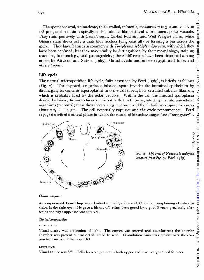
Life cycleThe normal microsporidian life cycle, fully described by Petri (i969), is briefly as follows(Fig. 2). The ingested, or perhaps inhaled, spore invades the intestinal epithelium bydischarging its contents (sporoplasm) into the cell through its extruded tubular filament,which is probably fired by the polar vacuole. Within the cell the injected sporoplasmdivides by binary fission to form a schizont with 2 to 6 nuclei, which splits into unicellularorganisms (meronts); these then secrete a rigid capsule and the fully-formed spore measuresabout 2 5 X I *5 jm. The cell eventually ruptures and the cycle recommences. Petri(I969) described a sexual phase in which the nuclei of binuclear stages fuse ("autogamy").
5chi7o,on,vSpor o,,onv
Spore Sporop1is/ll e ?
Plasnmodium '
It 1^w FIG. 2 Life cycle of Nosema bombycis(Q V ( jJ Z (adaptedfrom Fig. 3: Petri, 1969)
Autogamy
Case report
An ux-year-old Tamil boy was admitted to the Eye Hospital, Colombo, complaining of defectivevision in the right eye. He gave a history of having been gored by a goat 6 years previously afterwhich the right upper lid was sutured.
Clinical examination
RIGHT EYE
Visual acuity was perception of light. The cornea was scarred and vascularized; the anteriorchamber was present but no details could be seen. Granulation tissue was present over the con-junctival surface of the upper lid.
LEFT EYE
Visual acuity was 6/6. Follicles were present in both upper and lower conjunctival fornices.
670copyright.
on April 24, 2020 by guest. P
rotected byhttp://bjo.bm
j.com/
Br J O
phthalmol: first published as 10.1136/bjo.57.9.669 on 1 S
eptember 1973. D
ownloaded from
Encephalitozoonosis (Nosematosis) of the cornea
BLOOD PRESSURE
Normal.
Laboratory investigationsBLOOD
Examination for Brucellosis and Microfilaria-negative.
BIOPSY
Granulation tissue from the right upper lid showed follicular hyperplasia and reactive proliferation ofconnective tissue. The appearances suggested trachoma.
Operation3 weeks later a right penetrating keratoplasty was performed and the corneal disc was sent to theInstitute of Ophthalmology, London, for report.
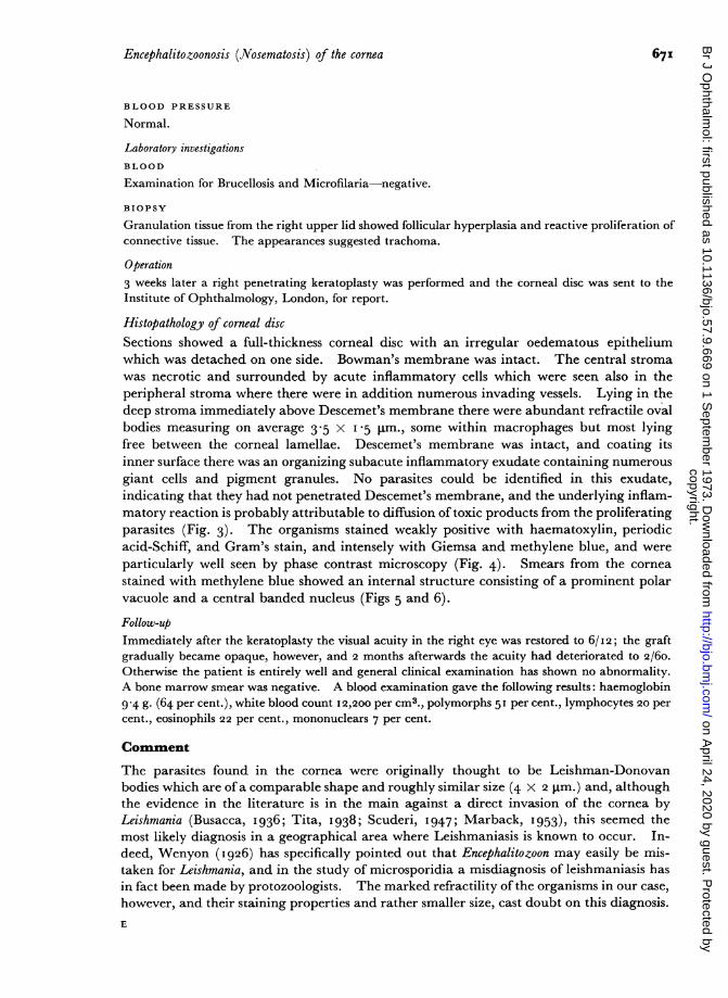
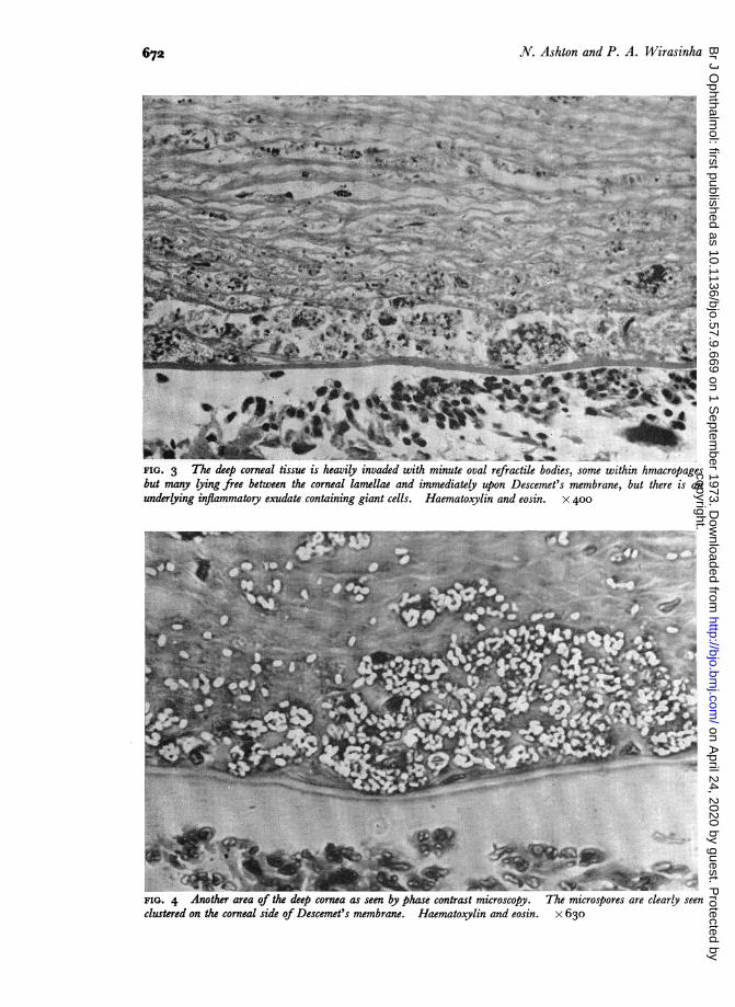
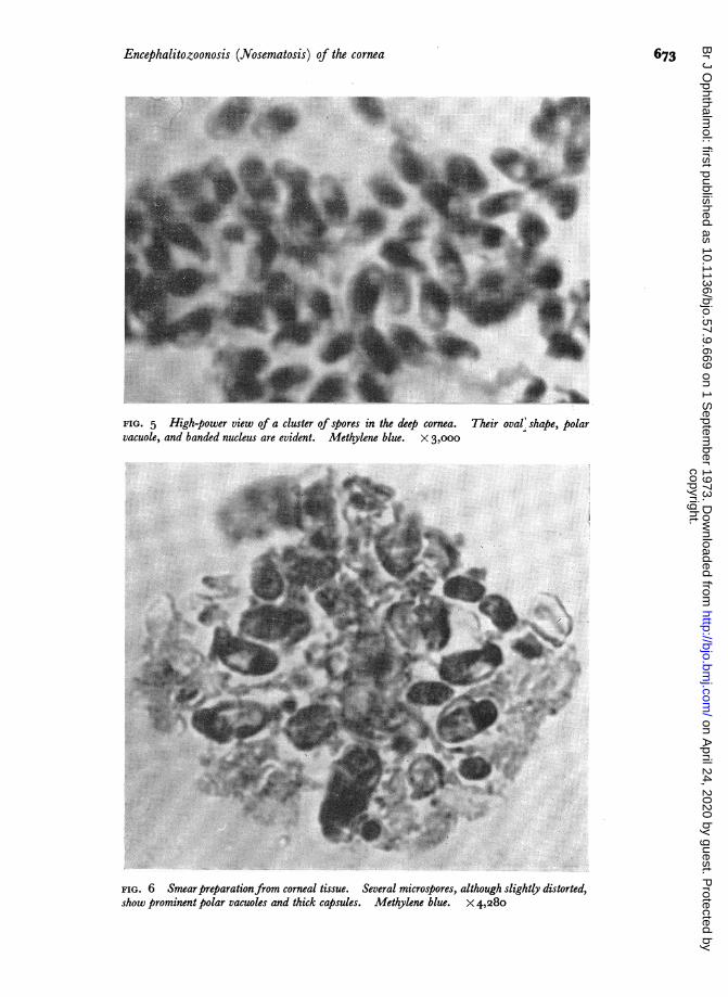
Histopathology of corneal discSections showed a full-thickness corneal disc with an irregular oedematous epitheliumwhich was detached on one side. Bowman's membrane was intact. The central stromawas necrotic and surrounded by acute inflammatory cells which were seen also in theperipheral stroma where there were in addition numerous invading vessels. Lying in thedeep stroma immediately above Descemet's membrane there were abundant refractile ovalbodies measuring on average 3-5 X I5 gim., some within macrophages but most lyingfree between the corneal lamellae. Descemet's membrane was intact, and coating itsinner surface there was an organizing subacute inflammatory exudate containing numerousgiant cells and pigment granules. No parasites could be identified in this exudate,indicating that they had not penetrated Descemet's membrane, and the underlying inflam-matory reaction is probably attributable to diffusion of toxic products from the proliferatingparasites (Fig. 3). The organisms stained weakly positive with haematoxylin, periodicacid-Schiff, and Gram's stain, and intensely with Giemsa and methylene blue, and wereparticularly well seen by phase contrast microscopy (Fig. 4). Smears from the corneastained with methylene blue showed an internal structure consisting of a prominent polarvacuole and a central banded nucleus (Figs 5 and 6).
Follow-upImmediately after the keratoplasty the visual acuity in the right eye was restored to 6/I2; the graftgradually became opaque, however, and 2 months afterwards the acuity had deteriorated to 2/60.Otherwise the patient is entirely well and general clinical examination has shown no abnormality.A bone marrow smear was negative. A blood examination gave the following results: haemoglobin9 4 g. (64 per cent.), white blood count I2,200 per cm3., polymorphs 51 per cent., lymphocytes 20 percent., eosinophils 22 per cent., mononuclears 7 per cent.
Comment
The parasites found in the cornea were originally thought to be Leishman-Donovanbodies which are ofa comparable shape and roughly similar size (4 X 2 jim.) and, althoughthe evidence in the literature is in the main against a direct invasion of the cornea byLeishmania (Busacca, 1936; Tita, I938; Scuderi, I947; Marback, I953), this seemed themost likely diagnosis in a geographical area where Leishmaniasis is known to occur. In-deed, Wenyon (1926) has specifically pointed out that Encephalitozoon may easily be mis-taken for Leishmania, and in the study of microsporidia a misdiagnosis of leishmaniasis hasin fact been made by protozoologists. The marked refractility of the organisms in our case,however, and their staining properties and rather smaller size, cast doubt on this diagnosis.E
671
copyright. on A
pril 24, 2020 by guest. Protected by
http://bjo.bmj.com
/B
r J Ophthalm
ol: first published as 10.1136/bjo.57.9.669 on 1 Septem
ber 1973. Dow
nloaded from
672 N. Ashton and P. A. Wirasinha
s..XSXe^q<RO.ist-l~~~~~~~~~~~~~~~~~~~~~~~~.<D . .....gse
ntf,,, i iEN; r& f 6 ^
FIG. 3 T-he deep corneal tissue is heavily invaded with minute oval refractile bodies, some within hmacropagesbut many lying free between the corneal lamellae and immediately upon Descemet's membrane, but there is anunderlying inflammatory exudate containing giant cells. Haematoxylin and eosin. x 400
FIG. 4 Another area of the deep cornea as seen by phase contrast microscopy. The microspores are clearly seenclustered on the corneal side of Descemet's membrane. Haematoxylin and eosin. x 630
copyright. on A
pril 24, 2020 by guest. Protected by
http://bjo.bmj.com
/B
r J Ophthalm
ol: first published as 10.1136/bjo.57.9.669 on 1 Septem
ber 1973. Dow
nloaded from
Encephalitozoonosis (Nosematosis) of the cornea
FIG. 5 High-power view of a cluster of spores in the deep cornea. Their oval' shape, polarvacuole, and banded nucleus are evident. Methylene blue. x 3,ooo
FIG. 6 Smearpreparationfrom corneal tissue. Several microspores, although slightly distorted,show prominent polar vacuoles and thick capsules. Methylene blue. X 4,280
673
AL
A...
N
9W
copyright. on A
pril 24, 2020 by guest. Protected by
http://bjo.bmj.com
/B
r J Ophthalm
ol: first published as 10.1136/bjo.57.9.669 on 1 Septem
ber 1973. Dow
nloaded from
6N. Ashton and P. A. Wirasinha
Prof. A. S. Dissanaike of the Department of Parasitology, University of Ceylon, who in thepast has especially studied microsporidian parasites of tapeworms of sheep and goats(Dissanaike, 1957; Dissanaike and Canning, I957), expressed the view that the parasiteswere not L-D bodies but more probably microsporidian spores. Sections were then sent toDr. L. E. Zimmerman of the Armed Forces Institute of Pathology, Washington, whoconsulted his colleagues (Drs. Ronald Neafie and A. J. Strano) in the Geographic Pathologyand Infectious Disease Branch. They were firmly of the opinion that the organisms weremicrosporidia belonging to the family Nosematidae. Interestingly Drs. Neafie and Stranohad recently studied a fatal case in an immunologically abnormal infant (Margileth, Strano,Chandra, Neafie, Blum, and McCully, I973). Prof. Dissanaike was informed of thisdevelopment and he put forward the suggestion that, in view of the history of the eye beingoriginally injured by a goat, it was just possible that the spores were those of Nosemahelminthorum, the microsporidian parasite of tapeworms of sheep and goats. He recom-mended that the sections be sent for the opinion of Prof. P. C. C. Garnham, F.R.S.,Emeritus Professor ofMedical Protozoology, University of London. In Professor Garnham'sview the parasite was undoubtedly a microsporidan and he thought it reasonable toclassify it as Nosema cuniculi (or rather Encephalitozoon cuniculi); he felt the diagnosis ofNosema helminthorum to be rather unlikely.We are grateful to Dr. D. S. R. Gunawardana, Medical Superintendent, Eye Hospital, Colombo, for permis-sion to report this case, and to Dr. D. Chanmugam of The Faculty of Medicine, University of Ceylon, forproviding the results of his investigations of the patient.
It is a pleasure to acknowledge our indebtedness to Prof. A. S. Dissanaike, Dr. L. E. Zimmerman, Dr.Ronald Neafie, Dr. A. J. Strano, and Prof. P.C.C. Garnham, F.R.S., for their valuable opinions. Our thanksare also due to Mr. G. E. Knight for technical assistance and to Miss E. FitzGerald for secretarial help.
References
ATTWOOD, H. D., and SUTTON, R. D. (I965) J. Path. Bact., 89, 735BUSACCA, A. (1936) In "Trabalhos do Primiero Congresso Braziliero de Ophthalmologica, Sao
Paulo, I935", vol. I, p. 26IDISSANAIKE, A. S. (I957) Parasitology, 47, 335
and CANNING, E. U. (I957) Ibid., 47, 92INNES, J. R. M., ZEMAN, W., FRENKEL, J. K., and BORNERS, G. (I962) J. Neuropath. exp. Neurol., 21, 519LAINSON, R., GARNHAM, P. C. C., KILLICK-KENDRICK, R., and BIRD, R. G. (I964) Brit. med. J., 2, 470LEVADITI, C., NICOLAU, S., and SCH6EN, R. (1923) C. R. Acad. Sci. (Paris), 177, 985MARBACK, H. (1953) "Lesoes Oculares da Leishmaniose Tegumentar Americana." Oficina
Tipografica Manu., Bahia, BrazilMARCUS P. B., WAIT, J. J. VAN DER, and BURGER, P. J. (1973) Arch. Path., 95, 341MARGILETH, A. M., STRANO, A. J., CHANDRA, R., NEAFIE, R., BLUM, M., and MCCULLY, R. M. (1973) Ibid.,
95, 145MATSUBAYASHI, H., KOIKE, T., MIKATA, I., TAKEI, H., and HAGIWARA, S. (I959) Ibid., 67, i8iPETRI, M. (I969) Acta path. microbiol. scand., Suppl. 204, pp. 1-91SHADDUCK, J. A., and PAKES, S. P. (197I) Amer. J. Path., 64, 657SCUDERI, G. (I947) Rass. ital. Ottal., I6, 335SPRAGUE, V., and VERNICK. S. H. (1971) J. Protozool., I8, 56oTITA, C. (1938) Arch. Ottal., 45, 86WENYON, C. M. (I926) "Protozoology", vol. I, p. 756. Bailliere, Tindall and Cox, LondonWRIGHT, J. H., and CRAIGHEAD, E. M. (1922) J. exp. Med., 36, 135
Since this paper was submitted a further human case has been reported in which theparasites were found in a pancreatic adenocarcinoma (Marcus, Wait and Burger, I973).
674copyright.
on April 24, 2020 by guest. P
rotected byhttp://bjo.bm
j.com/
Br J O
phthalmol: first published as 10.1136/bjo.57.9.669 on 1 S
eptember 1973. D
ownloaded from