Embed Size (px)
Citation preview
ciclo deconferencias y debates en
cienciasmonografía
FUNDACIÓN RAMÓN ARECESSPRINGER NATURE
Células madre y organoidesDesvelando su potencial para avanzar
hacia nuevos tratamientos
STEM CELLS AND ORGANOIDSUnlocking their potential to understand and treat disease
ciclo deconferencias y debates en
ciencias
monografía
FUNDACIÓN RAMÓN ARECESSPRINGER NATURE
Madrid, 8 de febrero de 2018
FUNDACIÓN RAMÓN ARECES
C/ Vitruvio, 5 • 28006 Madrid
CÉLULAS MADRE Y ORGANOIDES
STEM CELLS AND ORGANOIDSDesvelando su potencial para avanzar hacia nuevos tratamientos
Unlocking their potential to understand and treat disease
© Fundación Ramón Areces Vitruvio, 5 - 28006 Madrid www.fundacionareces.es
© 2018 Springer Healthcare Ibérica, part of Springer Nature groupRosario Pino, 14 - 4ª planta28020 Madrid (España)Tel.: +34 91 555 40 62www.springerhealthcare.comwww.springernature.com
Depósito legal: M-13104-2018 Impreso en España – Printed in Spain
3
Células madre y organoidespresentaciónFederico Mayor Zaragoza ___________________________________________________________ 7Soledad Santos ___________________________________________________________________ 8
introducciónErika Pastrana __________________________________________________________________ 11
conferenciasOrganoides y células madre de hígado y páncreas: presente y futuro de su utilidad en biomedicinaDra. Meritxell Huch ______________________________________________________________ 15
Utilización de organoides cerebrales para comprender mejor las enfermedades neurológicasProf. Guo-Li Ming _______________________________________________________________ 19
Implicaciones terapéuticas de las células madre del cáncerProf. Cédric Blanpain _____________________________________________________________ 23
Senescencia y reprogramación: una visión integradora de la reparación tisularDr. Manuel Serrano ______________________________________________________________ 27
debateMeritxell Huch, Guo-Li Ming, Cédric Blanpain, Manuel Serrano ______________________________ 33
Stem cells and organoidspresentationFederico Mayor Zaragoza __________________________________________________________ 39Soledad Santos __________________________________________________________________ 40
introductionErika Pastrana __________________________________________________________________ 43
lecturesAdult liver and pancreas organoids and stem cells: Present and future of their biomedical utilityDr Meritxell Huch _______________________________________________________________ 47
Using brain organoids to understand neurological diseasesProf. Guo-Li Ming _______________________________________________________________ 51
Therapeutic implications of Cancer Stem Cells Prof. Cédric Blanpain _____________________________________________________________ 55
Senescence and reprogramming: An integrated view of tissue repairDr Manuel Serrano _______________________________________________________________ 59
discussionMeritxell Huch, Guo-Li Ming, Cédric Blanpain, Manuel Serrano _______________________________ 65
ÍNDICE

5
Cél
ula
s m
adre
y o
rgan
oid
es
pre
sent
ació
n
Federico Mayor ZaragozaPresidente del Consejo Científico
de la Fundación Ramón Areces
Soledad SantosDirectora Editorial España y Portugal,
Springer Healthcare, a Springer Nature Business

7
E n esta décima edición de “Ciclos de conferencias y debates”, organizada conjun-tamente con una entidad del prestigio de Springer Nature, y bajo el título Células madre y organoides. Desvelando su potencial para avanzar hacia nuevos
tratamientos, nos acercamos a grandes temas que constituyen la base fundamental de algo que debería llevarnos a todos los ciudadanos, pero especialmente a los científicos, a recla-mar una mayor atención hacia la investigación, sobre todo en biomedicina.
Cuando hablamos de fisiopatología el problema fundamental es que cada ser humano es único y, por eso, nos interesa pensar progresivamente en acercarnos a cada paciente. La personalización progresiva de la medicina es el camino del éxito.
Cuando hace algunos años se empezó a hablar de “reprogramación genética” ya se aludió a este tema, y se convirtió en uno de los términos fundamentales, porque cualquier célula adulta se puede reprogramar y, por lo tanto, se puede hacer que sea una célula pluripotente, una célula madre, a partir de la célula concreta de un paciente determinado.
Por esta razón, los organoides, aparte de su descubrimiento o de su utilización como herramienta para la investigación biomédica, van a beneficiar extraordinariamente no solo al diagnóstico sino a la terapéutica, por los avances que se consigan en la reprogra-mación genética. Existe también la posibilidad de terapia celular directa, y por todos estos motivos, en el Consejo Científico de la Fundación Ramón Areces hemos considerado que esta era una de las oportunidades importantes para contribuir a la divulgación de los últimos avances en el conocimiento y en la fisiopatología.
En ocasiones como esta me gusta repetir lo que decía D. Ramón Areces: “La ciencia es para aliviar o evitar el sufrimiento humano”. La prevención es fundamental, pero a falta de ella, al menos, se ha de mitigar, paliar y aliviar el sufrimiento. Creo que esta es nuestra gran vocación y nuestro gran objetivo.
Por todo ello, insto a Springer Nature y a su directora editorial para España y Portugal, Soledad Santos Suárez, a que sigamos trabajando juntos, para que la investigación y la ciencia ocupen, en nuestra sociedad, el espacio que les corresponde. ❙
Federico Mayor ZaragozaPresidente del Consejo Científico de la Fundación Ramón Areces
Presentación
CÉLULAS MADRE Y ORGANOIDES
B ienvenidos a la décima conferencia-debate, fruto de la colaboración entre la Fundación Ramón Areces y el grupo Springer Nature, dedicada a las células madre y los organoides.
Fue hace una década cuando comenzó esta fructífera andadura entre la Fundación Ramón Areces y Springer Nature. Para nosotros es un gran privilegio tener cada año la oportunidad de colaborar con una entidad tan comprometida como nosotros en el objetivo de servir a la comunidad científica y de divulgar la ciencia en nuestro país. Agradecemos especialmente al comité científico, representado en esta mesa por los profesores Federico Mayor Zaragoza y José María Medina, que nos acogen cada año y colaboran siempre con gran criterio en la elección de un tema relevante para el pro-greso científico y de interés para el público en general. Al director general de la fun-dación, el Sr. Raimundo Pérez-Hernández y Torra, y al Sr. Manuel Azcona, director de comunicación, les agradecemos que cada año nos abran las puertas de la fundación para realizar estas jornadas.
Esta tarde abordamos un tema que hace 10 años podría haber parecido ciencia fic-ción; reprogramar células adultas para generar células pluripotentes o “madre”, y órga-nos in vitro u organoides. El estudio de estas células ha permitido conocer la respuesta de los tejidos al daño y su papel en el proceso reparativo, además de abrir nuevas vías en el descubrimiento de su función en la progresión del tumor, la metástasis y la resis-tencia a la terapia. Gracias al desarrollo de los organoides el estudio de la regeneración de tejidos complejos, como el hígado y el páncreas, se puede abordar hoy teniendo en cuenta la estructura tisular y las interacciones entre sus distintos tipos celulares. Ni siquiera las complejas estructuras cerebrales han podido escapar a este avance, permi-tiendo los organoides de cerebro estudiar las infecciones por el virus Zika, y la pato-logía asociada al desarrollo de graves trastornos cerebrales en bebés.
Este año los Dres. Meritxell Huch, Guo-Li Ming, Cédric Blanpain y Manuel Serrano nos desvelarán el potencial de las células madre y los organoides para avanzar hacia nuevos tratamientos. Es para nosotros un honor que hayan aceptado participar en estas conferencias; les estamos muy agradecidos y esperamos que sea para ellos una experiencia tan interesante como para nosotros.
Por último, quisiera dar las gracias a Erika Pastrana, editora ejecutiva de Nature Research, que este año repite en la moderación de este evento. Es siempre un placer poder contar con tu buen hacer, disponibilidad y compromiso en la preparación de esta jornada.
Muchas gracias a todos, espero que disfruten de las conferencias y de su debate posterior. ❙
Soledad SantosDirectora Editorial España y Portugal,
Springer Healthcare, a Springer Nature Business
Sín
dro
me
de
Do
wn
Cél
ula
s m
adre
y o
rgan
oid
es
9
intr
od
ucci
ón
Erika PastranaEditora ejecutiva de Nature Research,
Nueva York, EE.UU.
Erika es licenciada en bioquímica y biología molecular
por la Universidad Autónoma de Madrid y obtuvo su
doctorado en la misma universidad, de la mano del
Dr. Javier Díaz-Nido, investigando los mecanismos ce-
lulares y moleculares responsables de promover la re-
generación de axones dañados en el sistema nervioso
central de los mamíferos. Posteriormente se trasladó
a Nueva York, donde realizó estudios posdoctorales
en la Universidad de Columbia, en el laboratorio de la
Dra. Fiona Doetsch, donde estudió la forma en la que se
crean nuevas neuronas y se incorporan a los circuitos
de ciertas áreas del cerebro de los mamíferos adultos.
En 2010 se unió a la revista Nature Methods como edi-
tora responsable de neurociencias y en 2014 se tras-
ladó a Nature Communications como editora jefe de la
sección de neurociencias. En la actualidad, Erika es la
editora ejecutiva de las revistas Nature en el área de las
ciencias aplicadas y químicas, incluidas Nature Biotech-
nology, Nature Methods y Nature Chemistry, entre otras.

11
Q uisiera agradecer, en primer lugar, a la Fundación Ramón Areces por permi-tirnos por décima vez organizar esta
conferencia; estamos muy satisfechos de poder ser los anfitriones de un evento de este calibre. Como ya han adelantado el profesor Federico Mayor y Soledad Santos, hemos elegido el tema de células madre y orga-noides por los avances que ha habido en este campo durante la última década. Así, estudios recientes han mostrado que las células madre tienen un beneficio terapéutico en los seres humanos, en enfermedades reales y en pacientes reales, por lo que estamos en un momento crítico, tanto para el progreso científico como para la salud humana. Las células madre, aunque no creo que requieran de mucha presentación, son desde mi perspectiva unas de las células más mágicas de nuestro organismo; como saben, son las primeras células que aparecen en el embrión y tienen el potencial de dar lugar a cualquier célula del organismo, pueden convertirse en neuronas, células del corazón, cutáneas, musculares, etc. Uno de los hitos en el estudio de las células madre probablemente fue el descubrimiento, hace aproximadamente 10 años de Shin’ya Yamanaka y colaboradores, por el cual pudieron retroceder el reloj de una célula diferenciada del organismo del adulto, y reprogramarla a un estado previo de célula madre, lo que se denominó reprogramación.
Como todos sabéis, estos investigadores indu-jeron células madre propias, convirtiéndose en una herramienta muy importante para el descubrimiento
científico, ya que han ayudado a comprender la fisio-logía y el desarrollo humano.
Además, han permitido no solo ahondar en nuestro conocimiento sobre las enfermedades, sino que se han postulado como una herramienta efectiva por sus aplicaciones terapéuticas.
Otro gran descubrimiento de los últimos 10 años comenzó en el laboratorio de Hans Clevers en Holanda, de Yoshiki Sasai en Japón y otros. Se descubrió que cuando las células madre se dejaban solas en la placa, sorprendentemente se autoensam-blaban en unas estructuras tridimensionales simi-lares a nuestros tejidos, que más adelante se llamaron organoides o mini-tejidos. En los últimos tiempos hemos visto el desarrollo de mini-cerebros, mini-páncreas, hígados, estómagos, etc., lo que ha abierto las puertas al estudio de los órganos humanos gracias a esta técnica. Es un honor tener hoy, aquí con noso-tros, a dos de los mejores expertos en organoides a nivel mundial; Meritxell Huch, que estuvo en el laboratorio de Hans Clevers y más tarde se unió a la Universidad de Cambridge, donde creó su propio laboratorio. Meritxell ha participado en descubri-mientos muy interesantes en la aplicación de los organoides para desarrollar no solamente estómago, donde ella comenzó, sino también hígado y páncreas. Más recientemente, su laboratorio ha mostrado cómo la aplicación de estos organoides de células del tejido adulto pueden utilizarse para comprender los esta-dios tempranos del desarrollo del cáncer. También
Introducción
CÉLULAS MADRE Y ORGANOIDES
contamos con la presencia de la Dra. Guo-Li Ming de la Universidad de Pensilvania, la cual ha trabajado durante mucho tiempo en el campo de las neurocien-cias y ha aportado muchos resultados interesantes para la comprensión de las enfermedades del desarrollo neuronal. Recientemente, ella y sus colaboradores se han centrado en el potencial de los organoides, en particular los organoides cerebrales, para comprender las enfermedades que afectan a las fases tempranas del desarrollo humano. Una de las claves o hallazgos más llamativos de su laboratorio ha sido mostrar el enorme poder de los organoides del cerebro cuando se utilizan para estudiar las fases tempranas de la infección por el virus Zika en el cerebro. Este virus provoca microcefalia en los bebés, y en este momento es una enfermedad muy difícil de estudiar, ya que se produce durante el desarrollo humano dentro del útero. El laboratorio de la Dra. Guo-Li Ming ha estu-diado fases tempranas de la infección, cómo el virus mata a las células, qué células son las que mata y qué tratamientos serían eficaces en esta infección. Por otro lado, y antes del descubrimiento de la reprogramación celular y de los organoides, hubo otro gran hito en el campo de las células madre que fue mostrar que estas no solamente existen en el embrión, sino que también están presentes en todos los seres humanos adultos: el cerebro, el corazón, el estómago, el hígado o la piel tienen células madre a lo largo de toda nuestra vida. Este descubrimiento es crítico porque sugiere que estas células contribuyen a la regeneración de alguno de los tejidos y también a la fisiología normal; así, contribuyen en los principales procesos fisioló-gicos como el aprendizaje y la memoria, por ejemplo. Hoy nos acompaña Cédric Blanpain de la Univer-sidad Libre de Bruselas; Cédric ha trabajado también durante muchos años intentando comprender estas
células madre endógenas que viven en determinados tejidos de nuestro cuerpo y, en particular, en la piel y las glándulas mamarias. Uno de los lados oscuros de las células madre es que también pueden dar lugar a enfermedades y ahora, gracias al laboratorio de Cédric y sus colaboradores, sabemos que a veces las células madre pueden contribuir al desarrollo del cáncer. Cuando Cédric y otros investigadores han anali-zado diversos cánceres, encuentran que existe una enorme heterogeneidad y la presencia de células que se asemejan a las células madre, a las que se ha deno-minado células madre de cáncer. El Dr. Blanpain nos hablará de los resultados de su laboratorio, que han contribuido a entender los mecanismos moleculares que regulan estas células, y aún más importante, cómo estas células desempeñan un papel clave en el inicio, desarrollo y progresión del tumor y la metástasis.
Por último, pero no menos importante, me alegra presentar a Manuel Serrano, al que todos conocen muy bien, porque no solamente es un investigador de renombre mundial, sino porque ha hecho grandes aportaciones a la ciencia aquí, en Madrid. Manuel estuvo en el Centro Nacional de Biotecnología, poste-riormente en el Centro Nacional del Cáncer y ahora ha venido desde el Institute for Research in Biomedi-cine (IRB), en Barcelona. Hay muchas cosas que yo podría destacar sobre el trabajo de Manuel a lo largo de los años; ha trabajado en supresores tumorales y en procesos como el envejecimiento y la senescencia, pero creo que hoy describirá un descubrimiento muy importante realizado en su laboratorio.
Sus resultados han mostrado cómo se relacionan varios procesos entre sí durante la regeneración de tejidos y, en particular, cómo la regeneración se ve afectada por la senescencia y la reprogramación en el animal in vivo. ❙
Cél
ula
s m
adre
y o
rgan
oid
es
13
conf
eren
cias
Organoides y células madre de hígado y páncreas: presente y futuro de su utilidad en biomedicinaDra. Meritxell HuchWellcome Trust/Cancer Research UK Gurdon Institute, Universidad de Cambridge, Cambridge, Reino Unido
Utilización de organoides cerebrales para comprender mejor las enfermedades neurológicasProf. Guo-Li MingDepartamento de Neurociencias, Mahoney Institute for Neurosciences, Facultad de Medicina Perelman, Universidad de Pensilvania, Filadelfia, EE. UU.
Implicaciones terapéuticas de las células madre del cáncerProf. Cédric BlanpainWELBIO, Laboratorio Células Madre y Cáncer, Universidad Libre de Bruselas, Bruselas, Bélgica
Senescencia y reprogramación: una visión integradora de la reparación tisularDr. Manuel Serrano Cellular Plasticity and Disease Group, Institute for Research in Biomedicine (IRB), Barcelona, España
•
•
•
•

15
La pregunta inicial que habría que plantearse es si podemos generar tejidos ex vivo que se parezcan a un tejido determinado y cuál podría
ser su utilidad. Para ello, primero habría que saber cuál es la célula de origen de la que debemos partir. En principio, podríamos comenzar a partir de una célula pluripotente embrionaria o inducida (iPSC), debido a la capacidad de ambas de dar lugar a los diferentes linajes, y de este modo obtener organoides gástricos, intestinales, cerebrales, etc.
Sin embargo, en los últimos años se ha demos-trado que las células madre (SCs) también existen en los tejidos adultos y que podemos aislarlas y marcarlas. Partiendo de este conocimiento se han podido derivar tejidos ex vivo a partir de varios órganos, expan-diéndolos y generando estructuras organoides que conservan algunas de sus funciones principales.
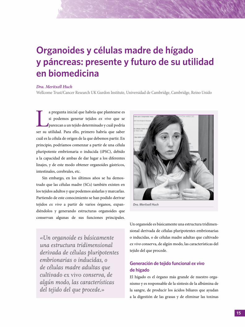
Organoides y células madre de hígado y páncreas: presente y futuro de su utilidad en biomedicinaDra. Meritxell HuchWellcome Trust/Cancer Research UK Gurdon Institute, Universidad de Cambridge, Cambridge, Reino Unido
Un organoide es básicamente una estructura tridimen-sional derivada de células pluripotentes embrionarias o inducidas, o de células madre adultas que cultivado ex vivo conserva, de algún modo, las características del tejido del que procede.
Generación de tejido funcional ex vivo de hígado El hígado es el órgano más grande de nuestro orga-nismo y es responsable de la síntesis de la albúmina de la sangre, de producir los ácidos biliares que ayudan a la digestión de las grasas y de eliminar las toxinas
Dra. Meritxell Huch
«Un organoide es básicamente una estructura tridimensional derivada de células pluripotentes embrionarias o inducidas, o de células madre adultas que cultivado ex vivo conserva, de algún modo, las características del tejido del que procede.»
CÉLULAS MADRE Y ORGANOIDES
procedentes de la sangre. Estas funciones las realizan, en concreto, células muy especializadas denomi-nadas hepatocitos. Para desempeñar correctamente su función, los hepatocitos requieren de la colabora-ción de otras células que forman el tejido, que son las células epiteliales denominadas colangiocitos o células ductales. Además, intervienen células endoteliales, mesenquimales y macrófagos.
A partir de hígados de ratón (tanto sanos como dañados), nuestro laboratorio consiguió aislar células que, tras añadirles un cóctel de factores de crecimiento presentes en el proceso de regeneración de este órgano in vivo, formaban estructuras quísticas que pudimos expandir en cultivo durante más de un año. Las células de estas estructuras mostraban funciones hepáticas, ya que eran capaces de sintetizar albúmina y absorber colesterol. Por otro lado, pudimos implantar estas estructuras en modelos de ratón con enfermedades hepáticas, corroborando así su funcionalidad.
Posteriormente, comprobamos si podíamos obtener resultados similares a partir de hígados humanos. Para ello, utilizamos biopsias de hígados de donantes que, con un protocolo similar al utilizado en los modelos murinos, pudimos mantener en cultivo durante más de seis meses. Para comprobar si estos organoides humanos se comportaban como un tejido hepático normal analizamos sus marcadores celulares, y observamos que se correspondían con los de células progenitoras y células ductales del hígado.
Las células ductales que componían los orga-noides derivados de tejido humano eran capaces de diferenciarse hacia un fenotipo similar al hepatocito y, en este estadio, también secretaban albúmina al medio de cultivo. Igualmente, presentaban actividad citocromo (CYP3A4) requerida por el hígado para el proceso de destoxificación, de hecho, eran capaces de metabolizar el antidepresivo midazolam. Finalmente, también detectamos que las estructuras poseían habi-lidad para captar colesterol. Esta fue la primera vez
que se consiguió crecer ex vivo un tejido procedente de hígado humano, y creemos que este descubrimiento puede ser esencial en el estudio de las enfermedades hepáticas y los trasplantes.
Uso de organoides como modelo de estudio de enfermedades hepáticas: cáncer de hígado A partir de los organoides hepáticos generados de biopsias de pacientes, por ejemplo, con tumores hepá-ticos o deficiencia de alfa-1 antitripsina, podemos reproducir esta enfermedad en el laboratorio y utili-zarlos, entre otros fines, para el cribado de fármacos.
Existen tres tipos principales de cáncer primario de hígado: el carcinoma hepatocelular (HCC), el colan-giocarcinoma (CC) y un subtipo intermedio (mixto). Si pudiéramos mantener en cultivo estos tumores y conservar sus características específicas, podríamos realizar una medicina personalizada acorde con cada paciente. Cuando analizamos organoides originados a partir de tejido sano y de los tres subtipos tumorales mencionados anteriormente, observamos que cada uno presentaba una morfología histológica diferente, tanto con respecto al tejido sano como a los otros subtipos. Incluso dentro del mismo subtipo tumoral podíamos identificar el cultivo que pertenecía a cada paciente.
En los organoides que procedían de tejido sano se observaba un crecimiento en monocapa similar al del epitelio ductal, pero los organoides tumorales
«Si pudiéramos mantener en cultivo estos tumores y conservar sus características específicas, podríamos realizar una medicina personalizada acorde con cada paciente.»
17
Organoides y células madre de hígado y páncreas: presente y futuro de su utilidad en biomedicina | Dra. Meritxell Huch
formaban estructuras muy diferentes, sólidas y rellenas de células que retenían la arquitectura histo-lógica original, pese a haber crecido todos en simi-lares condiciones de cultivo y con los mismos factores exógenos. Por ejemplo, se observó únicamente en los organoides HCC un dominio pseudoglandular que es específico de este subtipo. Es importante saber hasta qué punto estas estructuras están estrechamente rela-cionadas con el tumor de origen, porque si no es así, en vez de mimetizar el tejido estaríamos generando un artefacto ex vivo.
Cuando comparamos los perfiles de expre-sión génica entre los organoides y los tejidos origi-nales encontramos que existe una alta correlación entre los del mismo subtipo, pero no con los del resto de subtipos, lo que indica que los organoides retienen de manera intrínseca el perfil de expresión del tejido del que derivan. Por ejemplo, los orga-noides tumorales del tipo HCC presentan un grado de expresión de alfa-fetoproteína (marcador especí-fico de HCC) mayor que el de los organoides tumo-rales del tipo CC. Del mismo modo, cuando se analiza la expresión de S100 (marcador específico del subtipo CC) encontramos una mayor expresión en los orga-noides tumorales derivados de este subtipo tumoral.
A continuación, analizamos mediante secuencia-ción completa del exoma la similitud entre el tejido tumoral de origen y los organoides tras tres meses
en cultivo. El 84% de las variaciones genéticas del tumor original eran retenidas por los organoides. Por ejemplo, beta-catenina (CTNNB1) en HCC y KRAS en los subtipos CC y mixto. Además, estos organoides no presentaban mutaciones adicionales, lo que demuestra que son genéticamente estables.
Finalmente, trasplantamos en ratones los orga-noides tumorales y analizamos los tumores que se formaban. La estructura histológica del nuevo tumor correspondía con la del subtipo tumoral del que procedía. La tecnología de organoides permite que estas estructuras retengan la memoria de lo que eran originalmente.
Uso de organoides hepáticos para el cribado de fármacosLa tecnología de organoides tumorales se podría utilizar para identificar nuevos biomarcadores que nos puedan permitir en un futuro entender mejor el cáncer e identificar nuevos fármacos antitumorales. A partir de tumores de hígado provenientes de pacientes hemos generado una plataforma de organoides para el cribado de fármacos. De esta manera, analizamos la resistencia/sensibilidad de 21 fármacos antitumo-rales bien conocidos y, curiosamente, los organoides fueron resistentes a la mayoría de ellos. Estos resul-tados difieren mucho de los que se obtienen cuando se utilizan líneas celulares en cultivo, y podrían ser muy relevantes ya que, debido a la estructura tridi-mensional de los organoides, consideramos que su comportamiento se asemeja más a lo que realmente ocurre en un paciente.
«Es importante saber hasta qué punto estas estructuras están estrechamente relacionadas con el tumor de origen, porque si no es así, en vez de mimetizar el tejido estaríamos generando un artefacto ex vivo.»
«La tecnología de organoides permite que estas estructuras retengan la memoria de lo que eran originalmente.»
CÉLULAS MADRE Y ORGANOIDES
De los fármacos identificados nos hemos centrado en estudiar el efecto del inhibidor de ERK (SH772984) en modelos in vivo, ya que actualmente se emplea para el tratamiento de muchos cánceres, pero no en el cáncer de hígado. Cuando implantamos subcutáneamente los organoides tumorales en ratones y los tratamos con este fármaco observamos una reducción del volumen tumoral. A nivel histológico detectamos un gran número de células necróticas, lo que estaba en concordancia con los resultados previos obtenidos en la plataforma de cribado.
Generación de tejido funcional ex vivo de páncreasEl páncreas es un órgano muy similar al hígado, con conductos que almacenan las enzimas producidas por las células acinares (función exocrina); también es responsable de la producción de insulina, esencial para la regulación de los niveles de azúcar en sangre (función endocrina). Como todos sabéis, existen diversas enfer-medades comunes asociadas a este órgano como, por ejemplo, la diabetes y el cáncer pancreático.
Cuando intentamos generar organoides a partir de páncreas murinos conseguimos estructuras que se
podían expandir en cultivo durante largos periodos de tiempo y que retenían el fenotipo ductal. Además, en un ensayo de trasplante combinado de organoides con células embrionarias de páncreas observamos que sus células ductales eran capaces de secretar insulina.
En el caso de la generación de organoides de páncreas humanos, hasta el momento solo habían podido crecer en cultivo durante dos o tres semanas. Sin embargo, en estos momentos acabamos de esta-blecer nuevas condiciones de cultivo que nos permi-tirán la expansión de tejido pancreático humano sano durante largos periodos de tiempo.
La tecnología de organoides permite que tanto los tejidos sanos como los tumorales procedentes de hígado y páncreas se puedan expandir en cultivo de manera estable y conservando las características gené-ticas del tejido del que proceden. Esta tecnología nos permitirá entender mejor diversas enfermedades, pero también nos servirá para realizar una medicina perso-nalizada. Por ejemplo, a partir de la biopsia del tumor de un paciente, podremos generar organoides que se utilicen en el cribado de fármacos e identificar aque-llos que resulten efectivos para el tratamiento de ese tumor en un paciente en concreto.
19
Utilización de organoides cerebrales para comprender mejor las enfermedades neurológicas Prof. Guo-Li MingDepartamento de Neurociencias, Mahoney Institute for Neurosciences, Facultad de Medicina Perelman, Universidad de Pensilvania, Filadelfia, EE. UU.
Se han empleado muchos modelos animales para intentar estudiar la biología humana y sus enfer-medades, desde las bacterias hasta los primates
no humanos. Aunque hemos obtenido mucha infor-mación a partir de ellos, sabemos que existen nume-rosas diferencias entre estos modelos y el humano, siendo en el tejido cerebral donde se presentan mayores diferencias.
Es evidente que el cerebro del ratón tiene un tamaño muy diferente al humano. El cerebro humano contiene además un mayor número de células madre neuronales (NSCs) en comparación con el del ratón, y está compuesto por diferentes tipos de neuronas, lo que nos ha permitido desarrollar circuitos neuronales más complejos. Además, tenemos una población espe-cífica de NSCs que no se encuentra en el cerebro del ratón. La pregunta sería entonces, ¿cómo podemos estudiar la biología y el desarrollo del cerebro humano a partir de un modelo más apropiado?
En nuestro laboratorio utilizamos células madre pluripotentes inducidas de origen humano (hiPSCs) como modelo de estudio del desarrollo del cerebro. Estas células se pueden diferenciar de cualquier tipo de célula del organismo humano, pero ¿podemos utilizarlas para estudiar la organogénesis? El grupo de la Dra. Huch ha utilizado células madre adultas endógenas como modelo de estudio de tejidos como el hígado y el páncreas, sin embargo, nosotros no
podemos aislar estas células a partir de muestras de cerebro; por lo tanto, necesitamos comenzar a partir de un estadio anterior, como son las células madre pluripotentes.
Desarrollo de un mini-biorreactor para el estudio en organoidesUna de las tecnologías desarrollada para la genera-ción de organoides es el cultivo en suspensión de hiPSCs mediante el uso de spinners, donde las células pueden autoagregarse y formar estas estructuras
Prof. Guo-Li Ming
CÉLULAS MADRE Y ORGANOIDES
tridimensionales. Con estos dispositivos se pueden cultivar grandes volúmenes de células, pero este no es nuestro propósito. Además, este sistema permite utilizar únicamente entre seis a ocho spinners por incubador, puesto que requieren de una base para mantenerlos en agitación constante. Por otro lado, no se dispone comercialmente de spinners pequeños, con lo que esta tecnología resultaría muy costosa para el abordaje de nuestros estudios. Para reducir estos costes, en nuestro laboratorio hemos diseñado un sistema de mini-spinners (SpinΩ bioreactor) desarro-llado con tecnología de impresión 3D, donde todas las filas de una placa convencional de 12 pocillos están interconectadas mediante ruedas dentadas, permi-tiendo la agitación de todos los pocillos al unísono. De este modo, podemos hacer crecer organoides cere-brales con solo 3-5 mL de medio de cultivo por pocillo frente a los 100-200 mL que se requieren por cada spinner convencional.
Las primeras generaciones de organoides cere-brales se basaron en las condiciones intrínsecas del organoide per se, es decir, no se les añadía ningún tipo de factor exógeno para su inducción. Utilizando este sistema hemos conseguido minimizar gastos y hemos probado alrededor de 100 condiciones de crecimiento diferentes, en distintos estadios. El SpinΩ nos ha faci-litado encontrar una combinación de factores que ha permitido la generación de poblaciones de organoides más homogéneas.
Organoides de prosencéfaloEn nuestro laboratorio hemos desarrollado orga-noides que se asemejan a la parte anterior del cerebro durante la fase de desarrollo del embrión, de ahí que los hayamos denominado organoides de prosen-céfalo. Así, en el día 14 observamos que alrededor del 100% de las células presentes en el organoide eran todavía células madre de cerebro que expresan marcadores específicos del prosencéfalo (PAX6, OTX2). A medida que el organoide crece adquiere mayor complejidad, por ejemplo, en el día 28 se apreciaba una extensa capa de NSCs muy bien orga-nizada, y también se observó una estructura ventri-cular similar a los ventrículos cerebrales. Se percibía además cómo las neuronas emergían desde la capa de NSCs para constituir la placa cortical, del mismo modo que ocurre en el desarrollo. En estadios más avanzados del organoide no solo tenía lugar la expan-sión de las SCs, sino también la de las neuronas de la placa cortical. Los organoides de prosencéfalo pueden reproducir las propiedades características del cerebro humano.
En los organoides de prosencéfalo no solo encontramos las poblaciones tempranas de NSCs que se observan en modelos de ratón, sino también una capa de NSCs localizadas en la capa externa de la zona subventricular (oSVZ) con propiedades específicas de NSCs humanas. Pero ¿qué tipo de neuronas se pueden generar en estos organoides?
«El SpinΩ nos ha facilitado encontrar una combinación de factores que ha permitido la generación de poblaciones de organoides más homogéneas.»
«Los organoides de prosencéfalo pueden reproducir las propiedades características del cerebro humano.»
21
Utilización de organoides cerebrales para comprender mejor las enfermedades neurológicas | Prof. Guo-Li Ming
En el cerebro humano existe una gran diversidad de tipos de neuronas y seis capas diferentes de células en la corteza cerebral. ¿Es posible identificar, además, los tipos de neuronas que habitan en cada una de estas capas específicamente? Los organoides forman también estas seis capas y, lo que es más importante, el patrón de migración de las neuronas se asemeja a lo que ocurre en el cerebro humano durante el desarrollo. Así mismo, podemos encontrar neuronas que representan a cada una de las seis capas de la corteza. Estas neuronas resultan ser funcionales y eléctricamente activas. En los organoides de pros-encéfalo es posible registrar eventos de activación e inhibición sináptica, lo que demuestra que contienen neuronas excitatorias principales (glutamatérgicas) y neuronas inhibitorias o interneuronas (gabaérgicas). De acuerdo con la expresión de marcadores mole-culares detectados mediante secuenciación de ARN en organoides cultivados durante 100 días es posible confirmar, a nivel transcripcional, su similitud con la zona cortical del cerebro del feto al final del segundo trimestre del desarrollo (semana 20-22).
Organoides para el estudio de la infección por el virus ZikaDurante el año 2015 surgió un nuevo descubrimiento que sugería la vinculación de la infección por el virus Zika (ZIKV) con la aparición de microcefalia en fetos, anomalía en la que el cerebro está escasamente desarrollado, el cráneo se colapsa y la piel que lo cubre se arruga. Se pueden detectar partículas virales del ZIKV en el cerebro del paciente, pero no sabemos realmente qué hacen allí.
Para estudiar este fenómeno empleamos la tecno-logía de organoides. Nos preguntábamos si el ZIKV era capaz de infectar los organoides y si esta infección era generalizada o específica de algún tipo de célula.
Intentamos reproducir la infección por el virus en estadios muy tempranos del desarrollo, justo cuando comienza a formarse el sistema nervioso. Para ello, infectamos organoides al día 14 con el ZIKV durante 24 horas (cuando prácticamente todas las células son NSCs) y fuimos capaces de detectar proteínas virales a los 10 días postinfección. Esto sugiere que este virus es capaz de infectar NSCs, con consecuencias devasta-doras, ya que se observa la desintegración total de los organoides infectados. En esta misma línea, cuando se estudió este efecto en modelos murinos con animales infectados en estadios muy tempranos, se observó que los embriones no eran viables.
¿Qué ocurre cuando intentamos reproducir la infección en estadios tardíos del embarazo? Para ello infectamos organoides al día 28 de cultivo, donde no solamente existen NSCs, sino también neuronas. En este caso se encontró que el ZIKV era capaz de infectar únicamente NSCs. Muy pocas células progenitoras intermedias fueron infectadas y prácticamente ninguna neurona. Más adelante se observó la muerte de un gran número de células por apoptosis y una disminución de la tasa de proliferación de las células infectadas que permanecían vivas. Como consecuencia, disminuyó el número de NSCs y de las progenitoras derivadas de estas. Los organoides infectados por el ZIKV exhiben carac-terísticas muy similares a las observadas en pacientes con microcefalia.
«Los organoides infectados por el ZIKV exhiben características muy similares a las observadas en pacientes con microcefalia.»
CÉLULAS MADRE Y ORGANOIDES
Uso de organoides en el cribado de fármacosFinalmente, en nuestro laboratorio hemos utilizado los organoides también para identificar fármacos potenciales para su uso en clínica. Para ello, hicimos un cribado de 6.000 compuestos: 2.000 han sido aprobados por la Food and Drug Administration para su uso en el tratamiento de varias enferme-dades, otros 2.000 se están utilizando en ensayos clínicos y el resto son compuestos activos farmaco-lógicamente que actúan como activadores/inhibi-dores de enzimas celulares.
Lo que buscamos principalmente son compuestos neuroprotectores o antivirales. Por ejemplo, utilizando este sistema, identificamos un compuesto con propie-dades neuroprotectoras (emricasan). En organoides infectados por el ZIKV, al añadir este compuesto se observó una disminución de la apoptosis en las células infectadas, a pesar de que se seguía detectando la
presencia del virus, lo que demuestra las propiedades neuroprotectoras de emricasan frente a la infección por el ZIKV. Del mismo modo, también identificamos otros compuestos con propiedades antivirales y con alta capacidad de disminuir la carga viral celular.
A modo de resumen podemos concluir que el uso de organoides cerebrales no solo nos permite estu-diar las bases de la biología implicadas en el desa-rrollo neuronal, sino también su utilización como una herramienta para entender los mecanismos molecu-lares/celulares de las enfermedades neuronales; por ejemplo, el estudio de la infección por el ZIKV. Este sistema no solamente permite generar organoides de prosencéfalo, ya que mediante diferentes protocolos hemos conseguido generar organoides de mesencéfalo (región del cerebro afectada por enfermedades como el Parkinson), organoides que representan las regiones del hipotálamo y del hipocampo (zona cerebral impli-cada en el aprendizaje y la memoria).
23
N o todos los pacientes que padecen un mismo tipo de cáncer tienen el mismo pronóstico, esto es lo que se denomina
heterogeneidad intertumoral. Cuando analizamos un tejido tumoral a nivel microscópico, se observa además que no todas las células son idénticas, unas están proli-ferando, otras están diferenciadas, otras mueren y cada tipo celular expresa determinados marcadores; esto es lo que se conoce como heterogeneidad intratumoral. La pregunta inicial sería, ¿qué está promoviendo el creci-miento tumoral y su heterogeneidad? Una de las posi-bles teorías apunta a que el cáncer, al igual que el tejido normal, está gobernado por la presencia de células madre (SCs), en este caso, SCs del cáncer (CSCs), de las que derivarían los diferentes linajes celulares que componen el tumor. Desde el punto de vista terapéu-tico, esta teoría tiene implicaciones importantes. En primer lugar, para destruir el tumor ya no sería nece-sario eliminar todas las células tumorales, sino sola-mente las CSCs. En segundo lugar, podría explicarse la aparición de recidivas al utilizarse tratamientos que no actúan frente a las CSCs. Finalmente, según esta teoría, las CSCs son quienes determinan los procesos metastá-sicos que pueden derivar en la muerte del paciente.
Estudio de células madre del cáncer mediante ensayos de trasplante Históricamente el estudio de CSCs se ha realizado a partir de tumores primarios cuyas células son
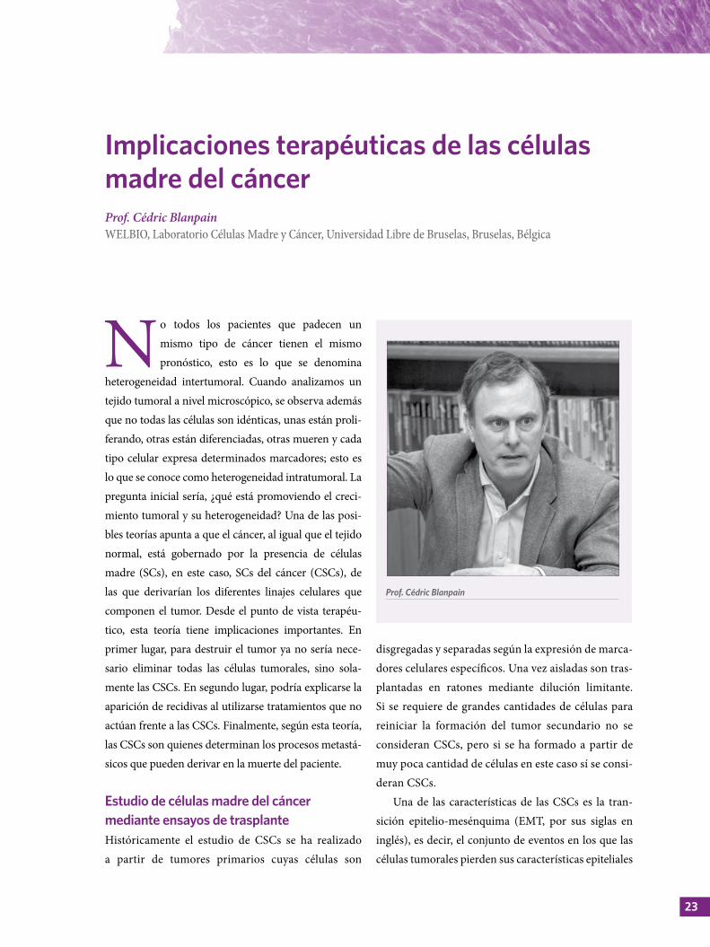
Implicaciones terapéuticas de las células madre del cáncer Prof. Cédric BlanpainWELBIO, Laboratorio Células Madre y Cáncer, Universidad Libre de Bruselas, Bruselas, Bélgica
disgregadas y separadas según la expresión de marca-dores celulares específicos. Una vez aisladas son tras-plantadas en ratones mediante dilución limitante. Si se requiere de grandes cantidades de células para reiniciar la formación del tumor secundario no se consideran CSCs, pero si se ha formado a partir de muy poca cantidad de células en este caso sí se consi-deran CSCs.
Una de las características de las CSCs es la tran-sición epitelio-mesénquima (EMT, por sus siglas en inglés), es decir, el conjunto de eventos en los que las células tumorales pierden sus características epiteliales
Prof. Cédric Blanpain
CÉLULAS MADRE Y ORGANOIDES
para adoptar un fenotipo mesenquimal y adquieren propiedades migratorias y potencial metastásico. Hemos observado que cuando se activan ciertos oncogenes en diferentes células de un tejido sano se obtienen células con diversos fenotipos tumorales, lo que demuestra que el origen celular puede influir en el tipo de tumor que se obtiene, pese a contener la misma mutación genética.
Estudios realizados in vitro sugieren que las células mesenquimales no constituyen una población bien definida de células, y que pueden existir diferentes estadios transicionales que van desde el epitelio hasta el mesénquima. Recientemente, hemos reali-zado un rastreo masivo de marcadores de superficie de células tumorales epiteliales y mesenquimales en modelos in vivo y hemos encontrado que la expre-sión de alguno de estos marcadores era heterogénea. Mediante técnicas de inmunofluorescencia identifi-camos al menos seis poblaciones diferentes durante la EMT. Al inicio, de una a dos poblaciones expresaban marcadores epiteliales y mesenquimales (población híbrida). Progresivamente, estas poblaciones iban perdiendo la expresión de los marcadores epiteliales y solo expresaban marcadores mesenquimales. Utili-zando ensayos de propagación de tumores obser-vamos que las diferentes poblaciones tenían la misma capacidad para generar nuevos tumores. Sin embargo, cuando estas poblaciones fueron trasplantadas y se analizó el fenotipo de célula que generaban, algunas de las poblaciones híbridas conservaban sus caracte-rísticas híbridas y la mayoría de las células mesenqui-males generaban poblaciones mesenquimatosas.
En relación con el potencial metastásico, se observó que las poblaciones más tempranas de la EMT (triple negativas) tenían mayor potencial que las poblaciones tardías. Esta población también era la más preponde-rante cuando se analizó la presencia de células tumo-rales circulantes en la sangre de los animales. Hasta aquí, y de acuerdo con nuestros resultados, podemos
concluir entonces que el periodo de transición de epitelio a mesénquima puede dividirse en tres estadios diferentes con distintas funciones. Alguno de ellos más metastásico que otros, por ejemplo, los triples nega-tivos. Por otro lado, estas poblaciones más tempranas muestran además mayor resistencia al tratamiento. Las poblaciones más tempranas de la EMT son poten-cialmente más metastásicas y resistentes al tratamiento en comparación con las tardías.
Estudio de células madre del cáncer mediante técnicas de rastreo de linajesEl rastreo de linajes permite detectar lo que ocurre realmente en el microambiente natural del tumor, hecho que representa una ventaja en comparación con los estudios de trasplante. Mediante esta técnica se activa la expresión de proteínas fluorescentes en el microambiente tumoral que sirven como marcadores irreversibles para toda la progenie de una célula. Por ejemplo, si marcamos una célula madre, todos sus linajes podrán ser detectados, pero si marcamos una célula progenitora solo los linajes que derivan de ella podrán ser rastreados.
Si hacemos un experimento en donde se marcan todos los tipos celulares de diferentes colores en un tumor en estadios iniciales del crecimiento y todas las células son igual de competentes/proliferativas, cuando el tumor crezca tendremos una combina-ción de colores en igual proporción que en el tumor de inicio. Sin embargo, si solo unas pocas células son
«Las poblaciones más tempranas de la EMT son potencialmente más metastásicas y resistentes al tratamiento en comparación con las tardías.»
25
Implicaciones terapéuticas de las células madre del cáncer | Prof. Cédric Blanpain
competentes, con el tiempo, solo estos tipos celu-lares compondrán la mayor parte del tumor, siendo esto lo que ha observado nuestro grupo de trabajo. Se ha demostrado, además, que en los estadios muy tempranos del desarrollo tumoral la jerarquía celular es idéntica a la de un tejido normal; sin embargo, en la medida en que el tumor progresa, las proporciones entre estas poblaciones se han ido haciendo diferentes.
El rastreo de linajes ha permitido contribuir al conocimiento de las CSCs. Así, se ha podido iden-tificar cómo ocurre una expansión rápida de clones dominantes Lgr5+ en ratones con adenoma intes-tinal (Clevers et al. 2012) e, incluso, cuantificar el número de clones funcionales de estas CSCs (Winton et al. 2013). El grupo de Jacco Van Rheenen (2013), mediante microscopia intravital, también ha obser-vado una rápida expansión de uno de los clones marcados en el tumor de mama. Por tanto, utilizando diferentes aproximaciones se ha podido demostrar que esta dinámica parece ser universal en una amplia variedad de tumores. Queda mucho por descubrir para entender completamente cuál es el significado de esta dinámica clonal: ¿por qué estas células crecen tan deprisa en el tumor?, ¿qué es lo que les confiere estas propiedades?, etc.
Estudio de las células madre del cáncer mediante técnicas de ablación de linajes Las implicaciones terapéuticas de las CSCs derivan de la siguiente hipótesis: 1. No sería necesario eliminar todas las células que componen el tumor, solo bastaría con eliminar las CSCs de las que derivan los diferentes linajes que lo componen. El tumor perdería la capa-cidad de generar nuevas células y terminaría dege-nerando. 2. Las CSCs podrían ser resistentes al trata-miento, y aunque disminuya el tamaño del tumor se produciría la recidiva del mismo.
En este sentido, nuestro grupo ha comenzado a utilizar una nueva aproximación genética que
permite eliminar exclusivamente unas pocas células del tumor (ablación de CSCs) y evaluar su repercu-sión en la regresión tumoral. Hemos identificado un marcador (SOX-2) con un alto nivel de expresión en pequeñas fracciones de cáncer de piel que tienen mayor capacidad de formar tumores en modelos de trasplante. Cuando eliminamos este pequeño porcen-taje de células que expresaban SOX-2 en el tumor, observamos la regresión de los mismos al cabo de una semana y no volvían a aparecer. La eliminación de unas pocas células tumorales específicas (CSCs) podría ser suficiente para inducir un gran impacto en el tumor y conseguir su regresión.
Otros grupos han mostrado resultados similares. Por ejemplo, se observó una regresión del tumor al eliminar una pequeña población de clones domi-nantes (Lgr5+) en tumores generados por xenotras-plante de organoides como modelo de cáncer de colon humano. Además, cuando realizaron la ablación de esta población, unida a quimioterapia, se observó mayor impacto en el tratamiento (Sato et al. 2017). Otro resultado interesante se observó al eliminar una población similar de CSCs Lgr5+ en un modelo de cáncer de colon. Los autores conseguían inhibir el crecimiento tumoral, pero cuando retiraban la presión selectiva el tumor volvía a crecer. A pesar de ello, estos ratones no desarrollaban metástasis en el
«Queda mucho por descubrir para entender completamente cuál es el significado de esta dinámica clonal: ¿por qué estas células crecen tan deprisa en el tumor?, ¿qué es lo que les confiere estas propiedades?, etc.»
CÉLULAS MADRE Y ORGANOIDES
hígado, lo que sugiere que alguna de estas CSCs resi-dentes en el tumor podrían ser las responsables de la diseminación del mismo.
Solución práctica del uso de las células madre del cáncerEl carcinoma basocelular (BCC) es un tumor que gene-ralmente se elimina por cirugía sin mayores conse-cuencias, pero en algunos casos los pacientes tardan demasiado en acudir al especialista y el tumor es extre-madamente grande para ser extirpado. En este caso, el BCC tiene que ser tratado con diferentes fármacos, por ejemplo, vismodegib (inhibidor de Smo), que permite la regresión del tumor antes de la cirugía. Algunos pacientes generan resistencia primaria al medicamento debido a mecanismos genéticos y el tumor continúa creciendo. Otros en pequeño número
responden completamente al tratamiento, pero en la mayoría de ellos la respuesta es parcial y el tumor no desaparece por completo.
Nuestro laboratorio ha desarrollado un modelo murino de BCC (Ptch1KO) al que hemos tratado con vismodegib, y hemos observado que a partir de la segunda semana el volumen del tumor comenzaba a disminuir, aunque no desaparece completamente. Estas lesiones resistentes no mostraban un ciclo de división celular muy activo y expresaban el marcador Lgr5+. Se conoce además que los cánceres resistentes a vismodegib, tanto en humanos como en ratón, tienen la vía de señalización Wnt activa (Lef1), una vía de señalización muy importante de las SCs en diferentes órganos y tejidos.
¿Cómo podríamos superar la resistencia a este tratamiento? Nuestro laboratorio probó a combinar fármacos que actúan sobre las vías de señalización de Wnt y Smo. Así, observamos que el tratamiento combinado de vismodegid + LGK-974 (inhibidor de Wnt) permitía erradicar la mayoría de las lesiones resistentes sin mostrar recidiva tras la retirada del tratamiento. Estos fármacos están disponibles en el mercado y, debido a que el BCC se manifiesta con lesiones externas, podrían ser administrados por vía tópica, evitando así la posible toxicidad del trata-miento sistémico con inhibidores de Wnt.
«La eliminación de unas pocas células tumorales específicas (CSCs) podría ser suficiente para inducir un gran impacto en el tumor y conseguir su regresión.»
27
Senescencia y reprogramación: una visión integradora de la reparación tisularDr. Manuel SerranoCellular Plasticity and Disease Group, Institute for Research in Biomedicine (IRB), Barcelona, España
Dr. Manuel Serrano
La plasticidad celular puede ser manipulada genética y farmacológicamenteCuando los tejidos envejecen acumulan muchas células dañadas y se reduce el número de células progenitoras. Con el propósito de regenerar tejidos afectados por determinadas patologías o por el enveje-cimiento, hemos intentado incrementar el número de células progenitoras en los mismos mediante terapia de reprogramación. Hace tiempo se consideraba que cuando la célula adquiría un fenotipo determinado durante el desarrollo, ese proceso era irreversible. Sin embargo, mediante el uso de los factores de transcrip-ción de Yamanaka (Oct4, Klf4, SOX-2 y Myc) se ha conseguido in vitro borrar toda la información conte-nida en una célula diferenciada y convertirla en una célula pluripotente embrionaria. Para estudiar si este proceso se podía realizar in vivo, hemos generado modelos murinos inducibles (ratones reprogramables i4F), en los que podemos inducir, de manera ubicua, la expresión de estos factores.
Cuando analizamos los ratones i4F en la primera semana tras la inducción, identificamos en varios tejidos la existencia de regiones celulares que habían perdido sus marcadores de diferenciación, perdiendo así su identidad. En la segunda semana los ratones enfermaban y se observaba la expresión de marca-dores de células pluripotentes, como Nanog (proteína característica del desarrollo embrionario). Finalmente, a la tercera semana, los animales fallecían debido a
que los tejidos perdían su funcionalidad. Sin embargo, si se retiraba previamente la inducción, los ratones sobrevivían y al mes se detectaban regiones comple-tamente reprogramadas. Estos animales desarrollaban teratomas (tumores característicos de células pluripo-tentes inducidas [iPSCs] o embrionarias [ESCs]), y a partir de su sangre pudimos aislar iPSCs con capa-cidad de formar colonias en cultivo en ausencia de inducción. El cuerpo es bastante más permisivo de lo que creíamos, admite un cierto grado de desdiferen-ciación y las células son capaces de sobrevivir en un estadio embrionario pluripotente.
CÉLULAS MADRE Y ORGANOIDES
En el intestino grueso de los animales reprogra-mados i4F detectamos la expresión del marcador CK19 (citoqueratina 19), típico de células epite-liales de intestino; pero también grupos de células que habían perdido su expresión y algunas de ellas expresaban Nanog. Además, se detectaron similares efectos en otros tejidos como el hígado, páncreas, riñón, intestino, estómago, etc. Debido a que una gran mayoría de las células eran incapaces de reprogra-marse, pensamos que los factores de Yamanaka podían estar induciendo daño celular y analizamos el grado de apoptosis y/o senescencia de estos. Aunque encontramos algunas células en apoptosis, principal-mente detectamos la presencia de senescencia (SAβG y p21). Curiosamente, cada vez que observábamos grupos de células senescentes encontrábamos células reprogramadas muy próximas a estas, lo que nos hizo pensar que quizás ambos procesos estaban estrecha-mente relacionados.
El daño tisular condiciona la plasticidad celularEn el pulmón, sin embargo, no fuimos capaces de detectar células reprogramadas ni senescentes. Teniendo en cuenta los resultados anteriores, pensamos que quizás los factores de Yamanaka no eran suficientes para inducir reprogramación in vivo y que se requería de un ambiente proinflamatorio, como el que generan las células senescentes. Por ello,
indujimos farmacológicamente este daño y bajo estas condiciones pudimos observar en el pulmón tanto células senescentes como reprogramadas.
La interleucina 6 desempeña un papel fundamental en la inducción de la plasticidad celularBasándonos en los trabajos de Helen Blau et al., deci-dimos analizar la función de la interleucina 6 (IL6) en la inducción de la reprogramación. En ratones IL6KO reprogramables no detectamos reprogramación en ninguno de los tejidos, había muy poco daño/displasia y no se detectó expresión de Nanog, por lo que IL6 parece ser esencial para este proceso. Los factores de Yamanaka no son capaces de inducir la reprograma-ción sin un microambiente proinflamatorio, en parti-cular requieren de la presencia de IL6, aunque no podemos excluir la intervención de otras citoquinas. La IL6 es parte de la familia del factor inhibitorio de la leucemia, un factor que se utiliza para mantener las iPSCs in vitro; por ello, estudiamos si la IL6 también era necesaria en nuestro caso. Al añadir medio condi-cionado de células senescentes con altos niveles de IL6, observamos un incremento en la eficiencia de reprogramación. En presencia de anticuerpos anti-IL6 no se obtuvo ninguna colonia de iPSCs, por lo que la IL6 parece ser necesaria también para el proceso de la reprogramación in vitro.
«El cuerpo es bastante más permisivo de lo que creíamos, admite un cierto grado de desdiferenciación y las células son capaces de sobrevivir en un estadio embrionario pluripotente.»
«Los factores de Yamanaka no son capaces de inducir la reprogramación sin un microambiente proinflamatorio, en particular requieren de la presencia de IL6, aunque no podemos excluir la intervención de otras citoquinas.»
29
Senescencia y reprogramación: una visión integradora de la reparación tisular | Dr. Manuel Serrano
En resumen, cuando se produce daño en el tejido in vivo, las células senescentes altamente proinflama-torias generan un microambiente rico en citoquinas, entre las que se encuentra IL6, la cual colabora con los factores de Yamanaka e induce la formación de iPSCs. Fisiológicamente hablando, solo cuando un tejido está dañado requiere de un mayor grado de plasticidad celular para repararse y los factores proinflamatorios que se secretan en respuesta al daño ayudarían a la inducción de esta plasticidad. Este modelo in vivo se puede manipular farmacológicamente. Por ejemplo, podemos provocar un ambiente proinflamatorio con palbociclib (inhibidor de CDK4/6 que potencia la senescencia) e incrementar la reprogramación, o podemos eliminar las células senescentes con navito-clax y reducir la reprogramación.
Estadios intermedios de plasticidad que no generan pluripotencia embrionariaMediante la conversión directa podemos obtener in vitro o in vivo una célula diferenciada a partir de otra, por ejemplo, una neurona a partir de un fibro-blasto. Con ello, evitamos la posibilidad de generar teratomas debido a la reprogramación completa. Por otro lado, existen evidencias de que quizás no es nece-sario regresar a estadios tan tempranos del desarrollo para llevar a cabo procesos de reparación tisular. Para averiguar si esto es posible examinamos los órganos de nuestros ratones i4F tras la primera semana de inducción y detectamos la existencia de células en un estadio primitivo intermedio y que no llegaban a ser pluripotentes.
Actualmente se sabe que la reprogramación en la que una célula somática diferenciada genera iPSCs presenta una primera fase de desdiferenciación (primera semana tras la inducción de la reprograma-ción in vitro) que es altamente estocástica y hetero-génea, y que induce la formación de múltiples tipos celulares (estadio intermedio de plasticidad). Solo
una minoría de estas células es capaz de encontrar la ruta correcta de reprogramación, la fase deter-minística, que se caracteriza por ser homogénea y predecible. Cuando analizamos in vivo el estado de las células del páncreas tras una semana de inducción observamos que sus marcadores no correspondían con ningún tipo celular pancreático (células progeni-toras embrionarias, endocrinas, acinares o ductales, etc.). Estas células presentaban algunos marcadores de células ductales (queratinas), pero no contenían otros marcadores ductales, por lo que las conside-ramos células ductales atípicas.
Los estadios intermedios de plasticidad conservan la capacidad de diferenciaciónLas células ductales atípicas podrían no tener utilidad alguna, pero observamos que cuando se retiraba la inducción el páncreas retornaba completamente a la normalidad. Por tanto, estas células podrían haberse rediferenciado o haber sido eliminadas por el sistema inmune. La Dra. Huch ha mostrado que no es posible generar organoides a partir de células acinares pancreáticas, pero nosotros nos pregun-tamos si las células pancreáticas ductales atípicas serían capaces de formar organoides. Cuando disgre-gamos páncreas de ratones i4F (no inducidos) y acti-vamos in vitro los factores de Yamanaka, pudimos efectivamente obtener organoides que se mostraban inicialmente como estructuras huecas y que daban lugar posteriormente a estructuras sólidas; pero cuando los factores de Yamanaka fueron activados in vivo y se retiró la inducción in vitro obtuvimos organoides de alta complejidad.
Estos organoides fueron secuenciados, obser-vándose un patrón de transcripción acorde con los organoides ductales pancreáticos, no expresaban marca-dores de queratina (Krt14) y sí expresaban SOX-9. Por tanto, las células ductales atípicas no son células
CÉLULAS MADRE Y ORGANOIDES
inútiles ni aberrantes, ya que pueden generar orga-noides ductales in vitro. Si se mantiene la inducción in vitro se observa la formación de organoides que presentan un fenotipo mixto de regiones ductales y ductales atípicas que pensamos que representan los cambios dinámicos entre ambos tipos celulares.
A continuación, analizamos a través de técnicas de rastreo de linaje el destino de estas células cuando se generan colonias de iPSCs in vitro. El rastreo de células Krt14+ mostró que aunque estas células permanecían en cultivo durante todo el proceso, no daban lugar a colonias de iPSCs. En resumen, las células ductales atípicas (Krt14+) son capaces de producir organoides ductales pero no generan colonias de iPSCs.
Los estadios intermedios de plasticidad son comunes entre múltiples tejidos Hemos encontrado células Krt14+ también en otros tejidos de ratones reprogramados, por ejemplo, en
intestino, riñón, estómago e hígado, por lo que consi-deramos que esta reprogramación parcial conduce a la célula a un estado que es común en los diferentes tejidos. Además, cuando reprogramamos hepato-citos o células epiteliales renales in vitro observamos nuevamente la presencia de células Krt14+ que no generaban iPSCs. Por tanto, las células en estadios intermedios de plasticidad o parcialmente repro-gramadas, que presentan características ductales atípicas, podrían tener la capacidad de producir células reparadoras de tejidos.
«En resumen, las células ductales atípicas (Krt14+) son capaces de producir organoides ductales, pero no generan colonias de iPSCs.»
Cél
ula
s m
adre
y o
rgan
oid
es
31
deb
ate Moderadora:
Erika Pastrana Mesa redonda: Meritxell Huch, Guo-Li Ming, Cédric Blanpain y Manuel Serrano

33
En relación con el protocolo de obtención de organoides a partir de SpinΩ, ¿es posible obtener otro tipo de población de neuronas, por ejemplo, organoides de astrocitos u oligodendrocitos?
Prof. Guo-Li Ming: Sí, es posible. En los organoides que generamos en nuestro laboratorio, las NSCs pueden cambiar su destino desde un perfil neurogénico a gliogénico y podemos obtener astrocitos en estadios tardíos, pero no hemos obtenido oligodendrocitos.
Acerca de los datos que muestran los diferentes estadios de la EMT, ¿tienen ustedes alguna evidencia sólida que demuestre que las célu-las tempranas del estadio mesen-quimal son realmente las células que dan lugar a todos los demás estadios intermedios, hasta adqui-rir el fenotipo mesenquimal?, ¿o es posible que los diferentes estadios intermedios provengan directa-mente de una célula epitelial?
Prof. Blanpain: No, no tenemos pruebas definitivas de esta progre-sión. Ciertamente no parece ser una progresión lineal, creemos que ocurre el primer estadio de transi-ción y luego hay, posiblemente, dos vías diferentes de progresión para alcanzar el estado mesenquimal. Hemos estudiado el paisaje trans-cripcional de estas células para analizar la progresión y, aunque estos diferentes estadios muestran
distintos grados de EMT, no tenemos claro cuál es la ruta por la que discurre esta progresión.
Acerca de los ratones IL6KO, ¿se conoce algo acerca de sus pro-piedades regenerativas cuando envejecen?
Dr. Serrano: Sí, de hecho estos datos han sido publicados hace varios años. Estos ratones tienen
Debate
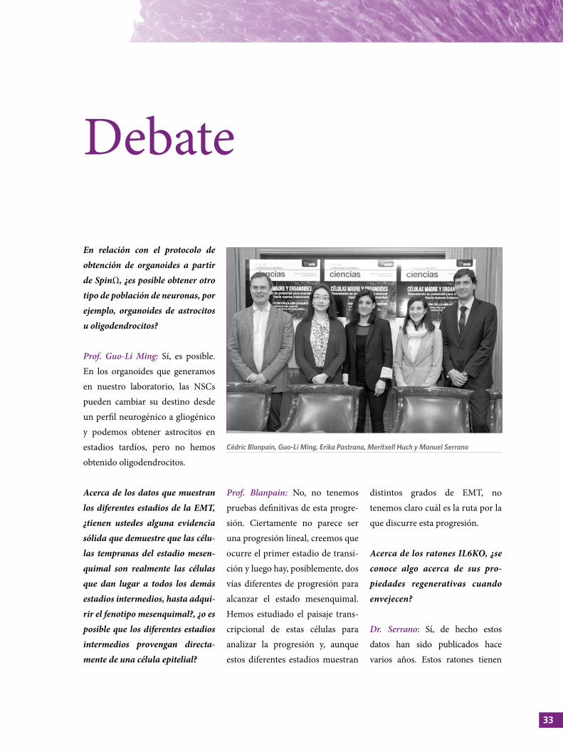
Cédric Blanpain, Guo-Li Ming, Erika Pastrana, Meritxell Huch y Manuel Serrano
CÉLULAS MADRE Y ORGANOIDES
problemas para la regeneración tisular, por ejemplo en el hígado se observa una regeneración alterada tras realizar una hepatectomía. La conexión de IL6 con regeneración es anterior incluso a los factores de Yamanaka.
El Prof. Blanpain ha presentado unos resultados muy interesantes acerca de cómo las células madre del cáncer responden a quimio-terapia y a tratamientos combi-nados, y quizás la audiencia po-dría estar interesada en saber la relación que puede tener esto con la inmunoterapia. Se está escu-chando cada vez más acerca de los avances de la inmunoterapia en cáncer y queríamos saber si las células madre del cáncer, en par-ticular, responden a este tipo de tratamientos.
Prof. Blanpain: Esta es una pregunta muy interesante, pero nosotros ahora mismo conocemos muy poco acerca de ello. Hay una muy buena correlación entre la respuesta a la inmunoterapia y la EMT, pero existe poco conocimiento acerca del mecanismo que permite que la inmunoterapia sea efectiva, lo que demuestra que la investigación es esencial, ya que si no investi-gamos no podemos avanzar en este conocimiento y no podemos racionalizar el tratamiento en el
futuro. Por lo tanto, pienso que es una buena pregunta que no tiene una buena respuesta por ahora.
¿Cómo influye la edad del ani-mal/donante en la capacidad de formar organoides?
Dra. Huch: Nuestro grupo ha reali-zado experimentos con ratones jóvenes y ratones muy viejos, de un año de edad, y observamos que la eficiencia es similar, no notamos diferencias en cuanto a la proce-dencia de los animales en términos de derivación, aunque sí en expan-sión. Si me preguntas cómo es esto posible, aún no tengo una respuesta, pero hemos observado el mismo efecto con células humanas. Tenemos donantes jóvenes (niños) y mayores (77 años) y hemos podido establecer organoides con ambos. La biopsia del niño se realizó porque tenía una enfer-medad y creció mucho mejor, pero no puedo descartar que sea debido a la enfermedad o a que no se podía considerar como tal un donante de órganos. La persona más joven que realmente se podía considerar como un donante de órganos tenía 35 años y no se observó diferen-cias en relación con el potencial de expansión o de derivación en comparación con el donante de 77 años, pero no tengo una expli-cación que justifique el porqué.
Prof. Guo-Li Ming: Es cierto que si se reprograman células y se remontan a un estado pluripotencial (iPSCs) se elimina el factor edad y da igual la edad de la persona a partir de la cual han sido generadas. Cuando cultivamos organoides a partir de iPSCs o ESCs se encuentran en una etapa de desarrollo, no en un estado maduro. Originalmente pensá-bamos que estos modelos tenían limitaciones si se utilizaban para estudiar enfermedades neurodege-nerativas. Afortunadamente, noso-tros trabajamos en enfermedades que tienen lugar durante el neuro-desarrollo, para lo cual es perfecto. Pero también hay publicaciones recientes que muestran que incluso utilizando estos organoides inma-duros es posible recapitular ciertas características de patologías de pacientes con edad avanzada, como por ejemplo el Alhzeimer. No sé cómo sucede esto, pero sugiere que estos organoides se podrían utilizar incluso como modelo de estudio de estas patologías, al menos a nivel molecular y celular.
Es interesante ver cómo el ZIKV infecta solamente células madre. Me gustaría saber si se conoce cuál podría ser la causa de esta infec-ción selectiva.
Prof. Guo-Li Ming: Obviamente, cualquier tipo de infección por
35
Debate
bacterias o virus ocurre a través de receptores celulares. Las NSCs expresan receptores específicos que podrían ser los que utiliza el virus para su internalización. Hay varias publicaciones que lo explican, por ejemplo, el grupo del Dr. Kriegstein mostró que el virus se une a la familia de recep-tores AXL que se expresa exclusi-vamente en las NSCs y astrocitos. Pero pienso que deben existir otros correceptores, ya que en estudios con modelos de ratones AXLKO se sigue produciendo la infección de estas células de manera espe-cífica. Esto sugiere que el virus requiere de receptores auxiliares o que existen otros tipos de recep-tores adicionales que median la infección por el ZIKV.
El Dr. Serrano nos ha mostrado que la única manera de reprogramar in vivo en el pulmón es mediante la inducción de daño, ¿qué ocurre en el resto de los tejidos donde ya hay eventos de reprogramación cuando se induce este daño?
Dr. Serrano: Si inducimos daño exógeno en el páncreas tendremos más eventos de reprogramación, siempre se produce una respuesta aditiva.
Dr. Serrano, ¿cree usted que la se-nescencia constituye la ruta mole-cular clave para el desarrollo de las enfermedades neurodegenerativas?
Dr. Serrano: No se ha publicado aún, pero en varios congresos se han mostrado resultados que indican que el Parkinson y el Alzheimer están asociados a la presencia de células senescentes. Por otro lado, se sabe que eliminando las células senescentes en modelos murinos de enferme-dades neurodegenerativas se consigue la mejora de estos animales. Estos no son resultados nuestros, pero creo que es algo que veremos publicado en breve en revistas de alto impacto.
Según indica el Dr. Serrano, las cé-lulas parcialmente reprogramadas no se parecen a ningún otro tipo celular. Para aseverar esto, ¿se ha basado en un estudio comparativo de la expresión de algunos mar-cadores?, ¿o ha realizado estu dios comparativos de expresión génica con los diferentes estadios del desa-rrollo para determinar, al menos, a cuál de estos estadios se parece?
Dr. Serrano: Realmente, estas células se parecen a las células ductales pancreáticas, pero no presentan todos los marcadores
correspondientes a este tipo celular. Hemos realizado un ensayo de expresión génica y efectivamente son bastante similares entre sí, pero no son exactamente iguales.
En relación con esta población estocástica de supuestos esta-dios intermedios de plasticidad, ¿funciona también en dirección contraria?, es decir, durante el de-sarrollo, ¿puede que no exista una ruta determinada? Porque para la diferenciación de algunos tejidos sabemos que es así, pero quizás este proceso pueda ocurrir en otros estadios del desarrollo. Dr. Serrano: No lo sé, la impresión que yo obtengo de los especialistas en desarrollo es que se trata de una ruta muy determinada, pero puedo estar equivocado.
Prof. Guo-Li Ming: Algunos estudios realizados en modelos murinos muestran que, si sufren algún tipo de daño, las células parenquimales pueden convertirse en SCs y dar lugar a neuronas. Eso sería debido a una especie de reprogramación intrínseca, pero no estoy segura de que esto pueda ocurrir de la misma manera en humanos o en otros tejidos.

pre
sent
atio
n
Stem
cel
ls a
nd
org
ano
ids
37
Federico Mayor ZaragozaChairman of the Scientific Council
of the Ramón Areces Foundation
Soledad SantosEditorial Director
Spain and Portugal, Springer Healthcare, a Springer Nature Business

39
I n this 10th edition of “Conferences and debates on science”, jointly organised with a prestigious entity such as Springer Nature, and under the title “stems cells and organoids. Revealing their potential to progress towards new treatments”, we
address important issues that constitute the fundamental basis of something that should lead all citizens, but especially scientists, to claim greater importance to research, especially in biomedicine.
When we speak of pathophysiology, the main issue is that every human being is unique and, therefore, we need to think about progressively getting closer to each patient. The progressive personalisation of medicine is the road to success.
When some years ago we began to talk about “genetic reprogramming” this notion was mentioned, and became one of the essential terms, because any adult cell can be re-coded, and, therefore, turned into a pluripotent cell, a stem cell from a specific cell of a particular patient.
For this reason, organoids, apart from their discovery or use as a tool for biomedical research, will be of extraordinary benefit not only in the diagnosis but also in the therapeutical approach, due to the advances achieved on genetic reprogramming. The possibility of direct cell therapy also exists, and for all these reasons, the Scientific Board of Ramón Areces Foundation has considered this a great opportunity to contribute to the dissemination of the latest advances in knowledge and pathophysiology.
On occasions such as this one, I like to repeat what D. Ramón Areces used to say: “Science is to relieve or prevent human suffering”. Prevention is essential, but in its absence, at least science has to mitigate, alleviate and relieve suffering. I think that this is our calling and our most important aim.
Therefore, I urge Springer Nature and its publishing Director for Spain and Portugal, Mrs. Soledad Santos Suárez, to continue our collaboration, so that research and science take the space they deserve in our society. ❙
Federico Mayor Zaragoza Chairman of the Scientific Council of the Ramón Areces Foundation
Presentation
STEM CELLS AND ORGANOIDS
W elcome to the 10th Conference-debate on stem cells and organoids, which is the result of a collaboration between Ramón Areces Foundation and Springer Nature group.
It was a decade ago when this fruitful journey of the Ramón Areces Foundation and Springer Nature began. For us, it is a great privilege to have the opportunity of collaborating annually with an organisation that is as committed as we are in serving the scientific community and divulging science in our country. We would particularly like to thank the Scientific Board, represented by professors Federico Mayor Zaragoza and José María Medina, who welcome us every year and always cooperate in wisely choosing a topic relevant for scientific progress and of public interest in general. We would also like to thank Mr Raimundo Pérez-Hernández y Torra, General Manager of the Foundation, and Mr Manuel Azcona, Director of Communications, for opening the doors of the Foundation to conduct these conferences.
This afternoon we will address an issue that 10 years ago would have seemed like science fiction: reprogramming adult cells to generate pluripotent or stem cells, and in vitro organs or organoids. The study of these cells has allowed us to know the tissue response to damage and their role in the reparative process, in addition to opening new avenues in the discovery of their role in tumour progression, metastasis and resistance to therapy. Thanks to the development of organoids, the study of complex tissue regeneration, such as the liver and pancreas, can be addressed nowadays taking into account the tissue structure and the interactions among its various cell types. Not even complex brain structures have been able to escape from this progress, since brain organoids have allowed the study of the Zika virus infection as well as the pathology associated with the development of severe brain disorders in infants.
This year Drs Meritxell Huch, Guo-Li Ming, Cédric Blanpain and Manuel Serrano will reveal the potential of stem cells and organoids for progressing towards new treatments. We are honoured that they have agreed to participate in these conferences. We are very grateful and hope that this experience is as interesting to them as it is to us.
Finally, I would like to thank Erika Pastrana, executive editor of Nature Research, who comes once more as the moderator of this year’s event. It is always a pleasure to have her excellent input, availability and commitment in the preparation of this Conference.
Thank you all. I hope you enjoy the conferences and subsequent debates. ❙
Soledad SantosEditorial Director Spain and Portugal,
Springer Healthcare, a Springer Nature Business

intr
od
ucti
on
Stem
cel
ls a
nd
org
ano
ids
41
Erika PastranaExecutive Editor for Nature Research,
New York, USA
Erika received her degree in Biochemistry and
Molecular Biology from the Universidad Autónoma
de Madrid and was granted a doctorate at the
same University with a thesis supervised by Dr
Javier Díaz-Nido, investigating the cellular and
molecular mechanisms responsible for promoting the
regeneration of damaged axons in the mammalian
central nervous system. Later, she moved to New York
where she did postdoctoral studies in the laboratory
of Dr Fiona Doetsch at Columbia University, where
she studied the way in which new neurons are
created and incorporated into the circuit of certain
areas of the adult mammalian brain. In 2010 she
joined “Nature Methods” as editor responsible for
neuroscience, and in 2014 she started working on
“Nature Communications” as Editor-in-Chief of
the neuroscience section. Currently, Erika is the
Executive Editor of the “Nature” journals in the area
of applied sciences and chemistry, including Nature
Biotechnology, Nature Methods and Nature Chemistry,
among others.

43
To begin, I would like to thank the Ramón Areces Foundation for allowing us to organise the tenth edition of this Conference.
We are very pleased to be the hosts of such an important event. As Professor Federico Mayor and Soledad Santos have already mentioned, the subject we have chosen is “stem cells and organoids” due to the advances that have taken place in this field over the past decade. Thus, recent studies have shown that stem cells have a therapeutic benefit in humans, in real diseases and in real patients; so we are at a critical moment for both scientific progress and human health. Although I do not think that stem cells require much presentation, they are, from my viewpoint, one of the most magical cells in our body since, as you know, they are the first cells that appear in the embryo and have the potential to develop into any cell type in the body, such as neurons or heart, skin, muscle cells, among others. One of the landmarks in the study of stem cells, was probably a discovery made by Shin'ya Yamanaka and colleagues, approximately ten years ago, in which they moved the clock of a differentiated adult body cell backwards and reprogramed it to a prior state, a stem cell, which was called reprogramming.
As you all know, these researchers induced their own stem cells, which has become a very important tool for scientific discovery, since they have helped to understand human physiology and development. In addition, this has not only allowed the deepening of
our knowledge about diseases but also provided an effective tool due to its therapeutic applications.
Another important discovery of the last ten years began in the laboratory of Hans Clevers in Holland, of Yoshiki Sasai in Japan and in others. It was discovered that when stem cells were left alone on the plate, surprisingly they self-assembled creating three-dimensional structures similar to our tissues, later called organoids or mini-tissues. Recently, we have witnessed the development of mini-brains, mini-pancreas, livers, stomachs, which has opened the doors to the study of human organs using this technique. It is an honour to have here with us, two of the best experts in organoids worldwide; Meritxell Huch who worked in the laboratory of Hans Clevers and later joined Cambridge University, where she created her own laboratory. Meritxell has been involved in very interesting discoveries regarding the application of organoids to develop not only a stomach, where she began, but also a liver and a pancreas. More recently, her laboratory has shown how the application of these organoids obtained from adult tissue cells can be used to understand the early stages of cancer development. We also have the presence of Dr Guo-Li Ming from the University of Pennsylvania, who has been working in neuroscience for a long time providing many interesting results for the understanding of neurodevelopmental disorders. Recently, she and her colleagues have focused on the
Introduction
STEM CELLS AND ORGANOIDS
potential of organoids, notably brain organoids, to understand the diseases affecting the early phases of human development. One of the key or most striking findings from her laboratory has been to show the enormous power of brain organoids when they are used to study the early stages of the Zika virus infection in the brain. These viruses cause microcephaly in infants and currently it is a very difficult disease to study since it occurs during human development in the uterus. The laboratory of Dr Guo-Li Ming has studied the early stages of infection, how the virus kills cells, which cells are killed and what treatments would be effective for this infection. On the other hand, and before the discovery of cell reprogramming and organoids, there was another major milestone in the field of stem cells that consisted of proving their existence not only in the embryo, but also in all adult human beings: our brain, heart, stomach, liver and skin have stem cells throughout our lives. This discovery is critical because it suggests that these cells contribute to the regeneration of some tissues and also to the normal physiology; thus, they contribute to the main physiological processes, such as learning and memory. Today, Dr Cédric Blanpain from the Université Libre de Bruxelles is also with us; Cédric has worked for many years trying to understand these endogenous stem cells, which reside in certain tissues of our body and, in particular, in the skin and mammary glands. One of the dark sides of stem cells is that they can also cause diseases and now, thanks
to the laboratory of Cédric and collaborators, we know that, sometimes, stem cells can contribute to the development of cancer. When Cédric and other researchers analysed several neoplasms, they found that there was a huge heterogeneity and the presence of cells resembling stem cells, which have been called cancer stem cells. Dr Blanpain will discuss the findings from his laboratory that have contributed to the understanding of the molecular mechanisms governing these cells, and more importantly, how these cells play a key role in the initiation, development and progression of tumour and metastasis.
Last of all, but not less important, I am delighted to introduce Manuel Serrano, who everyone knows very well, because not only is he a world-renowned researcher, but he has also made important contributions to science here in Madrid. Manuel worked in the National Centre of Biotechnology (CNB), then in the National Cancer Centre (CNIO) and now has come from the Institute for Research in Biomedicine (IRB) in Barcelona. There are many things I could mention about Manuel᾽s work over the years. He has worked in tumour suppressors and in processes such as aging and senescence, but I think that today he will describe a very important discovery he has made in his laboratory.
His findings have shown how several processes are interrelated during tissue regeneration and, in particular, how regeneration is affected by senescence and in vivo animal reprogramming. ❙
lect
ures
Adult liver and pancreas organoids and stem cells: Present and future of their biomedical utility Dr Meritxell HuchWellcome Trust/Cancer Research UK Gurdon Institute, Cambridge University, Cambridge, United Kingdom
Using brain organoids to understand neurological diseasesProf. Guo-Li MingDepartment of Neuroscience, Mahoney Institute for Neurosciences, Perelman School for Medicine, University of Pennsylvania, Philadelphia, USA
Therapeutic implications of Cancer Stem Cells Prof. Cédric BlanpainWELBIO, Laboratory of Stem Cells and Cancer, Université Libre de Bruxelles, Bruxelles, Belgium
Senescence and reprogramming: An integrated view of tissue repair Dr Manuel Serrano Cellular Plasticity and Disease Group, Institute for Research in Biomedicine (IRB), Barcelona, Spain
Stem
cel
ls a
nd
org
ano
ids
45
•
•
•
•

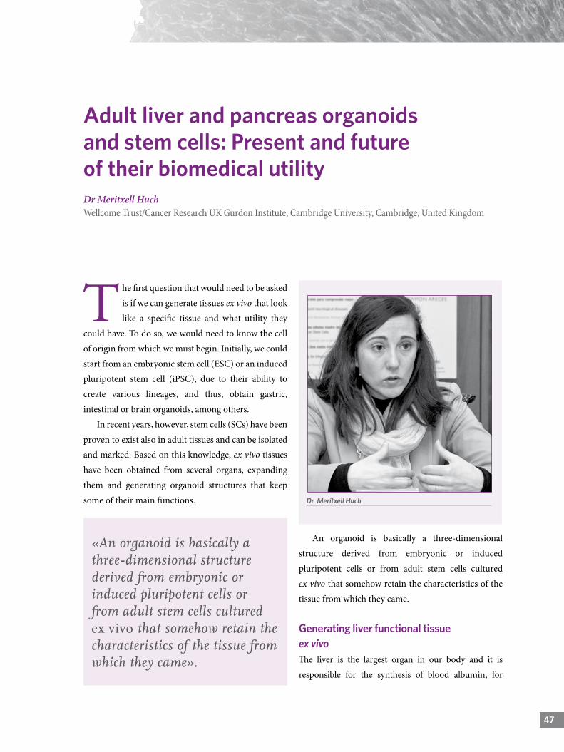
474747
T he first question that would need to be asked is if we can generate tissues ex vivo that look like a specific tissue and what utility they
could have. To do so, we would need to know the cell of origin from which we must begin. Initially, we could start from an embryonic stem cell (ESC) or an induced pluripotent stem cell (iPSC), due to their ability to create various lineages, and thus, obtain gastric, intestinal or brain organoids, among others.
In recent years, however, stem cells (SCs) have been proven to exist also in adult tissues and can be isolated and marked. Based on this knowledge, ex vivo tissues have been obtained from several organs, expanding them and generating organoid structures that keep some of their main functions.
Adult liver and pancreas organoids and stem cells: Present and future of their biomedical utilityDr Meritxell HuchWellcome Trust/Cancer Research UK Gurdon Institute, Cambridge University, Cambridge, United Kingdom
An organoid is basically a three-dimensional structure derived from embryonic or induced pluripotent cells or from adult stem cells cultured ex vivo that somehow retain the characteristics of the tissue from which they came.
Generating liver functional tissue ex vivoThe liver is the largest organ in our body and it is responsible for the synthesis of blood albumin, for
Dr Meritxell Huch
«An organoid is basically a three-dimensional structure derived from embryonic or induced pluripotent cells or from adult stem cells cultured ex vivo that somehow retain the characteristics of the tissue from which they came».
STEM CELLS AND ORGANOIDS
producing bile acids that help in the digestion of fats and for removing toxins from the blood. Very specialized cells, called hepatocytes, perform these functions. To properly perform their function, hepatocytes require the collaboration of other cells that form the tissue, which are epithelial cells called cholangiocytes or ductal cells. Endothelial and mesenchymal cells, as well as macrophages, are also involved.
Our laboratory isolated cells from mice livers (both healthy and damaged) that, after adding a cocktail of growth factors present in the regeneration process of this organ in vivo, created cystic structures that we could expand in a culture for more than a year. The cells of these structures showed liver functions, since they were able to synthesize albumin and absorb cholesterol. On the other hand, we could implant these structures in mouse models with liver disease, thus corroborating their functionality.
Subsequently, we tested if we could get similar results from human livers. To do this, we obtained liver biopsies from donors and used a similar protocol to that used in the murine models. We were able to keep these tissues in culture for more than six months. To test if these human organoids behaved like a normal liver tissue, we analysed their cell markers, and observed that they corresponded with liver stem and ductal cells.
Ductal cells in the organoids derived from human tissue were able to differentiate towards a phenotype similar to a hepatocyte and at this stage they also secreted albumin to the culture medium. Similarly, they showed cytochrome (CYP3A4) activity, required by the liver for the detoxification process, in fact, they could metabolize the antidepressant midazolam. Finally, we also detected that the structures had the ability to capture cholesterol. This was the first time that a tissue obtained from human liver tissue could be grown ex vivo; and
we believe that this discovery may be essential in the study of liver diseases and transplants.
Use of organoids as a model to study liver diseases: Liver cancer From the hepatic organoids created from biopsies taken from patients with, for example, liver tumours or Alpha-1 antitrypsin deficiency, we can reproduce these diseases in the laboratory and use them, among other purposes, for drug screening.
There are three main types of primary liver cancers: hepatocellular carcinoma (HCC), cholangiocarcinoma (CC) and an intermediate subtype (mixed). If we could keep these tumours in cultures and retain their specific features, we could administer a personalized treatment fitted to each patient. When we analyse organoids originated from healthy tissue and from the three tumour subtypes mentioned above, we can see that each one had a different histological morphology, both with respect to the healthy tissue and the other subtypes. Even within the same tumour subtype, we could identify the culture belonging to each patient.
In the organoids created from healthy tissue, a monolayer growth similar to that of the ductal epithelium was seen, but the tumour organoids created very different solid structures, filled with cells that retained the original histological architecture,
«If we could keep these tumours in cultures and retain their specific features, we could administer a personalized treatment fitted to each patient».
49
Using brain organoids to understand neurological diseases | Prof. Guo-Li Ming
49
Adult liver and pancreas organoids and stem cells: Present and future of their biomedical utility | Dr Meritxell Huch
despite having grown in similar culture conditions and with the same exogenous factors. For example, a pseudoglandular domain was observed only in the HCC organoid specific to this subtype. It is important to know the extent to which these structures are closely related to the original tumour, because if they are not, rather than mimic the tissue we would be generating an ex vivo artifact.
When we compare gene expression profiles of the organoids and the original tissues, we found that there is a high correlation between those of the same subtype, but not with the rest of the subtypes. This indicates that the organoid intrinsically retains the expression profile of the tissue from which it is derived. For example, organoids from the HCC tumour type have an Alpha fetoprotein expression (specific marker of HCC) greater than organoids from the CC tumour type. Similarly, when the expression of S100 (specific marker for CC subtype) is analysed, we find a greater expression in the tumour organoids derived from this tumour subtype.
Subsequently, we analysed the similarity between the original tumour tissue and the organoids that had been in culture for three months using whole-exome sequencing. Organoids retained 84% of the genetic variations of the original tumour; for example,
beta-catenin (CTNNB1) in HCC and KRAS in DC subtypes and mixed. In addition, these organoids had no additional mutations, showing that they are genetically stable.
Finally, we transplanted the tumour organoids in mice and then analysed the tumours that were formed. The histological structure of the new tumour corresponded to the tumour subtype from which it came. Organoid technology allows these structures to retain the memory of what they were originally.
Use of liver organoids for drug screeningTumour organoid technology could be used to identify new biomarkers that could allow us to better understand cancer and identify new anti-tumour drugs in the future. Starting from patients’ liver tumours we have generated an organoid platform for drug screening. This way, we can analyse the resistance/sensitivity of 21 well-known anti-tumour drugs and, curiously, organoids were resistant to most of them. These results greatly differ from those obtained when using cell lines in culture and could be very relevant since, due to the three-dimensional structure of organoids, we believe that their behaviour resembles more closely what really happens in a patient. Out of all the identified drugs, we focused on studying the effect of the ERK inhibitor (SH772984) in in vivo models, since it is currently used for the treatment of many cancers but liver cancer. When
«It is important to know the extent to which these structures are closely related to the original tumour, because if they are not, rather than mimic the tissue we would be generating an ex vivo artifact».
«Organoid technology allows these structures to retain the memory of what they were originally».
STEM CELLS AND ORGANOIDSSTEM CELLS AND ORGANOIDS
we subcutaneously implanted tumour organoids in mice and treated them with this drug, we observed a reduction in tumour volume. At the histological level, we detected a large number of necrotic cells, which was consistent with previous results obtained in the screening platform.
Generating pancreatic functional tissue ex vivoThe pancreas is an organ that is very similar to the liver, with ducts that store the enzymes produced by acinar cells (exocrine function). It is also responsible for insulin production, which is essential for the regulation of blood sugar levels (endocrine function). As you all know, there are several common diseases associated with this organ, such as diabetes and pancreatic cancer.
When we tried to create organoids from murine pancreas, we obtained structures that could expand in a culture for long periods of time retaining the
ductal phenotype. In addition, in a trial of a combined transplant of organoids and pancreatic embryonic cells, we saw that their ductal cells were able to secrete insulin.
As for the generation of human pancreatic organoids, they had only been able to grow in a culture for 2 or 3 weeks up till now. However, we have just established new culture conditions that will allow us the expansion of healthy human pancreatic tissue for long periods of time.
Organoid technology allows that both healthy and tumour tissue from liver and pancreas can expand in a culture stably and retain the genetic characteristics of the tissue they came from. This technology will not only allow us to better understand several diseases, but will also help us to create personalised medicine. For example, from a patient’s tumour biopsy we will create organoids that will be used for drug screening, allowing us to identify those drugs that will be effective in the treatment of the tumour of that specific patient.
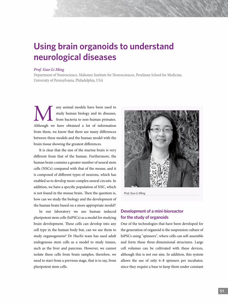
515151
Using brain organoids to understand neurological diseasesProf. Guo-Li MingDepartment of Neuroscience, Mahoney Institute for Neurosciences, Perelman School for Medicine, University of Pennsylvania, Philadelphia, USA
M any animal models have been used to study human biology and its diseases, from bacteria to non-human primates.
Although we have obtained a lot of information from them, we know that there are many differences between these models and the human model with the brain tissue showing the greatest differences.
It is clear that the size of the murine brain is very different from that of the human. Furthermore, the human brain contains a greater number of neural stem cells (NSCs) compared with that of the mouse, and it is composed of different types of neurons, which has enabled us to develop more complex neural circuits. In addition, we have a specific population of NSC, which is not found in the mouse brain. Then the question is, how can we study the biology and the development of the human brain based on a more appropriate model?
In our laboratory we use human induced pluripotent stem cells (hiPSCs) as a model for studying brain development. These cells can develop into any cell type in the human body but, can we use them to study organogenesis? Dr Huch’s team has used adult endogenous stem cells as a model to study tissues, such as the liver and pancreas. However, we cannot isolate these cells from brain samples; therefore, we need to start from a previous stage, that is to say, from pluripotent stem cells.
Development of a mini-bioreactor for the study of organoidsOne of the technologies that have been developed for the generation of organoid is the suspension culture of hiPSCs using "spinners", where cells can self-assemble and form these three-dimensional structures. Large cell volumes can be cultivated with these devices, although this is not our aim. In addition, this system allows the use of only 6–8 spinners per incubator, since they require a base to keep them under constant
Prof. Guo-Li Ming
STEM CELLS AND ORGANOIDS
agitation. On the other hand, small spinners are not commercially available, so this technology would be very costly to use in our studies. To reduce these costs, in our laboratory we have designed a set of mini-spinners (SpinΩ bioreactor) developed with 3D printing technology where all rows of a conventional 12-well plate are interconnected by means of dented wheels, allowing the agitation of all wells simultaneously. Thus, we can grow brain organoids with only 3–5 mL of culture medium per well versus 100–200 mL required for each conventional spinner.
The first generations of brain organoids were based on intrinsic conditions of the organoid per se; that is, no exogenous factor was added for their induction. Using this system we have managed to minimize costs and have tested around 100 different growing conditions at different stages. The SpinΩ has simplified the finding of a combination of factors that has enabled the generation of more homogeneous organoid populations.
Prosencephalon organoidsIn our laboratory we have developed organoids that resemble the front part of the brain during the developmental phase of the embryo, which we have called prosencephalon organoids. So, on day 14, we saw that around 100% of the cells present in the
organoid were still brain stem cells expressing specific markers of the prosencephalon (PAX6, OTX2). As the organoid grows it acquires greater complexity; for example, on day 28, a very well-organised extensive layer of NSCs could be seen, and ventricular structure similar to the cerebral ventricles could also be seen. The way neurons emerge from the NSCs layer to form the cortical plate, as happens during development, could also be seen. In more advanced stages of the organoid, not only the expansion of SCs, but also the expansion of the cortical plate neurons, occurred. Prosencephalon organoids can recreate the attributes characteristic of the human brain.
In prosencephalon organoids we found not only the early NSCs populations observed in mouse models, but also a layer of NSCs located in the outer layer of the subventricular zone (oSVZ) with specific properties of human NSCs. But, what kind of neurons can be generated in these organoids? There is a great diversity of neuron types in the human brain as well as six layers of different cells in the cerebral cortex. Is it possible to also identify the types of neurons that inhabit each of these layers specifically? Organoids also form these six layers and, more importantly, the migration pattern of neurons is similar to that occurring in the human brain during development. In addition, we can find neurons that represent each of the six layers of the cortex. These neurons are
«The SpinΩ has simplified the finding of a combination of factors that has enabled the generation of more homogeneous organoid populations».
«Prosencephalon organoids can recreate the attributes characteristic of the human brain».
53
Using brain organoids to understand neurological diseases | Prof. Guo-Li Ming
53
electrically active and functional. In prosencephalon organoids, it is possible to record synaptic inhibition and activation events which show that they contain primary excitatory neurons (glutamatergic) and inhibitory neurons or interneurons (GABAergic). Based on the expression of molecular markers detected by ARN sequencing in organoids cultivated for 100 days, it is possible to confirm, at a transcriptional level, their similarity with the cortical area of the foetus brain at the end of the second quarter of development (week 20–22).
Organoids to study the Zika virus infectionIn the year 2015, a new discovery suggesting a link between the Zika virus infection (ZIKV) and the emergence of microcephaly in foetuses (an anomaly in which the brain is sparsely developed, the skull collapses and the skin that covers it wrinkles) appeared. Although viral particles from the ZIKV can be detected in the patient’s brain, we do not really know what they are doing there. To study this phenomenon we employed organoid technology. We wondered if ZIKV was able to infect organoids and if this infection was widespread or specific to a cell type. We tried to reproduce the viral infection in the very early stages of development, just when the nervous system is starting to form. To that effect, we infected organoids at day 14 with the ZIKV for 24h (when virtually all cells are NSCs) and we were able to detect viral proteins 10 days post-infection. This suggests that this virus is capable of infecting NSCs, with very devastating consequences, since the total disintegration of the infected organoid is observed. In this same line, when we studied this effect in murine models with animals infected at very early stages, we observed that the embryos were not viable.
What happens when we try to recreate the infection in the late stages of pregnancy? For this purpose we infected organoids at day 28 of culture, where there are not only NSCs, but also neurons. In this case it was found that ZIKV was able to infect only NSCs. Very few intermediate stem cells were infected and practically no neurons. Subsequently, the death of a large number of cells by apoptosis, and a decrease in the rate of proliferation of infected cells that remained alive could be seen. As a result, the number of NSCs decreased as well as the stem cells derived from the former. Organoids infected by ZIKV exhibited characteristics very similar to those observed in patients with microcephaly.
Use of organoids in drug screeningFinally, in our laboratory we have also used organoids to identify potential drugs to be used in clinical practice. To do this, we screened 6,000 compounds: 2,000 approved by the FDA for use in the treatment of various diseases; another 2,000 are being used in clinical trials; and the rest are pharmacologically active compounds that act as activators/inhibitors of cellular enzymes.
We were mainly looking for neuroprotective or antiviral compounds. For example, using this system we identified a compound with neuroprotective properties, emricasan. When adding this compound
«Organoids infected by ZIKV exhibited characteristics very similar to those observed in patients with microcephaly».
STEM CELLS AND ORGANOIDS
to organoids infected by the ZIKV, we observed a decrease in the apoptosis of the infected cells, although the presence of the virus was still detected, which shows the neuroprotective properties of emricasan against the ZIKV infection. Likewise, we also identified other compounds with antiviral properties and high ability to decrease cell viral load.
To summarize, we can conclude that the use of brain organoids not only allows us to study the basis of the biology involved in neuronal development,
but also we can use it as a tool to understand the molecular/cellular mechanisms of neural diseases; for example, the study of the ZIKV infection. This system not only allows us to generate prosencephalon organoids, since we have also generated organoids of the mesencephalon (the region of the brain affected by diseases such as Parkinson’s), organoids representing regions of the hypothalamus and hippocampus (a brain area involved in learning and memory) using different protocols.
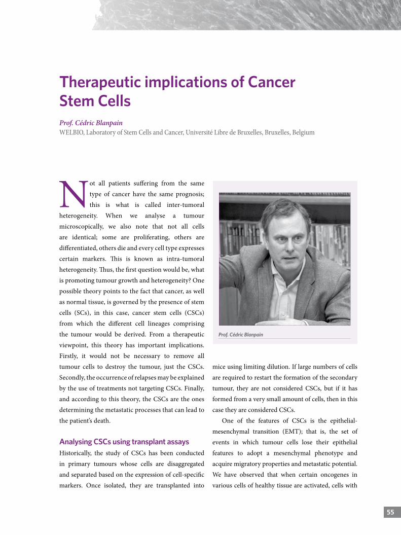
555555
Not all patients suffering from the same type of cancer have the same prognosis; this is what is called inter-tumoral
heterogeneity. When we analyse a tumour microscopically, we also note that not all cells are identical; some are proliferating, others are differentiated, others die and every cell type expresses certain markers. This is known as intra-tumoral heterogeneity. Thus, the first question would be, what is promoting tumour growth and heterogeneity? One possible theory points to the fact that cancer, as well as normal tissue, is governed by the presence of stem cells (SCs), in this case, cancer stem cells (CSCs) from which the different cell lineages comprising the tumour would be derived. From a therapeutic viewpoint, this theory has important implications. Firstly, it would not be necessary to remove all tumour cells to destroy the tumour, just the CSCs. Secondly, the occurrence of relapses may be explained by the use of treatments not targeting CSCs. Finally, and according to this theory, the CSCs are the ones determining the metastatic processes that can lead to the patient’s death.
Analysing CSCs using transplant assays Historically, the study of CSCs has been conducted in primary tumours whose cells are disaggregated and separated based on the expression of cell-specific markers. Once isolated, they are transplanted into
Therapeutic implications of Cancer Stem CellsProf. Cédric BlanpainWELBIO, Laboratory of Stem Cells and Cancer, Université Libre de Bruxelles, Bruxelles, Belgium
mice using limiting dilution. If large numbers of cells are required to restart the formation of the secondary tumour, they are not considered CSCs, but if it has formed from a very small amount of cells, then in this case they are considered CSCs.
One of the features of CSCs is the epithelial-mesenchymal transition (EMT); that is, the set of events in which tumour cells lose their epithelial features to adopt a mesenchymal phenotype and acquire migratory properties and metastatic potential. We have observed that when certain oncogenes in various cells of healthy tissue are activated, cells with
Prof. Cédric Blanpain
STEM CELLS AND ORGANOIDS
different tumour phenotypes are obtained, which shows that the cellular origin may influence the type of tumour obtained despite containing the same gene mutation.
In vitro studies suggest that mesenchymal cells do not constitute a well-defined cell population, and that there may be different transitional stages ranging from epithelial to mesenchymal. Recently, we made a massive screening of epithelial and mesenchymal cell surface markers in in vivo models and we have found that the expression of some of these markers was heterogeneous. Using immunofluorescence techniques, we identified at least six different populations during EMT. At the beginning, one to two populations expressed epithelial and mesenchymal markers (hybrid population). Progressively, these populations lost the expression of epithelial markers and only expressed mesenchymal markers. Using tumour dissemination assays, we observed that different populations had the same ability to generate new tumours. However, when these populations were transplanted, and the phenotype of the cell they created was analysed, some of the hybrid populations kept their hybrid features and most of the mesenchymal cells generated mesenchymal populations.
Regarding metastatic potential, the earliest EMT populations (triple negative) showed greater potential than the late populations. This population was also the most dominating when the presence of circulating tumour cells in animal blood was analysed. Thus far, and based on our findings, we can conclude that the epithelial to mesenchymal transition period can be divided into three different stages with different functions. Some are more metastatic than others; for example, the triple negative ones. On the other hand, these early populations are also more resistant to treatment. The earliest populations of EMT are
potentially more metastatic and resistant to treatment compared with the late ones.
Study of CSCs using lineage tracing techniquesLineage tracing allows the detection of what actually happens in the natural tumour microenvironment, which represents an advantage compared to transplantation studies. Using this technique, the expression of fluorescent proteins in the tumour microenvironment is activated, and they work as irreversible markers for all the progeny of a cell. For example, if we label a stem cell, all its lineages can be detected; however, if we label a progenitor cell, only the lineages derived from that cell can be tracked.
If we do an experiment where all cell types are labelled with different colours in the early stages of a tumour and all cells are equally competent/proliferative, once the tumour grows we will have the same proportion of colours than we had in the early stages of the tumour. However, if only a few cells are competent, only these cell types will be prominent in the tumour over time, and this is what our working group has observed. Furthermore, in the very early stages of tumour development, cell hierarchy is identical to that of normal tissue; however, as the tumour progresses the proportions between these populations become different.
«The earliest populations of EMT are potentially more metastatic and resistant to treatment compared with the late ones».
57
Therapeutic implications of Cancer Stem Cells | Prof. Cédric Blanpain
57
Lineage tracing has contributed to improve the knowledge on CSCs. Thus, it was possible to identify how a rapid spread of Lgr5+ dominant clones in mice with intestinal adenoma occurs (Clevers et al. 2012) and even quantify the number of functional clones of these CSCs (Winton et al. 2013). Jacco Van Rheenen’s team (2013) also observed a rapid spread of one of the clones labelled in breast tumour using intravital microscopy. Therefore, by using various approaches, this dynamic seems to be universal in a wide variety of tumours. Much remains to be discovered to fully understand what the meaning of this clonal dynamic is, why these cells grow so fast in the tumour, what gives them these properties, etc.
Study of CSCs using lineage ablation techniques The therapeutic implications of CSCs are derived from the following hypotheses: 1. It would not be necessary to remove all the cells that comprise the tumour, only those CSCs from which the various lineages present in the tumour are derived; the tumour would lose the ability to generate new cells and would eventually degenerate. 2. CSCs may be resistant to treatment and even though the tumour size decreases, tumour relapse would occur.
To address this issue, our team has started to use a new genetic approach that allows the deletion of only a few tumour cells (CSCs ablation) and the assessment of their impact on tumour regression. We have identified a marker (SOX-2) with a high level of expression in small fractions of skin cancer with a greater ability to form tumours in transplantation models. When we eliminated this small percentage of SOX-2-expressing cells from the tumour, we observed tumour regression within a week, as well as no recurrence. The elimination of a few specific tumour cells (CSCs) could be enough to cause a great impact on the tumour and allow its regression.
Other teams have obtained similar results. For example, a tumour regression was observed by eliminating a small population of dominant clones Lgr5+ in tumours generated by organoid xenotransplantation as a model of human colon cancer. In addition, when an ablation of this population combined with chemotherapy was performed, greater impact in the treatment was observed (Sato et al. 2017). Another interesting result was obtained when eliminating a similar population of CSCs Lgr5+ in a colon cancer model. The authors were able to inhibit tumour growth, but when they withdrew the selective pressure, the tumour started to grow again. Despite this, these mice did not develop liver metastases, suggesting that some of these CSCs in the tumour could be responsible for tumour spread.
Practical solution for using CSCs Basal cell carcinoma (BCC) is a tumour that is usually removed by surgery without greater consequences, but in some cases patients take too long to go to the specialist, and the tumour is too large to be removed. In this case, BCC has to be treated with different drugs; for example, vismodegib (Smo inhibitor), which allows
«Much remains to be discovered to fully understand what the meaning of this clonal dynamic is, why these cells grow so fast in the tumour, what gives them these properties, etc.».
STEM CELLS AND ORGANOIDS
tumour regression prior to surgery. Some patients generate primary resistance to the drug due to genetic mechanisms and the tumour continues growing. A few other patients respond fully to treatment, but most of them have a partial response and the tumour does not disappear completely.
Our laboratory has developed a mouse model of BCC (Ptch1KO), which we have treated with vismodegib and we noticed that from the second week, the tumour
volume began to diminish but did not isappear completely. These resistant lesions did not show a very active cell division cycle and expressed the Lgr5+ marker. It is also known that vismodegib-resistant cancers, both in human and mouse, have an active Wnt (Lef1) signalling pathway, which is a major signalling pathway of SCs in various organs and tissues.
How can we overcome the resistance to this treatment? Our laboratory tested a combination of drugs that act on the Wnt and Smo signalling pathways. Thus, we observed that the combined treatment of vismodegid+LGK-974 (Wnt inhibitor), allowed the eradication of the most resistant lesions without showing recurrence after treatment withdrawal. These drugs are commercialised, and since BCC manifests with external injuries, they could be administered topically, avoiding the possible toxicity of a systemic treatment with Wnt inhibitors.
«The elimination of a few specific tumour cells (CSCs) could be enough to cause a great impact on the tumour and allow its regression».
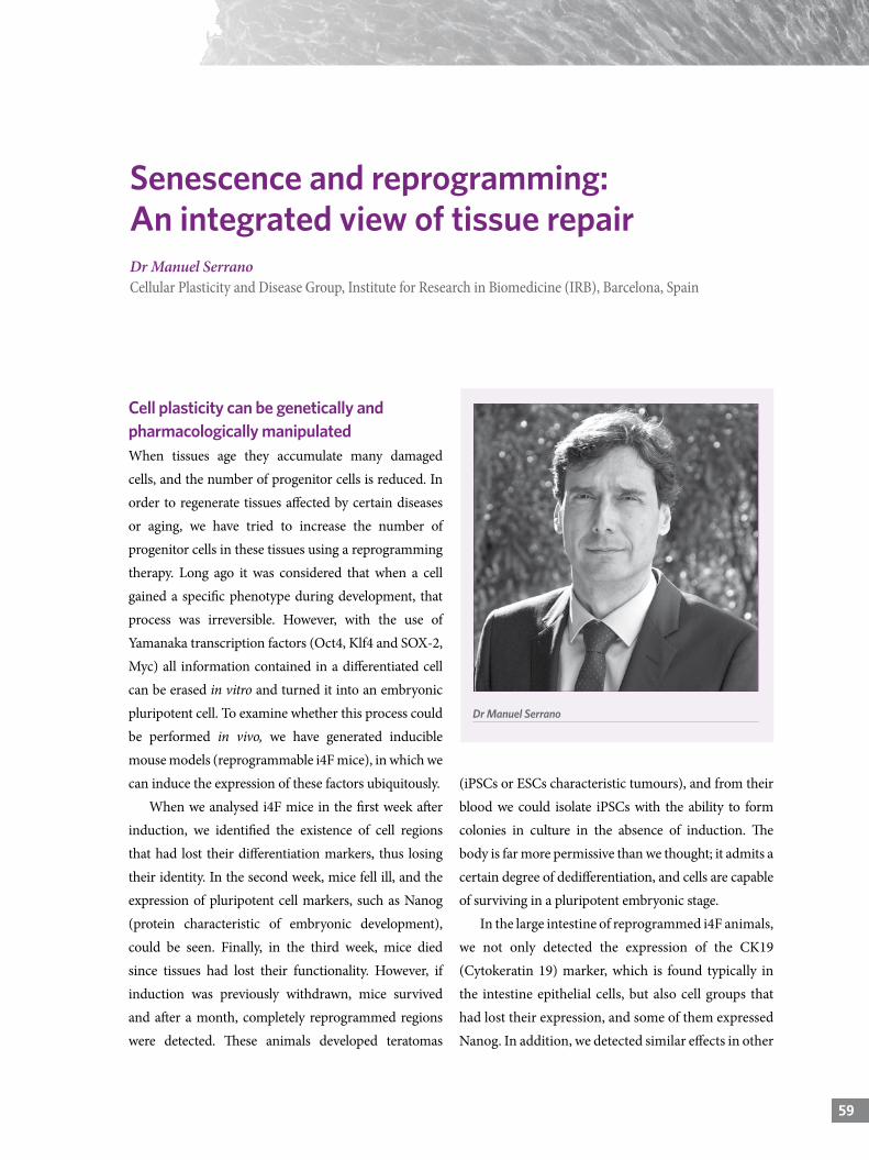
5959
Cell plasticity can be genetically and pharmacologically manipulatedWhen tissues age they accumulate many damaged cells, and the number of progenitor cells is reduced. In order to regenerate tissues affected by certain diseases or aging, we have tried to increase the number of progenitor cells in these tissues using a reprogramming therapy. Long ago it was considered that when a cell gained a specific phenotype during development, that process was irreversible. However, with the use of Yamanaka transcription factors (Oct4, Klf4 and SOX-2, Myc) all information contained in a differentiated cell can be erased in vitro and turned it into an embryonic pluripotent cell. To examine whether this process could be performed in vivo, we have generated inducible mouse models (reprogrammable i4F mice), in which we can induce the expression of these factors ubiquitously.
When we analysed i4F mice in the first week after induction, we identified the existence of cell regions that had lost their differentiation markers, thus losing their identity. In the second week, mice fell ill, and the expression of pluripotent cell markers, such as Nanog (protein characteristic of embryonic development), could be seen. Finally, in the third week, mice died since tissues had lost their functionality. However, if induction was previously withdrawn, mice survived and after a month, completely reprogrammed regions were detected. These animals developed teratomas
(iPSCs or ESCs characteristic tumours), and from their blood we could isolate iPSCs with the ability to form colonies in culture in the absence of induction. The body is far more permissive than we thought; it admits a certain degree of dedifferentiation, and cells are capable of surviving in a pluripotent embryonic stage.
In the large intestine of reprogrammed i4F animals, we not only detected the expression of the CK19 (Cytokeratin 19) marker, which is found typically in the intestine epithelial cells, but also cell groups that had lost their expression, and some of them expressed Nanog. In addition, we detected similar effects in other
59
Senescence and reprogramming: An integrated view of tissue repairDr Manuel SerranoCellular Plasticity and Disease Group, Institute for Research in Biomedicine (IRB), Barcelona, Spain
Dr Manuel Serrano
STEM CELLS AND ORGANOIDS
tissues, such as the liver, pancreas, kidney, intestine, stomach, etc. Since a large majority of cells were incapable of reprogramming, we think that Yamanaka factors could be inducing cell damage, and we analysed the degree of apoptosis and/or senescence of these cells. Although we found some cells on apoptosis, we mainly detected the presence of senescence (SAβG and p21). Oddly enough, whenever we watched groups of senescent cells were found reprogrammed cells very similar to the former, which made us think that perhaps both processes were closely related.
Tissue damage conditions cell plasticityHowever, in the lung we were unable to detect reprogrammed or senescent cells. Given the above results, we think that perhaps Yamanaka factors were not enough to induce in vivo reprogramming, and that a pro-inflammatory environment was required, such as the one created by senescent cells. Therefore we pharmacologically induced this damage, and under these conditions we saw both reprogrammed and senescent cells in the lung.
IL6 plays an important role in the induction of cell plasticityBased on the works of Helen Blau and others, we decided to analyse the role of IL6 in the induction
of reprogramming. In reprogrammable IL6KO mice, we did not detect reprogramming in any of the tissues that had very little damage/dysplasia and non-detectable Nanog expression; thus IL6 seems to be essential for this process. Yamanaka factors are not able to induce reprogramming without a pro-inflammatory microenvironment; in particular, the presence of IL6 is required, although we cannot exclude the involvement of other cytokines. IL6 is part of the LIF (leukemia inhibitory factor) family, a factor that is used to keep iPSCs in vitro; Therefore, we studied whether IL6 was also necessary in our case. When we added a conditioned medium of senescent cells with high levels of IL6, we observed an increase in the efficiency of reprogramming. In the presence of anti-IL6 antibodies, no colony of iPSCs was obtained; thus IL6 seems to be also necessary for the reprogramming process in vitro.
In summary, when tissue damage occurs in vivo, highly pro-inflammatory senescent cells generate in a microenvironment rich in cytokines, among which IL6 can be found, which collaborates with Yamanaka factors and induces iPSCs formation. Physiologically speaking, only when a tissue is damaged does it require a higher degree of cell plasticity to be repaired,
«The body is far more permissive than we thought; it admits a certain degree of dedifferentiation, and cells are capable of surviving in a pluripotent embryonic stage».
«Yamanaka factors are not able to induce reprogramming without a pro-inflammatory microenvironment; in particular, the presence of IL6 is required, although we cannot exclude the involvement of other cytokines».
61
Senescence and reprogramming: An integrated view of tissue repair | Dr Manuel Serrano
61
and pro-inflammatory factors secreted in response to that damage would help the induction of this plasticity. This in vivo model can be pharmacologically manipulated. For example, we can create a pro-inflammatory environment with palbociclib (CDK4/6 inhibitor which promotes senescence) and increase reprogramming; or we can remove senescent cells with navitoclax and reduce reprogramming.
Intermediate stages of plasticity that do not generate embryonic pluripotencyThrough direct conversion we can obtain a differentiated cell from another one in vitro or in vivo; for example, a neuron from a fibroblast. Thus, we avoid the possibility of generating teratomas due to the complete reprogramming. On the other hand, there is evidence that perhaps returning to very early developmental stages is not needed to conduct the tissue repair processes. To find out if this is possible, we examined the organs of our i4F mice after the first week of induction and detected the existence of cells in an intermediate primitive stage that were not yet pluripotent.
Currently, it is known that reprogramming where a differentiated somatic cell generates iPSCs has a first dedifferentiation phase (first week after in vitro reprogramming induction), which is highly stochastic and heterogeneous, and induces the formation of multiple cell types (intermediate stage of plasticity). Only a minority of these cells is capable of finding the correct reprogramming pathway, the deterministic phase, which is characterized for being predictable and homogeneous. When we analyse in vivo the stage of pancreatic cells after a week of induction, we observed that their markers did not correspond to any pancreatic cell type (embryonic, endocrine, acinar or ductal progenitor cells, etc.). These cells showed some
markers of ductal cells (keratins) but contained no other ductal markers, so we consider them atypical ductal cells.
Intermediate stages of plasticity retain the ability of differentiationAtypical ductal cells could have no use, but we observed that when induction was withdrawn, the pancreas would completely return to normal. Therefore, these cells may have been re-differentiated or eliminated by the immune system. Dr Huch has shown that it is not possible to generate organoids from pancreatic acinar cells, but we wonder if atypical ductal pancreatic cells would be able to form organoids. When we disaggregated i4F mice pancreas (not induced) and activated the Yamanaka factors in vitro, we could actually obtain organoids that were initially shown as hollow structures but subsequently turned into solid structures. However, when Yamanaka factors were activated in vivo, and the in vitro induction was withdrawn, we got highly complex organoids.
When these organoids were sequenced, a transcription pattern consistent with pancreatic ductal organoids not expressing keratin (Krt14) markers but expressing SOX-9 was seen. Therefore, atypical ductal cells are not useless or aberrant cells, since they can generate ductal organoid in vitro. If in vitro induction is maintained, the formation of organoids presenting a mixed phenotype of ductal and atypical ductal regions is present, which we think represents the dynamic changes between both cell types.
Subsequently, we analysed the destination of these cells when iPSCs colonies were generated in vitro using lineage-tracing techniques. Krt14+ cell tracing showed that although these cells remained in culture during the entire process, they did not result in iPSCs
STEM CELLS AND ORGANOIDS
colonies. In summary, atypical ductal cells (Krt14+) are capable of producing ductal organoids but do not generate iPSCs colonies.
Intermediate stages of plasticity are common in multiple tissues We have also found Krt14+ cells in other tissues of reprogrammed mice; for example, in the intestine, kidney, stomach and liver. Therefore, we consider that this partial reprogramming leads the cell to a status that is common in various tissues. In addition, when we reprogramed hepatocytes or renal epithelial
cells in vitro, we detected again the presence of Krt14+ cells not generating iPSCs. Therefore, cells in the intermediate stages of plasticity or partly reprogrammed, which have atypical ductal features, may be able to produce tissue repair cells.
«In summary, atypical ductal cells (Krt14+) are capable of producing ductal organoids but do not generate iPSCs colonies».
Stem
cel
ls a
nd
org
ano
ids
63
dis
cuss
ion
Moderator: Erika Pastrana
Panel discussion: Meritxell Huch, Guo-Li Ming, Cédric Blanpain and Manuel Serrano

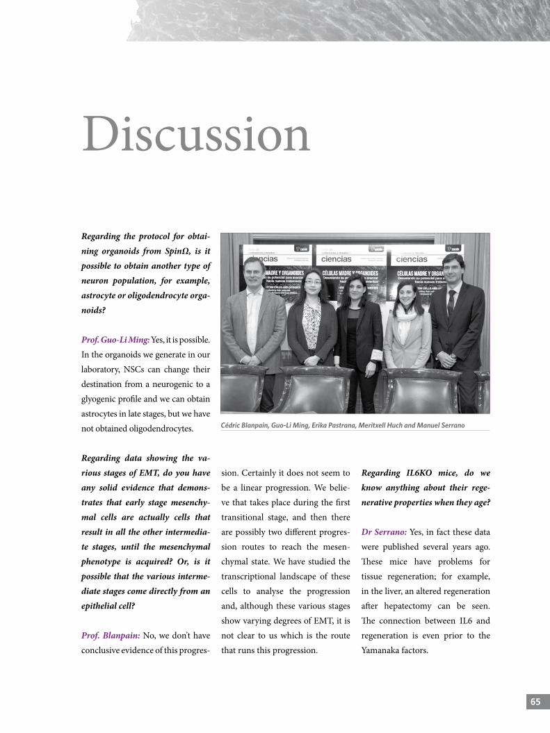
656565
Regarding the protocol for obtai-ning organoids from SpinΩ, is it possible to obtain another type of neuron population, for example, astrocyte or oligodendrocyte orga-noids?
Prof. Guo-Li Ming: Yes, it is possible. In the organoids we generate in our laboratory, NSCs can change their destination from a neurogenic to a glyogenic profile and we can obtain astrocytes in late stages, but we have not obtained oligodendrocytes.
Regarding data showing the va-rious stages of EMT, do you have any solid evidence that demons-trates that early stage mesenchy-mal cells are actually cells that result in all the other intermedia-te stages, until the mesenchymal phenotype is acquired? Or, is it possible that the various interme-diate stages come directly from an epithelial cell?
Prof. Blanpain: No, we don’t have conclusive evidence of this progres-
sion. Certainly it does not seem to be a linear progression. We belie-ve that takes place during the first transitional stage, and then there are possibly two different progres-sion routes to reach the mesen-chymal state. We have studied the transcriptional landscape of these cells to analyse the progression and, although these various stages show varying degrees of EMT, it is not clear to us which is the route that runs this progression.
Regarding IL6KO mice, do we know anything about their rege-nerative properties when they age?
Dr Serrano: Yes, in fact these data were published several years ago. These mice have problems for tissue regeneration; for example, in the liver, an altered regeneration after hepatectomy can be seen. The connection between IL6 and regeneration is even prior to the Yamanaka factors.
Discussion
Cédric Blanpain, Guo-Li Ming, Erika Pastrana, Meritxell Huch and Manuel Serrano
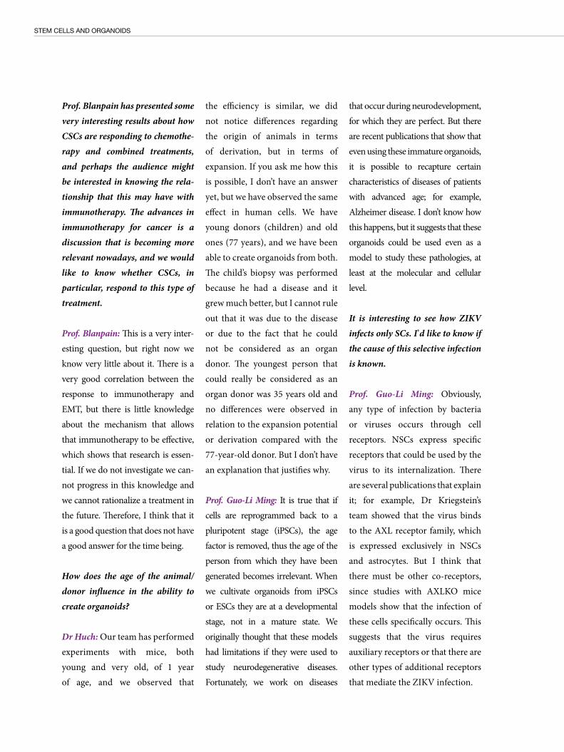
STEM CELLS AND ORGANOIDS
Prof. Blanpain has presented some very interesting results about how CSCs are responding to chemothe-rapy and combined treatments, and perhaps the audience might be interested in knowing the rela-tionship that this may have with immunotherapy. The advances in immunotherapy for cancer is a discussion that is becoming more relevant nowadays, and we would like to know whether CSCs, in particular, respond to this type of treatment.
Prof. Blanpain: This is a very inter-esting question, but right now we know very little about it. There is a very good correlation between the response to immunotherapy and EMT, but there is little knowledge about the mechanism that allows that immunotherapy to be effective, which shows that research is essen-tial. If we do not investigate we can-not progress in this knowledge and we cannot rationalize a treatment in the future. Therefore, I think that it is a good question that does not have a good answer for the time being.
How does the age of the animal/donor influence in the ability to create organoids?
Dr Huch: Our team has performed experiments with mice, both young and very old, of 1 year of age, and we observed that
the efficiency is similar, we did not notice differences regarding the origin of animals in terms of derivation, but in terms of expansion. If you ask me how this is possible, I don’t have an answer yet, but we have observed the same effect in human cells. We have young donors (children) and old ones (77 years), and we have been able to create organoids from both. The child’s biopsy was performed because he had a disease and it grew much better, but I cannot rule out that it was due to the disease or due to the fact that he could not be considered as an organ donor. The youngest person that could really be considered as an organ donor was 35 years old and no differences were observed in relation to the expansion potential or derivation compared with the 77-year-old donor. But I don’t have an explanation that justifies why.
Prof. Guo-Li Ming: It is true that if cells are reprogrammed back to a pluripotent stage (iPSCs), the age factor is removed, thus the age of the person from which they have been generated becomes irrelevant. When we cultivate organoids from iPSCs or ESCs they are at a developmental stage, not in a mature state. We originally thought that these models had limitations if they were used to study neurodegenerative diseases. Fortunately, we work on diseases
that occur during neurodevelopment, for which they are perfect. But there are recent publications that show that even using these immature organoids, it is possible to recapture certain characteristics of diseases of patients with advanced age; for example, Alzheimer disease. I don’t know how this happens, but it suggests that these organoids could be used even as a model to study these pathologies, at least at the molecular and cellular level.
It is interesting to see how ZIKV infects only SCs. I’d like to know if the cause of this selective infection is known.
Prof. Guo-Li Ming: Obviously, any type of infection by bacteria or viruses occurs through cell receptors. NSCs express specific receptors that could be used by the virus to its internalization. There are several publications that explain it; for example, Dr Kriegstein’s team showed that the virus binds to the AXL receptor family, which is expressed exclusively in NSCs and astrocytes. But I think that there must be other co-receptors, since studies with AXLKO mice models show that the infection of these cells specifically occurs. This suggests that the virus requires auxiliary receptors or that there are other types of additional receptors that mediate the ZIKV infection.
67
Discussion
67
Dr Serrano, has shown us that the only way to reprogram in vivo in the lung is by inducing damage. What happens when this damage is indu-ced in the rest of the tissues where there are already reprogramming events?
Dr Serrano: If we induce exogenous damage in the pancreas we will ob-tain more reprogramming events. There is always an additive response.
Dr Serrano, do you think that se-nescence is the key molecular path for the development of neurodege-nerative diseases?
Dr Serrano: Although they have not yet been published, results shown in several congresses in-dicate that Parkinson and Alzhe-imer diseases are associated with the presence of senescent cells. On the other hand, it is known that improvement is achieved when
removing senescent cells in mu-rine models of neurodegenerative diseases. These are not our results, but I think it is something that we will see posted soon in high-im-pact journals. According to Dr Serrano, partly re-programmed cells do not resemble any other cell type. To assert this, have you made a comparative study of the expression of some markers? Or have you performed compara-tive studies of gene expression with the different stages of development to determine, at least, which of these stages they look like?
Dr Serrano: Actually, these cells resemble pancreatic ductal cells, but do not have all the markers of this cell type. We have conducted a gene expression study, and the cells are actually quite similar among themselves, but are not exactly identical.
Regarding this stochastic popula-tion of alleged intermediate sta-ges of plasticity, does it also work in the opposite direction; that is, during development? Is it possible that there is no determined route? Because for the differentiation of some tissues, we know that it is so, but maybe this process occurs at other stages of development.
Dr Serrano: I don’t know, the im-pression that I get from develop-ment specialists is that it is a very specific route, but I could be wrong.
Prof. Guo-Li Ming: Some stu-dies conducted in murine models show that if they suffer any type of damage, parenchymal cells can become SCs and create neurons. That would be due to a kind of intrinsic reprogramming, but I’m not sure that this can happen in the same way in humans or in other tissues.

STEM CELLS AND ORGANOIDS Unlocking their potential to understand and treat disease
The development, now more than 10 years ago, of genetic strategies that allow “reprogramming” of adult cells to pluripotent stem cells brought to the biomedicine arena new means to study and treat diseases. This technology allows for the development of cellular models of disease by using human cells derived directly from patients, which allows the advancement of both basic disease knowledge and the development of specific therapies. Reprogrammed cells also have the potential to be used as cell therapy and they are being investigated for diseases such as Parkinson’s or immune diseases. In recent years, stem cell culture techniques (embryonic, adult or reprogrammed) have been refined to allow for the development of structures that resemble organs in vitro. Nowadays, “organoids” similar to the human intestine, heart, eye, or brain have been developed and are currently allowing the study of both the basic processes involved in the development of these organs and the dysfunctions associated with developmental diseases.
El desarrollo, hace ahora algo más de 10 años, de estrategias genéticas que permiten “reprogramar” células adultas a células pluripotentes o “madre” ha puesto a disposición de la biomedicina nuevos medios para estudiar y tratar enfermedades. Esta tecnología permite desarrollar modelos celulares de la enfermedad usando células humanas derivadas directamente del paciente y permite avanzar tanto en el conocimiento básico de la enfermedad como en el desarrollo de terapias específicas. Las células reprogramadas también tienen potencial como terapia celular y su uso se está investigando para enfermedades como el Parkinson o enfermedades inmunológicas. En los últimos años, las técnicas de cultivo de células madre (embrionarias, adultas o reprogramadas) se ha refinado hasta permitir el desarrollo de estructuras que asemejan órganos in vitro. En la actualidad, se han creado “organoides” similares al intestino, el corazón, el ojo o el cerebro humano que están permitiendo estudiar, tanto los procesos básicos involucrados en el desarrollo de estos órganos, como las disfunciones asociadas a enfermedades del desarrollo.
CÉLULAS MADRE Y ORGANOIDES Desvelando su potencial para avanzar hacia nuevos tratamientos