Embed Size (px)
Citation preview
Revista Mexicana de Ciencias Agrícolas Pub. Esp. Núm. 5 16 de mayo - 29 de junio, 2013 p. 913-924
Rutas de la penetración foliar en la fertilización de la orquídea Cymbidium sp. (Orchidaceae)*
Foliar penetration routes for fertilizing the Orchid Cymbidium sp. (Orchidaceae)
Víctor García Gaytán1, Guadalupe Valdovinos Ponce2, María de la Nieves Rodríguez Mendoza1, Martha E. Pedraza Santos3, Libia I. Trejo Tellez1 y Marcos Soto Hernández4
1Colegio de Postgraduados. Posgrado en Edafología. Carretera México-Texcoco, km 36.5, Montecillo, Texcoco, Estado de México. C. P. 56230. Tel. 01 595 95 2 02 00 ext. 1262. ([email protected], [email protected], [email protected]). 2Posgrado en Fitosanidad. Colegio de Postgraduados. Tel. 595 95 2 02 00 ext. 1612 ([email protected]). 3Facultad de Agrobiología “Pte. Juárez”, Uruapan, Michoacán, México. Paseo Gral. Lázaro Cárdenas y Berlín S/N, Colonia Viveros, C. P. 60170. Tel. 01 4521126710. ([email protected]). 4Posgrado en Botánica. Colegio de Postgraduados. Tel. 595 95 2 02 00. Ext. 1361. ([email protected]). §Autora para correspondencia:[email protected].
* Recibido: diciembre de 2012
Aceptado: marzo de 2013
Resumen
Los objetivos de la presente investigación fueron caracterizar la anatomía foliar e identificar las rutas de penetración de un fertilizante foliar10-10-10 (NPK) en la orquídea (Cymbidium sp.), especie con potencial comercial por alto su costo como flor de maceta y de corte. Por las características morfológicas de la planta, el precisar los sitios de absorción de nutrientes permite optimizar la técnica y los insumos a aplicar. Las aplicaciones foliares y cortes histológicos se llevaron a cabo en el laboratorio de Histopatología Vegetal del Colegio de Postgraduados en Ciencias Agrícolas, Campus Montecillo, durante los meses de abril a agosto de 2012. El grosor de la cutícula a lo largo de la hoja no fue uniforme, en la porción media fue más gruesa (29.90 µm) que en las porciones apical (26.91 µm) y basal (25.75 µm). El número de estomas a lo largo de la hoja fue constante. En la región media de la hoja, el grosor del tejido epidérmico (63.045 µm) y el diámetro de lo haces de las fibras subepidérmicas (184.55 µm) mostraron los mayores valores, mientras que el mayor diámetro del haz vascular (1 031 µm) y el mayor grosor del mesófilo (3 777.91 µm) se presentaron en la porción basal de las hojas. La ruta de penetración del fertilizante foliar fue vía cuticular
Abstract
The objectives of this study were to characterize the leaf anatomy and identify the routes of penetration of a foliar fertilizer 10-10-10 (NPK) on the Orchid (Cymbidium sp.), a species with high profitable potential for its high prices as a pot and cutting flower. Considering the plant´s morphological characteristics, to precise the nutrient absorption sites concede to optimize the technique and the inputs to be applied. Foliar applications and histological sections were conducted in the laboratory of Plant Histopathology of the Postgraduate College in Agricultural Sciences, Campus Montecillo, during the months April to August, 2012. The thickness of the cuticle along the leaf was not even; in the middle portion was thicker (29.90 µm) than in the apical (26.91 µm) and basal portions (25.75 µm). The number of stomata throughout the leaf was uniform. In the middle region of the leaf, the epidermal tissue thickness (63 045 µm), the diameter of the subepidermal fiber bundles (184.55 µm) showed the highest values, while the larger diameter of the vascular bundle (1 031 µm) and the thicker mesophyll (3 777.91 µm) occurred in the basal portion of the leaves. The route of penetration of the foliar fertilizer was via cuticular and stomatal from 30 min after making the
Víctor García Gaytán et al.914 Rev. Mex. Cienc. Agríc. Pub. Esp. Núm. 5 16 de mayo - 29 de junio, 2013
y estomática a partir de los 30 min después de haber hecho la aplicación y a los 120 min se observó en los pseudobulbos, lo que sugiere que fertilizante fluye hacia esta zona evitando su pérdida. Éstos resultados indican que la fertilización foliar es una alternativa viable que puede implementarse en las prácticas de producción de esta ornamental.
Palabras clave: Cymbidium sp., orquídea, pseudobulbos.
Introducción
La orquídea Cymbidium, considerada como una de las flores de maceta de mayor importancia, tiene un potencial ornamental con alto valor económico debido a que su demanda ha crecido en las últimas décadas tanto en diversidad de especies como en producción de flores (Huang et al., 2011). Es una orquídea perennifolia con grandes inflorescencias de donde brotan las flores llevadas por breves peciolos, las flores son color verde, rosa, amarillo o blanca. Las hojas son provistas por una sola nervadura central y pueden alcanzar el largo de hasta un metro en las especies más grandes y crecen de 5 hasta 15 por pseudobulbo. Los pseudobulbos que quedan revestidos por las vainas foliares de las hojas, dispuestos uno contra los otros a nivel del suelo y unidos entre ellos por cortos rizomas subterráneos. Las raíces parten de la parte inferior de los pseudobulbos y son carnosas y algo ramificadas revestidas por un velamen blanquecino.
La aplicación de nutrientes foliares puede ser un método que contribuya a reducir el impacto ambiental asociado con la fertilización al suelo; sin embargo, la respuesta a menudo es variable y no reproducible debido a factores climáticos, de especie y formulación (Fernández y Eichert, 2009). La fertilización foliar es una práctica común en los sistemas de producción de orquídeas, pero en Cymbidium se tiene poca información sobre la absorción de nutrientes a nivel foliar y radical (Hew y Wan, 2004). Durante años, ha habido controversia sobre las características estructurales y químicas de la cutícula que se relacionan con la absorción de los fertilizantes foliares.
Las ceras epicuticulares son el componente más externo e hidrofóbico y su estructura limita la penetración de agua y de iones a través de la membrana (Marschner, 1995; Wójcik, 2004), lo que afecta de manera significativa la permeabilidad cuticular, adicionalmente, cutículas con estructuras completamente reticuladas son más permeables a ciertas
application and 120 min later it was visually observed in the pseudobulbs, suggesting that the fertilizer flows into this area preventing loss. These results indicate that foliar feeding is a viable alternative that can be implemented in this ornamental production.
Keywords: Cymbidium sp., Orchid, pseudobulbs.
Introduction
The Orchid Cymbidium, considered one of the most important pot flowers has a high ornamental potential of economic value because its demand has grown in recent decades in both, species diversity and production of flowers (Huang et al. 2011). It is a large evergreen orchid inflorescences where the flowers bloom carried by short petioles, the flowers are green, pink, yellow or white. The leaves are provided by a single midrib and can reach up to a meter long in the largest species and grow 5-15 per pseudobulb. The pseudobulbs coated by the leaf sheaths of the leaves are arranged one against the other at ground level and joined together by short underground rhizomes. The roots start from the bottom of the pseudobulbs and are fleshy and slightly branched covered by a white canopy.
The foliar application of nutrients can be a method to help reducing the environmental impact associated with soil fertilization; however, the response is often variable and not reproducible due to climatic factors, species and formulation (Fernández and Eichert, 2009). Foliar feeding is a common practice in the production systems of Cymbidium orchids, but there is little information on nutrient absorption on leaf and root level (Hew and Wan, 2004). For years, there has been controversy over the structural and chemical characteristics of the cuticle that is related to the absorption of foliar fertilizers.
Epicuticular waxes are the most external and hydrophobic component and its structure limits the penetration of water and ions across the membrane (Marschner, 1995; Wojcik, 2004), significantly affecting the cuticular permeability, additionally, fully crosslinked cuticles structures are more permeable to certain substances than those with external lamellar regions (Ray, 2006). It has been suggested that, the absorption of ions from the leaves can be completed in three stages, in the first one, the substances penetrate the cuticle and the cell wall through free diffusion, in the second one, these substances pass through the plasma membrane, and in the third stage
Rutas de la penetración foliar en la fertilización de la orquídea Cymbidium sp. (Orchidaceae) 915
sustancias que aquellas con una región laminosa externa (Ray, 2006). Se ha sugerido que la absorción de iones por las hojas puede ser completada en tres etapas, en la primera, las sustancias penetran la cutícula y la pared celular a través de difusión libre; en la segunda, estas sustancias atraviesan la membrana plasmática, y en la tercera etapa las sustancias son llevadas al citoplasma en un proceso que requiere energía derivada del metabolismo (Fageria et al., 2009). Dada la importancia de la fertilización en plantas de Cymbidium, el objetivo de esta investigación fue caracterizar la anatomía foliar de la orquídea e identificar las rutas de penetración de un fertilizante foliar.
Materiales y métodos
Tres plantas de orquídeas (Cymbidium sp.) de 5 años de edad, cultivadas en invernadero en en la Universidad Michoacana de San Nicolás de Hidalgo, Facultad de Agrobiología “Pte. Juárez”, Uruapan, Michoacán, se trasladaron al laboratorio de Histopatología Vegetal del Campus Montecillo del Colegio de Postgraduados en Ciencias Agrícolas para en una primera etapa, caracterizar su estructura foliar y en la segunda etapa determinar la ruta de penetración del fertilizante y la tercera y última verificar la translocación del fertilizante a los órganos de almacenamiento. La investigación se hizo durante los meses de abril a agosto de 2012.
Primera etapa; caracterización de la estructura foliar
De cada planta se disectaron fragmentos foliares de aproximadamente 2 a 3 cm2. Los fragmentos se obtuvieron de las porciones apical, media y basal y se cortaron en un micrótomo de congelación (American Optical, modelo 880) a 25 µm de grosor, las secciones obtenidas se montaron en agua y se midieron el grosor de cutícula y mesófilo, y el diámetro de las células epidérmicas, fibras subepidérmicas, y haces vasculares, Sandoval (2005). La densidad estomática se hizo con impresiones de la lamina foliar y su lectura en microscopio con diferentes campos visuales (Rodés y Collazo, 2006).
Para medir el grosor de la cutícula, las secciones obtenidas se tiñeron con Sudan IV durante 25 min y se montaron en glicerol (35 secciones por cada sección). El sudan IV se caracteriza porque dan color a los lípidos al solubilizarse en ellos (Martínez y Gragera, 2008). Se tomaron fotografías con una cámara digital integrada a un microscopio de luz marca Velab modelo Ve-B6, y la medición de la cutícula se hizo
the substances are brought to the cytoplasm in a process that requires energy from the metabolism (Fageria et al., 2009). Given the importance of plant fertilization, the objective of this investigation was to characterize the orchid´s foliar anatomy and identify the penetration routes for foliar fertilizers.
Materials and methods
Three five-year of age plants (Cymbidium sp.) grown in the Universidad Michoacana de San Nicolás de Hidalgo, Faculty of Agrobiology "Presidente Juárez", Uruapan, Michoacán, were moved to the Plant Histopathology Laboratory, Campus Montecillo of the Postgraduate College in Agricultural Sciences to characterize the leaf structure, to determine the route of penetration of the fertilizer and, finally to verify the translocation of the fertilizer into the storing organs. The research was made during the months April to August, 2012.
First stage; leaf structure characterization
From each plant, leaf fragments were dissected from about 2 to 3 cm2. The fragments were obtained from apical and basal portions and were cut on a freezing microtome (American Optical, model 880) at 25 µm thick, the sections obtained were placed in water and the thickness of cuticle, the mesophyll and, the diameter of the epidermal cells, subepidermal fibers and vascular bundles were measured, Sandoval (2005). The stomatal density was made with lamellar leaf impressions and its reading in a microscope with different visual fields (Rodes and Collazo, 2006).
In order to measure the thickness of the cuticle, the sections obtained were stained with Sudan IV for 25 min and were placed in glycerol (35 sections per section). The Sudan IV is characterized by coloring the lipids by being solubilized within them (Martínez and Gragera, 2008). Photographs were taken with a digital camera integrated with a light microscope Velab brand Ve-B6 model, and the measurement of the cuticle was done with the software Motic Images Plus 2.0 ML considering four visual fields per cross section of the leaf section, the software has been very useful in digital measurement (Guerrero et al., 2008; Alejo-Plata et al., 2011).
Mesophyll thickness and the diameter of the epidermal cells, subepidermal fibers and vascular bundles were measured (Motic Images Plus 2.0 ml) from the images obtained four visual fields of 35 cuts, photographs were taken at 40x.
Víctor García Gaytán et al.916 Rev. Mex. Cienc. Agríc. Pub. Esp. Núm. 5 16 de mayo - 29 de junio, 2013
con el software Motic Imágenes Plus 2.0 Ml considerando cuatro campos visuales por corte transversal y sección de la hoja, el software ha sido de gran utilidad en la medición digital, para diversas investigaciones (Guerrero et al., 2008; Alejo-Plata et al., 2011).
El grosor del mesófilo y el diámetro de las células epidérmicas, fibras subepidérmicas y haces vasculares se midieron (software Motic Imágenes Plus 2.0 ml) a partir de las imágenes obtenidas de cuatro campos visuales de 35 cortes, las fotografías se tomaron a 40 x con el equipo indicado anteriormente.
La densidad estomática se cuantificó en las porciones apical, media y basal de cuatro hojas, en la superficie adaxial de cada muestra se aplicó una gota de la mezcla de Exactoden (silicón fluido) y solución endurecedora, a los 10 min se retiró y se aplicó esmalte de uñas a la zona de contacto, 5 min después las impresiones fueron retiradas con unas pinzas, y colocados en portaobjetos para ser observadas en el microscopio de luz simple (marca Carl Zeiss), se contaron el número de estomas por mm2 en cinco campos visuales de 10 impresiones (Rodés y Collazo, 2006).
Para determinar el peso seco de las hojas, se disectaron 50 fragmentos de 1 cm2 de las porciones apical, media y basal de 2 hojas, las muestras se colocaron en una estufa de aire forzado a temperatura de 70 °C marca Riossa Mod. Hof-125 a 70 °C durante 72 h (Alcantar y Sandoval, 1999; Rodríguez y Rodríguez, 2011). Posteriormente, cada segmento se pesó en una balanza digital marca House.
Con los resultados obtenidos se hizo un análisis de varianza y comparación de medias mediante la prueba de Tukey (p≤ 0.05) con el paquete estadístico SAS 9.3. (SAS, 2010).
Segunda etapa; identificación de las rutas de penetración del fertilizante foliar
El fertilizante foliar 10-10-10 (N P K), a base de urea y fosfato monopotásico al 1%, complementada con DAP-PLUS® como agente tensoactivo, se preparó independientemente con los colorantes verde rápido + azul negro de naftol (F-VR+ANN) a una concentración de 0.05% y con calcoflúor (F-C) al 0.01%. Se asperjaron individualmente con F-VR+ANN y F-C como testigo se asperjó una planta con agua; después de los 30, 60, 90 y 120 min de haber aplicado el fertilizante se muestreo la hoja y de ella se disectaron fragmentos de 0.6 x 0.4 cm (Sandoval, 2005), se cortaron transversalmente con
Stomatal density was quantified in apical, middle and basal portions in four leaves on the adaxial surface of each sample applying a drop of the mixture of Exactoden (silicone fluid) and hardening solution at 10 min was removed and applied nail polish to the contact zone, 5 min later, the impressions were removed with tweezers and placed on slides for observation using simple light microscope (Carl Zeiss brand), we counted the number of stomata per mm2 in five fields in 10 visual impressions (Rodes and Collazo, 2006).
In order to determine dry weight of the leaves, 50 1 cm2-long fragments were dissected from the apical portion, 2 from the middle and basal leaves, the samples were placed in a forced air oven at 70 °C Riossa Hof-Mod 125-70 °C for 72 h (Alcantar and Sandoval, 1999, Rodríguez and Rodríguez, 2011). Then, each segment was weighed on a digital scale, House brand.
With the results obtained an analysis of variance and comparison of means by Tukey test (p≤ 0.05) with the statistical package SAS 9.3. (SAS, 2010) were made.
Second stage; identification of the routes of penetration of the foliar fertilizer
The foliar fertilizer 10-10-10 (NPK), based on urea and 1% potassium dihydrogen phosphate, supplemented with DAP-PLUS® as surfactant was prepared independently with fast green dyes & naphthol blue black (F-VR + ANN) at a concentration of 0.05% and with calcofluor (FC) to 0.01%.
We sprayed them individually with F-VR + ANN and FC, as the control a plant was sprayed with water after 30, 60, 90 and 120 min after the fertilizer was applied, sampling the leaf and dissecting fragments 0.6 x 0.4 cm (Sandoval, 2005), cutting them transversely with a freezing microtome (American optical 880 model) 25 µm thick, the sections were placed between slides and cover slips, the samples were treated with F-VR + ANN observed under a microscope light and sprinkled with F-C.
Third stage; translocation of the foliar fertilizer
At this stage of the investigation, the entire plant was sprayed with FC Cymbidium. Two hours after application were hand cut with a razor about 1 mm thick from the base of the sheet to half pseudobulge the obtained sections were mounted in water and observed under a fluorescence microscope.
Rutas de la penetración foliar en la fertilización de la orquídea Cymbidium sp. (Orchidaceae) 917
micrótomo de congelación (American optical modelo 880) a 25 µm de grosor, los cortes se montaron entre portaobjetos y cubreobjetos, las muestras tratadas con F-VR+ANN se observaron en un microscopio de luz marca Velab modelo VE-B y las asperjadas con F-C en un microscopio de fluorescencia Carl Zeiss.
Tercera etapa: translocación de fertilizante foliar
En esta etapa de la investigación, se asperjó la planta completa de Cymbidium con F-C. Dos horas después de la aplicación se hicieron cortes a mano con una navaja de afeitar de aproximadamente 1 mm de grosor desde la base de la hoja a la mitad del pseudobulbo, las secciones obtenidas se montaron en agua y se observaron en el microscopio de fluorescencia.
Resultados y discusión
Caracterización de la estructura foliar
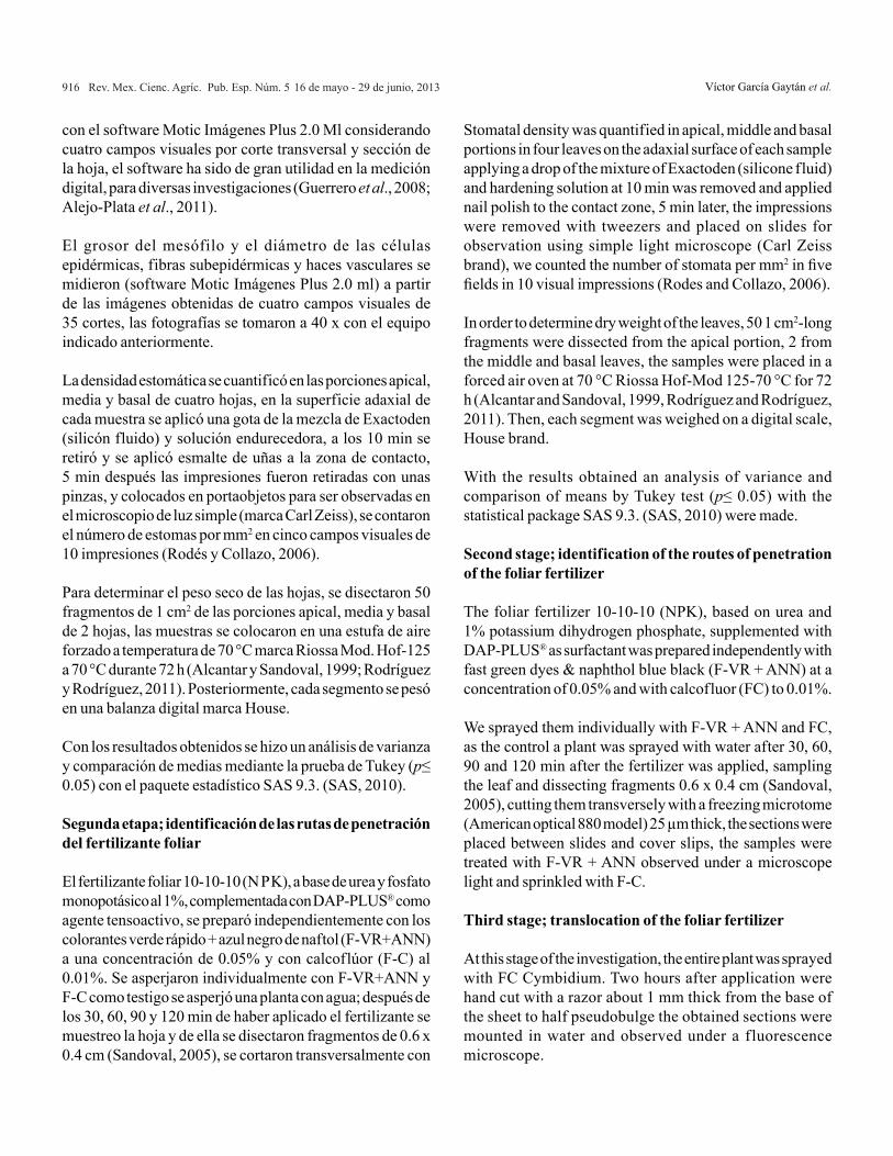
Las hojas de la plantas de Cymbidium estudiada son conduplicadas, rígidas y coriáceas. El tejido epidérmico, está formado por un estrato de células de forma cuadrada a circular y con una cutícula gruesa (Figuras 1A y 1B) que se proyecta hacia las paredes anticlinales. Las células guarda, son superficiales (Figura 1C) y presenta poros fusiformes en la parte central. En C. ensifolium, la cutícula cubre los estomas y parte de las células guarda se eleva por encima de la cutícula. Éstas características pueden ser una modificación xerofítica (Du Puy y Cribb, 2007).
Por debajo de la epidermis se presentan de uno a tres estratos de células hipodérmicas entre las que se agrupan alternadamente haces de tejido esclerenquimatoso (fibras) (Figuras 1A y 1B). La distribución de este tejido, se observa en las superficies adaxial y abaxial de la hoja (Figura 6A). El mesófilo está constituido por un sólo tipo de células de clorénquima de forma redonda y pared delgada entre las cuales se distribuyen los haces vasculares (Figura 1A). En las células del mesófilo se presentan rafidios de gran tamaño (Fig. 1D). Algunas de las células parenquimatosas del mesófilo cercanas a la nervadura central se alargan en dirección perpendicular a la superficie de la hoja.
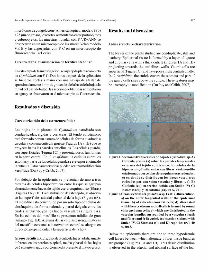
Grosor de cutícula. El grosor de la cutícula fue estadísticamente diferente en las porciones apical, media y basal de las hojas de Cymbidium sp. La porción media presentó el mayor grosor
Results and discussion
Foliar structure characterization
The leaves of the plants studied are conduplicate, stiff and leathery. Epidermal tissue is formed by a layer of square and circular cells with a thick cuticle (Figures 1A and 1B) projecting towards the anticlines walls. Guard cells are superficial (Figure 1C), and have pores in the central spindle. In C. ensifolium, the cuticle covers the stomata and part of the guard cells rises above the cuticle. These features may be a xerophytic modification (Du Puy and Cribb, 2007).
Below the epidermis there are one to three hypodermic cell layers between which alternately fiber tissue bundles are grouped (Figures 1A and 1B). This tissue distribution is observed in the adaxial and abaxial surface of the leaf
Figura 1. Secciones transversales de hoja de Cymbidium sp. A) Cutícula gruesa (a) sobre las paredes tangenciales externas del tejido epidérmico; b) células de la hipodermis; d) alternadas con fibras; c) el mesófilo está formado por células clorenquimatosas redondas; e) en donde se distribuyen los haces vasculares rodeados por una vaina vascular y fibras; y f). B) Cutícula (cu) en sección teñida con Sudán IV; C) Estomas (es); y D) rafidios (ra). 40 X, 2013.
Figure 1. Cross-sections of Cymbidium sp. Leaf: a) thick cuticle. a) on the outer tangential walls of the epidermal tissue; b) of subcutaneous fat cells; d) alternated with fibers; c) the mesophyll cells is formed by round chlorenchyma cells; e) which are distributed in the vascular bundles surrounded by a vascular sheath and fiber; and f) B) cuticle (cu) section stained with Sudan IV, C) Stomata (s); and D) raphides (ra). 40 x, 2013.
Víctor García Gaytán et al.918 Rev. Mex. Cienc. Agríc. Pub. Esp. Núm. 5 16 de mayo - 29 de junio, 2013
(29.90 µm), seguida por las porciones apical (26.91 µm) y basal (25.75 µm) (Figura 2A). Es posible que el mayor grosor en la parte media de las hojas se asocie con su posición, ya que esta sección requiere de mayor resistencia, minimizando la perdida de agua, así como el flujo de transpiración y solutos (Riederer y Schreiber, 2001).
Densidad estomática. En Cymbidium sp., no se encontraron diferencias estadísticas significativas en el número de estomas por mm2 a lo largo de la hoja, ya que en la porción apical se cuantificaron en promedio 7.5, en la media 7 y en la parte basal 7.3 (Figura 2B), éstos resultados no coinciden con lo mencionado por (Bird y Gray, 2003), que el número de los estomas varia en diferentes partes de la misma hoja y en diferentes hojas de la misma planta, este hecho está influenciado por factores ambientales como niveles de luz y CO2. Por la cantidad de impresiones y observaciones que se hicieron en Cymbidium se afirma que el número de estomas es constante a lo largo de la hoja.
Grosor del tejido epidérmico. El grosor de las células epidérmicas fue estadísticamente mayor en la porción media de la hoja (63.045 µm) que en las porciones basal (58.71 µm) y apical (57.738 µm) (Figura 2C). Por las características de las hojas y su longitud, la parte media de estas soporta el dobles de ésta por lo que es entendible el que sea más gruesa. Los cambios en el grosor de las células epidérmicas, están relacionadas por el hábitat donde se desarrollan las especies (Cutler et al., 2007). Éstas características le confieren mayor soporte mecánico y rigidez, protección al mesófilo de la radiación UV-B, almacenamiento de algunos productos metabólicos, minimiza la pérdida de agua así como la aireación del tejido interno a través de los estomas (Dietz et al., 1994; Bilger et al., 2001; Ray, 2006).
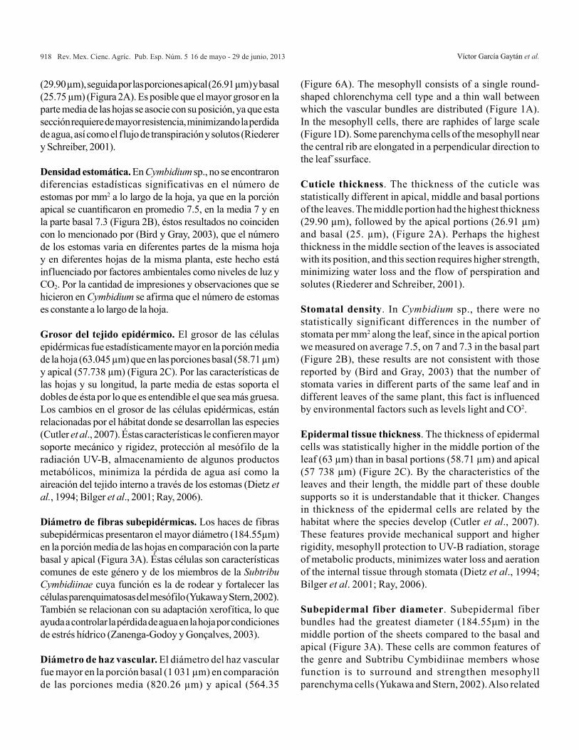
Diámetro de fibras subepidérmicas. Los haces de fibras subepidérmicas presentaron el mayor diámetro (184.55µm) en la porción media de las hojas en comparación con la parte basal y apical (Figura 3A). Éstas células son características comunes de este género y de los miembros de la Subtribu Cymbidiinae cuya función es la de rodear y fortalecer las células parenquimatosas del mesófilo (Yukawa y Stern, 2002). También se relacionan con su adaptación xerofítica, lo que ayuda a controlar la pérdida de agua en la hoja por condiciones de estrés hídrico (Zanenga-Godoy y Gonçalves, 2003).
Diámetro de haz vascular. El diámetro del haz vascular fue mayor en la porción basal (1 031 µm) en comparación de las porciones media (820.26 µm) y apical (564.35
(Figure 6A). The mesophyll consists of a single round-shaped chlorenchyma cell type and a thin wall between which the vascular bundles are distributed (Figure 1A). In the mesophyll cells, there are raphides of large scale (Figure 1D). Some parenchyma cells of the mesophyll near the central rib are elongated in a perpendicular direction to the leaf´ssurface.
Cuticle thickness. The thickness of the cuticle was statistically different in apical, middle and basal portions of the leaves. The middle portion had the highest thickness (29.90 µm), followed by the apical portions (26.91 µm) and basal (25. µm), (Figure 2A). Perhaps the highest thickness in the middle section of the leaves is associated with its position, and this section requires higher strength, minimizing water loss and the flow of perspiration and solutes (Riederer and Schreiber, 2001).
Stomatal density. In Cymbidium sp., there were no statistically significant differences in the number of stomata per mm2 along the leaf, since in the apical portion we measured on average 7.5, on 7 and 7.3 in the basal part (Figure 2B), these results are not consistent with those reported by (Bird and Gray, 2003) that the number of stomata varies in different parts of the same leaf and in different leaves of the same plant, this fact is influenced by environmental factors such as levels light and CO2.
Epidermal tissue thickness. The thickness of epidermal cells was statistically higher in the middle portion of the leaf (63 µm) than in basal portions (58.71 µm) and apical (57 738 µm) (Figure 2C). By the characteristics of the leaves and their length, the middle part of these double supports so it is understandable that it thicker. Changes in thickness of the epidermal cells are related by the habitat where the species develop (Cutler et al., 2007). These features provide mechanical support and higher rigidity, mesophyll protection to UV-B radiation, storage of metabolic products, minimizes water loss and aeration of the internal tissue through stomata (Dietz et al., 1994; Bilger et al. 2001; Ray, 2006).
Subepidermal fiber diameter. Subepidermal fiber bundles had the greatest diameter (184.55μm) in the middle portion of the sheets compared to the basal and apical (Figure 3A). These cells are common features of the genre and Subtribu Cymbidiinae members whose function is to surround and strengthen mesophyll parenchyma cells (Yukawa and Stern, 2002). Also related
Rutas de la penetración foliar en la fertilización de la orquídea Cymbidium sp. (Orchidaceae) 919
µm) (Figura 3B). Se tienen numerosos reportes que señalan la diferencia en tamaño del tejido vascular entre especies de Cymbidium, aunque en ninguna de ellas midió el diámetro. En C. ensifolium, los haces vasculares son de diferente tamaño y se localizan más cerca de las superficies adaxial y basal de las hojas; en otras especies el tejido vascular es adyacente a la superficie abaxial (Yukawa y Stern, 2002).
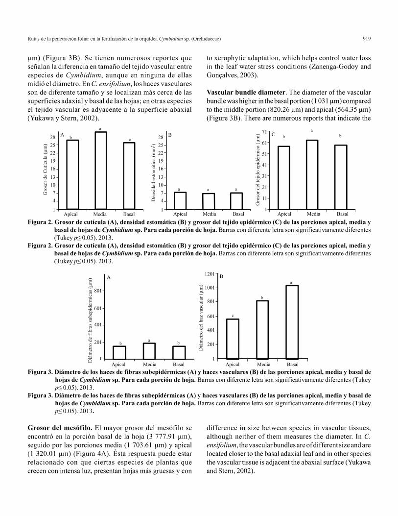
Grosor del mesófilo. El mayor grosor del mesófilo se encontró en la porción basal de la hoja (3 777.91 µm), seguido por las porciones media (1 703.61 µm) y apical (1 320.01 µm) (Figura 4A). Ésta respuesta puede estar relacionado con que ciertas especies de plantas que crecen con intensa luz, presentan hojas más gruesas y con
to xerophytic adaptation, which helps control water loss in the leaf water stress conditions (Zanenga-Godoy and Gonçalves, 2003).
Vascular bundle diameter. The diameter of the vascular bundle was higher in the basal portion (1 031 µm) compared to the middle portion (820.26 µm) and apical (564.35 µm) (Figure 3B). There are numerous reports that indicate the
difference in size between species in vascular tissues, although neither of them measures the diameter. In C. ensifolium, the vascular bundles are of different size and are located closer to the basal adaxial leaf and in other species the vascular tissue is adjacent the abaxial surface (Yukawa and Stern, 2002).
Figura 2. Grosor de cutícula (A), densidad estomática (B) y grosor del tejido epidérmico (C) de las porciones apical, media y basal de hojas de Cymbidium sp. Para cada porción de hoja. Barras con diferente letra son significativamente diferentes (Tukey p≤ 0.05). 2013.
Figura 2. Grosor de cutícula (A), densidad estomática (B) y grosor del tejido epidérmico (C) de las porciones apical, media y basal de hojas de Cymbidium sp. Para cada porción de hoja. Barras con diferente letra son significativamente diferentes (Tukey p≤ 0.05). 2013.
Gro
sor d
e C
utíc
ula
(µm
)
Den
sida
d es
tom
átic
a (m
m2 )
Gro
sor d
el te
jido
epid
érm
ico
(µm
) A B C2825221916131074
1
2825221916131074
1
71
61
51
41
31
21
11
1Apical Media Basal Apical Media Basal Apical Media Basal
a
b c
a a a
a b b
Figura 3. Diámetro de los haces de fibras subepidérmicas (A) y haces vasculares (B) de las porciones apical, media y basal de hojas de Cymbidium sp. Para cada porción de hoja. Barras con diferente letra son significativamente diferentes (Tukey p≤ 0.05). 2013.
Figura 3. Diámetro de los haces de fibras subepidérmicas (A) y haces vasculares (B) de las porciones apical, media y basal de hojas de Cymbidium sp. Para cada porción de hoja. Barras con diferente letra son significativamente diferentes (Tukey p≤ 0.05). 2013.
Diá
met
ro d
e fib
ras s
ubep
ider
mic
as (µ
m)
Diá
met
ro d
el h
az v
ascu
lar (
µm)
Apical Media Basal Apical Media Basal
A B
801
601
401
201
1
1201
1001
801
601
401
201
1
b a b
a
b
c
Víctor García Gaytán et al.920 Rev. Mex. Cienc. Agríc. Pub. Esp. Núm. 5 16 de mayo - 29 de junio, 2013
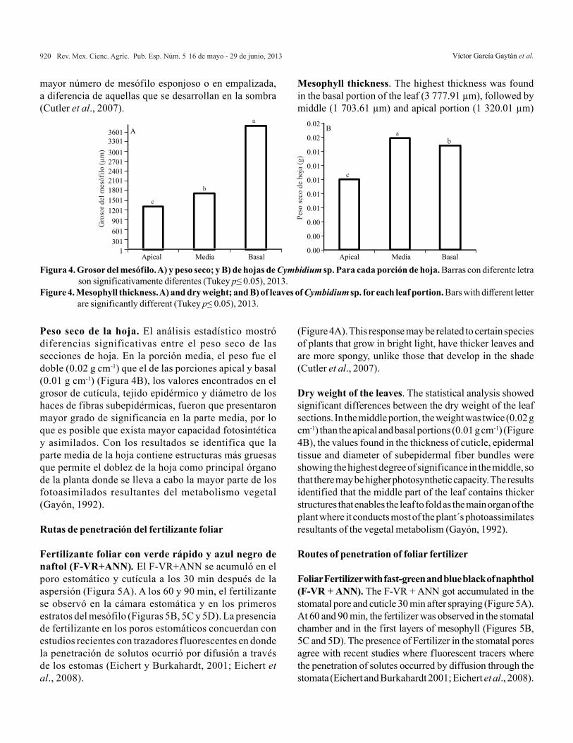
Mesophyll thickness. The highest thickness was found in the basal portion of the leaf (3 777.91 µm), followed by middle (1 703.61 µm) and apical portion (1 320.01 µm)
(Figure 4A). This response may be related to certain species of plants that grow in bright light, have thicker leaves and are more spongy, unlike those that develop in the shade (Cutler et al., 2007).
Dry weight of the leaves. The statistical analysis showed significant differences between the dry weight of the leaf sections. In the middle portion, the weight was twice (0.02 g cm-1) than the apical and basal portions (0.01 g cm-1) (Figure 4B), the values found in the thickness of cuticle, epidermal tissue and diameter of subepidermal fiber bundles were showing the highest degree of significance in the middle, so that there may be higher photosynthetic capacity. The results identified that the middle part of the leaf contains thicker structures that enables the leaf to fold as the main organ of the plant where it conducts most of the plant´s photoassimilates resultants of the vegetal metabolism (Gayón, 1992).
Routes of penetration of foliar fertilizer
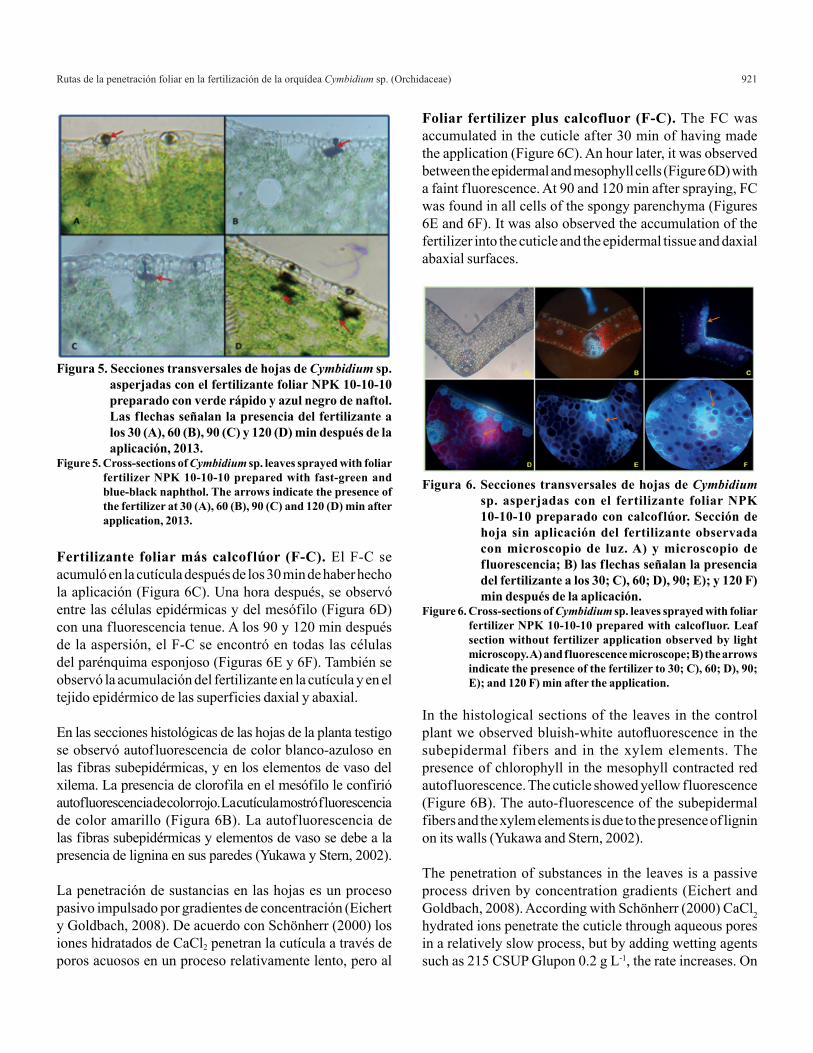
Foliar Fertilizer with fast-green and blue black of naphthol (F-VR + ANN). The F-VR + ANN got accumulated in the stomatal pore and cuticle 30 min after spraying (Figure 5A). At 60 and 90 min, the fertilizer was observed in the stomatal chamber and in the first layers of mesophyll (Figures 5B, 5C and 5D). The presence of Fertilizer in the stomatal pores agree with recent studies where fluorescent tracers where the penetration of solutes occurred by diffusion through the stomata (Eichert and Burkahardt 2001; Eichert et al., 2008).
mayor número de mesófilo esponjoso o en empalizada, a diferencia de aquellas que se desarrollan en la sombra (Cutler et al., 2007).
Peso seco de la hoja. El análisis estadístico mostró diferencias significativas entre el peso seco de las secciones de hoja. En la porción media, el peso fue el doble (0.02 g cm-1) que el de las porciones apical y basal (0.01 g cm-1) (Figura 4B), los valores encontrados en el grosor de cutícula, tejido epidérmico y diámetro de los haces de fibras subepidérmicas, fueron que presentaron mayor grado de significancia en la parte media, por lo que es posible que exista mayor capacidad fotosintética y asimilados. Con los resultados se identifica que la parte media de la hoja contiene estructuras más gruesas que permite el doblez de la hoja como principal órgano de la planta donde se lleva a cabo la mayor parte de los fotoasimilados resultantes del metabolismo vegetal (Gayón, 1992).
Rutas de penetración del fertilizante foliar
Fertilizante foliar con verde rápido y azul negro de naftol (F-VR+ANN). El F-VR+ANN se acumuló en el poro estomático y cutícula a los 30 min después de la aspersión (Figura 5A). A los 60 y 90 min, el fertilizante se observó en la cámara estomática y en los primeros estratos del mesófilo (Figuras 5B, 5C y 5D). La presencia de fertilizante en los poros estomáticos concuerdan con estudios recientes con trazadores fluorescentes en donde la penetración de solutos ocurrió por difusión a través de los estomas (Eichert y Burkahardt, 2001; Eichert et al., 2008).
Figura 4. Grosor del mesófilo. A) y peso seco; y B) de hojas de Cymbidium sp. Para cada porción de hoja. Barras con diferente letra son significativamente diferentes (Tukey p≤ 0.05), 2013.
Figure 4. Mesophyll thickness. A) and dry weight; and B) of leaves of Cymbidium sp. for each leaf portion. Bars with different letter are significantly different (Tukey p≤ 0.05), 2013.
Apical Media Basal
A360133013001270124012101180115011201901601301
1
a
b
c
Gro
sor d
el m
esóf
ilo (µ
m)
Apical Media Basal
B a
b
c
0.02
0.02
0.01
0.01
0.01
0.01
0.01
0.00
0.00
0.00
Peso
seco
de
hoja
(g)
Rutas de la penetración foliar en la fertilización de la orquídea Cymbidium sp. (Orchidaceae) 921
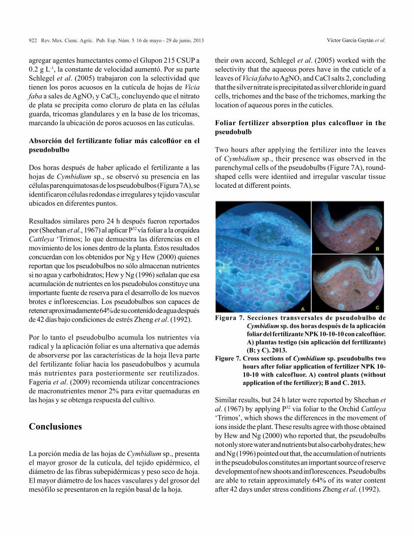
Fertilizante foliar más calcof lúor (F-C). El F-C se acumuló en la cutícula después de los 30 min de haber hecho la aplicación (Figura 6C). Una hora después, se observó entre las células epidérmicas y del mesófilo (Figura 6D) con una fluorescencia tenue. A los 90 y 120 min después de la aspersión, el F-C se encontró en todas las células del parénquima esponjoso (Figuras 6E y 6F). También se observó la acumulación del fertilizante en la cutícula y en el tejido epidérmico de las superficies daxial y abaxial.
En las secciones histológicas de las hojas de la planta testigo se observó autofluorescencia de color blanco-azuloso en las fibras subepidérmicas, y en los elementos de vaso del xilema. La presencia de clorofila en el mesófilo le confirió autofluorescencia de color rojo. La cutícula mostró fluorescencia de color amarillo (Figura 6B). La autof luorescencia de las fibras subepidérmicas y elementos de vaso se debe a la presencia de lignina en sus paredes (Yukawa y Stern, 2002).
La penetración de sustancias en las hojas es un proceso pasivo impulsado por gradientes de concentración (Eichert y Goldbach, 2008). De acuerdo con Schönherr (2000) los iones hidratados de CaCl2 penetran la cutícula a través de poros acuosos en un proceso relativamente lento, pero al
Foliar fertilizer plus calcofluor (F-C). The FC was accumulated in the cuticle after 30 min of having made the application (Figure 6C). An hour later, it was observed between the epidermal and mesophyll cells (Figure 6D) with a faint fluorescence. At 90 and 120 min after spraying, FC was found in all cells of the spongy parenchyma (Figures 6E and 6F). It was also observed the accumulation of the fertilizer into the cuticle and the epidermal tissue and daxial abaxial surfaces.
In the histological sections of the leaves in the control plant we observed bluish-white autofluorescence in the subepidermal fibers and in the xylem elements. The presence of chlorophyll in the mesophyll contracted red autofluorescence. The cuticle showed yellow fluorescence (Figure 6B). The auto-fluorescence of the subepidermal fibers and the xylem elements is due to the presence of lignin on its walls (Yukawa and Stern, 2002).
The penetration of substances in the leaves is a passive process driven by concentration gradients (Eichert and Goldbach, 2008). According with Schönherr (2000) CaCl2 hydrated ions penetrate the cuticle through aqueous pores in a relatively slow process, but by adding wetting agents such as 215 CSUP Glupon 0.2 g L-1, the rate increases. On
Figura 5. Secciones transversales de hojas de Cymbidium sp. asperjadas con el fertilizante foliar NPK 10-10-10 preparado con verde rápido y azul negro de naftol. Las flechas señalan la presencia del fertilizante a los 30 (A), 60 (B), 90 (C) y 120 (D) min después de la aplicación, 2013.
Figure 5. Cross-sections of Cymbidium sp. leaves sprayed with foliar fertilizer NPK 10-10-10 prepared with fast-green and blue-black naphthol. The arrows indicate the presence of the fertilizer at 30 (A), 60 (B), 90 (C) and 120 (D) min after application, 2013.
Figura 6. Secciones transversales de hojas de Cymbidium sp. asperjadas con el fertilizante foliar NPK 10-10-10 preparado con calcoflúor. Sección de hoja sin aplicación del fertilizante observada con microscopio de luz. A) y microscopio de fluorescencia; B) las flechas señalan la presencia del fertilizante a los 30; C), 60; D), 90; E); y 120 F) min después de la aplicación.
Figure 6. Cross-sections of Cymbidium sp. leaves sprayed with foliar fertilizer NPK 10-10-10 prepared with calcofluor. Leaf section without fertilizer application observed by light microscopy. A) and fluorescence microscope; B) the arrows indicate the presence of the fertilizer to 30; C), 60; D), 90; E); and 120 F) min after the application.
Víctor García Gaytán et al.922 Rev. Mex. Cienc. Agríc. Pub. Esp. Núm. 5 16 de mayo - 29 de junio, 2013
agregar agentes humectantes como el Glupon 215 CSUP a 0.2 g L-1, la constante de velocidad aumentó. Por su parte Schlegel et al. (2005) trabajaron con la selectividad que tienen los poros acuosos en la cutícula de hojas de Vicia faba a sales de AgNO3 y CaCl2, concluyendo que el nitrato de plata se precipita como cloruro de plata en las células guarda, tricomas glandulares y en la base de los tricomas, marcando la ubicación de poros acuosos en las cutículas.
Absorción del fertilizante foliar más calcoflúor en el pseudobulbo
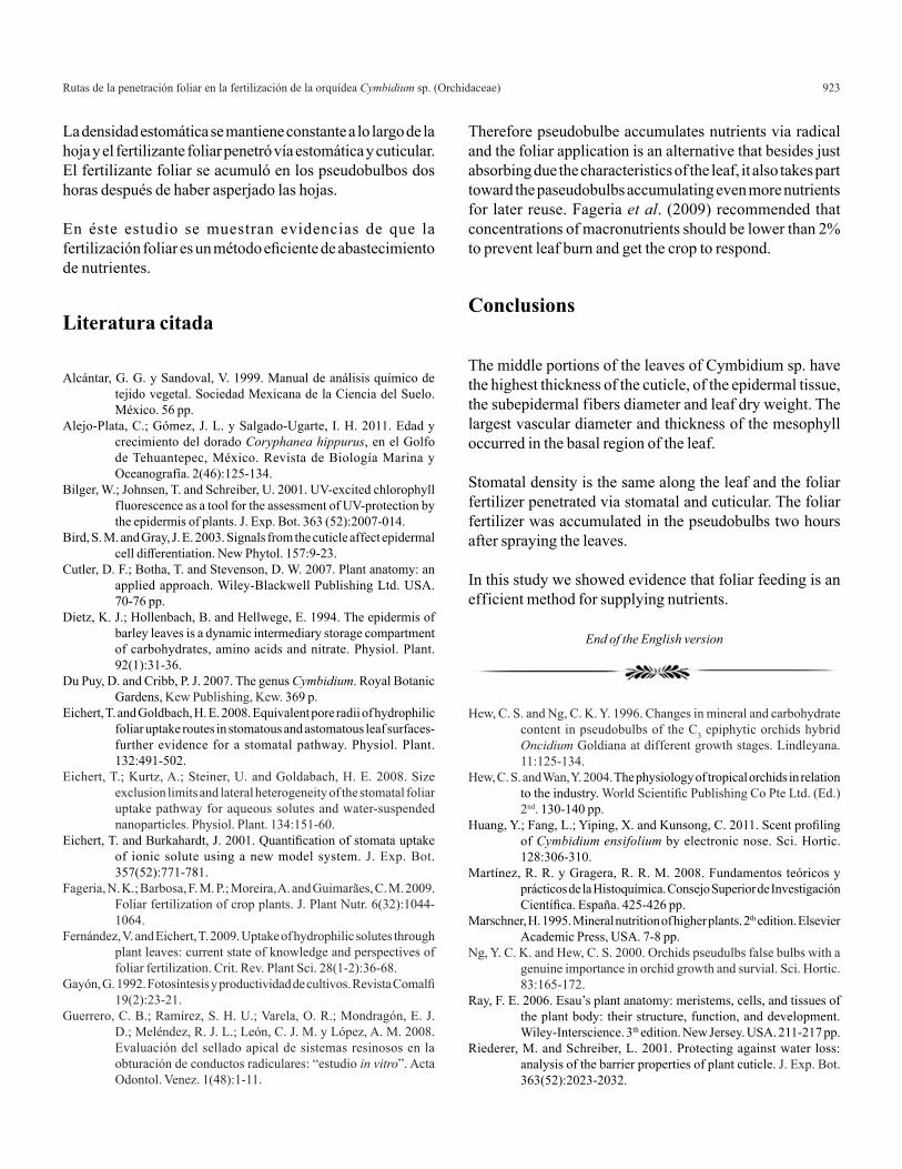
Dos horas después de haber aplicado el fertilizante a las hojas de Cymbidium sp., se observó su presencia en las células parenquimatosas de los pseudobulbos (Figura 7A), se identificaron células redondas e irregulares y tejido vascular ubicados en diferentes puntos.
Resultados similares pero 24 h después fueron reportados por (Sheehan et al., 1967) al aplicar P32 vía foliar a la orquídea Cattleya ‘Trimos; lo que demuestra las diferencias en el movimiento de los iones dentro de la planta. Éstos resultados concuerdan con los obtenidos por Ng y Hew (2000) quienes reportan que los pseudobulbos no sólo almacenan nutrientes si no agua y carbohidratos; Hew y Ng (1996) señalan que esa acumulación de nutrientes en los pseudobulos constituye una importante fuente de reserva para el desarrollo de los nuevos brotes e inflorescencias. Los pseudobulbos son capaces de retener aproximadamente 64% de su contenido de agua después de 42 días bajo condiciones de estrés Zheng et al. (1992).
Por lo tanto el pseudobulbo acumula los nutrientes vía radical y la aplicación foliar es una alternativa que además de absorverse por las características de la hoja lleva parte del fertilizante foliar hacia los paseudobulbos y acumula más nutrientes para posteriormente ser reutilizados. Fageria et al. (2009) recomienda utilizar concentraciones de macronutrientes menor 2% para evitar quemaduras en las hojas y se obtenga respuesta del cultivo.
Conclusiones
La porción media de las hojas de Cymbidium sp., presenta el mayor grosor de la cutícula, del tejido epidérmico, el diámetro de las fibras subepidérmicas y peso seco de hoja. El mayor diámetro de los haces vasculares y del grosor del mesófilo se presentaron en la región basal de la hoja.
their own accord, Schlegel et al. (2005) worked with the selectivity that the aqueous pores have in the cuticle of a leaves of Vicia faba to AgNO3 and CaCl salts 2, concluding that the silver nitrate is precipitated as silver chloride in guard cells, trichomes and the base of the trichomes, marking the location of aqueous pores in the cuticles.
Foliar fertilizer absorption plus calcofluor in the pseudobulb
Two hours after applying the fertilizer into the leaves of Cymbidium sp., their presence was observed in the parenchymal cells of the pseudobulbs (Figure 7A), round-shaped cells were identiied and irregular vascular tissue located at different points.
Similar results, but 24 h later were reported by Sheehan et al. (1967) by applying P32 via foliar to the Orchid Cattleya ‘Trimos’, which shows the differences in the movement of ions inside the plant. These results agree with those obtained by Hew and Ng (2000) who reported that, the pseudobulbs not only store water and nutrients but also carbohydrates; hew and Ng (1996) pointed out that, the accumulation of nutrients in the pseudobulos constitutes an important source of reserve development of new shoots and inflorescences. Pseudobulbs are able to retain approximately 64% of its water content after 42 days under stress conditions Zheng et al. (1992).
Figura 7. Secciones transversales de pseudobulbo de Cymbidium sp. dos horas después de la aplicación foliar del fertilizante NPK 10-10-10 con calcoflúor. A) plantas testigo (sin aplicación del fertilizante) (B; y C). 2013.
Figure 7. Cross sections of Cymbidium sp. pseudobulbs two hours after foliar application of fertilizer NPK 10-10-10 with calcofluor. A) control plants (without application of the fertilizer); B and C. 2013.
Rutas de la penetración foliar en la fertilización de la orquídea Cymbidium sp. (Orchidaceae) 923
Therefore pseudobulbe accumulates nutrients via radical and the foliar application is an alternative that besides just absorbing due the characteristics of the leaf, it also takes part toward the paseudobulbs accumulating even more nutrients for later reuse. Fageria et al. (2009) recommended that concentrations of macronutrients should be lower than 2% to prevent leaf burn and get the crop to respond.
Conclusions
The middle portions of the leaves of Cymbidium sp. have the highest thickness of the cuticle, of the epidermal tissue, the subepidermal fibers diameter and leaf dry weight. The largest vascular diameter and thickness of the mesophyll occurred in the basal region of the leaf.
Stomatal density is the same along the leaf and the foliar fertilizer penetrated via stomatal and cuticular. The foliar fertilizer was accumulated in the pseudobulbs two hours after spraying the leaves.
In this study we showed evidence that foliar feeding is an efficient method for supplying nutrients.
Hew, C. S. and Ng, C. K. Y. 1996. Changes in mineral and carbohydrate content in pseudobulbs of the C3 epiphytic orchids hybrid Oncidium Goldiana at different growth stages. Lindleyana. 11:125-134.
Hew, C. S. and Wan, Y. 2004. The physiology of tropical orchids in relation to the industry. World Scientific Publishing Co Pte Ltd. (Ed.) 2nd. 130-140 pp.
Huang, Y.; Fang, L.; Yiping, X. and Kunsong, C. 2011. Scent profiling of Cymbidium ensifolium by electronic nose. Sci. Hortic. 128:306-310.
Martínez, R. R. y Gragera, R. R. M. 2008. Fundamentos teóricos y prácticos de la Histoquímica. Consejo Superior de Investigación Científica. España. 425-426 pp.
Marschner, H. 1995. Mineral nutrition of higher plants. 2th edition. Elsevier Academic Press, USA. 7-8 pp.
Ng, Y. C. K. and Hew, C. S. 2000. Orchids pseudulbs false bulbs with a genuine importance in orchid growth and survial. Sci. Hortic. 83:165-172.
Ray, F. E. 2006. Esau’s plant anatomy: meristems, cells, and tissues of the plant body: their structure, function, and development. Wiley-Interscience. 3th edition. New Jersey. USA. 211-217 pp.
Riederer, M. and Schreiber, L. 2001. Protecting against water loss: analysis of the barrier properties of plant cuticle. J. Exp. Bot. 363(52):2023-2032.
La densidad estomática se mantiene constante a lo largo de la hoja y el fertilizante foliar penetró vía estomática y cuticular. El fertilizante foliar se acumuló en los pseudobulbos dos horas después de haber asperjado las hojas.
En éste estudio se muestran evidencias de que la fertilización foliar es un método eficiente de abastecimiento de nutrientes.
Literatura citada
Alcántar, G. G. y Sandoval, V. 1999. Manual de análisis químico de tejido vegetal. Sociedad Mexicana de la Ciencia del Suelo. México. 56 pp.
Alejo-Plata, C.; Gómez, J. L. y Salgado-Ugarte, I. H. 2011. Edad y crecimiento del dorado Coryphanea hippurus, en el Golfo de Tehuantepec, México. Revista de Biología Marina y Oceanografía. 2(46):125-134.
Bilger, W.; Johnsen, T. and Schreiber, U. 2001. UV-excited chlorophyll fluorescence as a tool for the assessment of UV-protection by the epidermis of plants. J. Exp. Bot. 363 (52):2007-014.
Bird, S. M. and Gray, J. E. 2003. Signals from the cuticle affect epidermal cell differentiation. New Phytol. 157:9-23.
Cutler, D. F.; Botha, T. and Stevenson, D. W. 2007. Plant anatomy: an applied approach. Wiley-Blackwell Publishing Ltd. USA. 70-76 pp.
Dietz, K. J.; Hollenbach, B. and Hellwege, E. 1994. The epidermis of barley leaves is a dynamic intermediary storage compartment of carbohydrates, amino acids and nitrate. Physiol. Plant. 92(1):31-36.
Du Puy, D. and Cribb, P. J. 2007. The genus Cymbidium. Royal Botanic Gardens, Kew Publishing, Kew. 369 p.
Eichert, T. and Goldbach, H. E. 2008. Equivalent pore radii of hydrophilic foliar uptake routes in stomatous and astomatous leaf surfaces-further evidence for a stomatal pathway. Physiol. Plant. 132:491-502.
Eichert, T.; Kurtz, A.; Steiner, U. and Goldabach, H. E. 2008. Size exclusion limits and lateral heterogeneity of the stomatal foliar uptake pathway for aqueous solutes and water-suspended nanoparticles. Physiol. Plant. 134:151-60.
Eichert, T. and Burkahardt, J. 2001. Quantification of stomata uptake of ionic solute using a new model system. J. Exp. Bot. 357(52):771-781.
Fageria, N. K.; Barbosa, F. M. P.; Moreira, A. and Guimarães, C. M. 2009. Foliar fertilization of crop plants. J. Plant Nutr. 6(32):1044-1064.
Fernández, V. and Eichert, T. 2009. Uptake of hydrophilic solutes through plant leaves: current state of knowledge and perspectives of foliar fertilization. Crit. Rev. Plant Sci. 28(1-2):36-68.
Gayón, G. 1992. Fotosíntesis y productividad de cultivos. Revista Comalfi 19(2):23-21.
Guerrero, C. B.; Ramírez, S. H. U.; Varela, O. R.; Mondragón, E. J. D.; Meléndez, R. J. L.; León, C. J. M. y López, A. M. 2008. Evaluación del sellado apical de sistemas resinosos en la obturación de conductos radiculares: “estudio in vitro”. Acta Odontol. Venez. 1(48):1-11.
End of the English version
Víctor García Gaytán et al.924 Rev. Mex. Cienc. Agríc. Pub. Esp. Núm. 5 16 de mayo - 29 de junio, 2013
Statistical Analysis System (SAS Institute Inc.) 2010. SAS/STAT®User’s Guide. Version 9.0 SAS Institute Inc., Cary. NC. USA.
Schlegel, K.; Shönherr, T. J. and Schreiber, L. 2005. Size selectivity of aqueous pores in stomatous cuticles of Vicia faba leaves. Planta. 221:648-655.
Wójcik, P. 2004. Uptake of mineral nutrients from foliar fertilization (Review). J. Fruit Ornam, Plant Res. 12(2004):202-218.
Yukawa, T. and Stern, W. 2002. Comparative vegetative anatomy and systematic of Cymbidium (Cymbidieae: Orchidaceae). Bot. J. Linnean Soc. 138:383-419.
Zanenga-Godoy, R. y Gonçalves, C. C. 2003. Anatomía foliar de cuatro especies de género de CattleyaLindl. (ORCHIDACEAE) Do Planalto Central. Act. Bot. Bras. 17(1):101-118.
Zheng, X. N.; Wen, Z. Q. and Hew, C. S. 1992. Response of Cymbidium sinense to drought stree. J. Hort. Sci. 67:295-299.
Ródes, G. G. y Collazo, O. M. 2006. Manual de prácticas de fotosíntesis. Universidad Nacional Autónoma de México (UNAM). Facultad de Ciencias. México, D. F. 8-10 pp.
Rodríguez, H. F. y Rodríguez, J. A. 2011. Métodos de análisis de suelo y planta criterios de interpretación. Universidad Autónoma de Nuevo León. Ed. Trillas.2nd. México. 146-157 pp.
Sandoval, Z. E. 2005. Técnicas aplicadas al estudio de la anatomía vegetal. Instituto de Biología. Colección: Cuadernos del Instituto de Biología. Universidad Nacional Autónoma de México. 64-66 pp.
Schönherr, J. 2000. Calcium chloride penetrates plant cuticles via aqueous pores. Planta. 212:212-118.
Sheehan, T. J.; Joiner, J. N. and Cowart, J. K. 1967. Absoption of P32 by Cattleya ‘Trimos’ from foliar and root applications. Florida State Horticultural Society. Florida Agricultural Experiment Stations Journal Series No. 2812.