CRIBADO DEL CÁNCER DECRIBADO DEL CÁNCER DE PULMÓNJOSÉ CERVERA DEVAL
Servicio de Diagnóstico por ImagenServicio de Diagnóstico por ImagenFUNDACIÓN INSTITUTO VALENCIANO DE ONCOLOGÍA
IELCAP Valencia
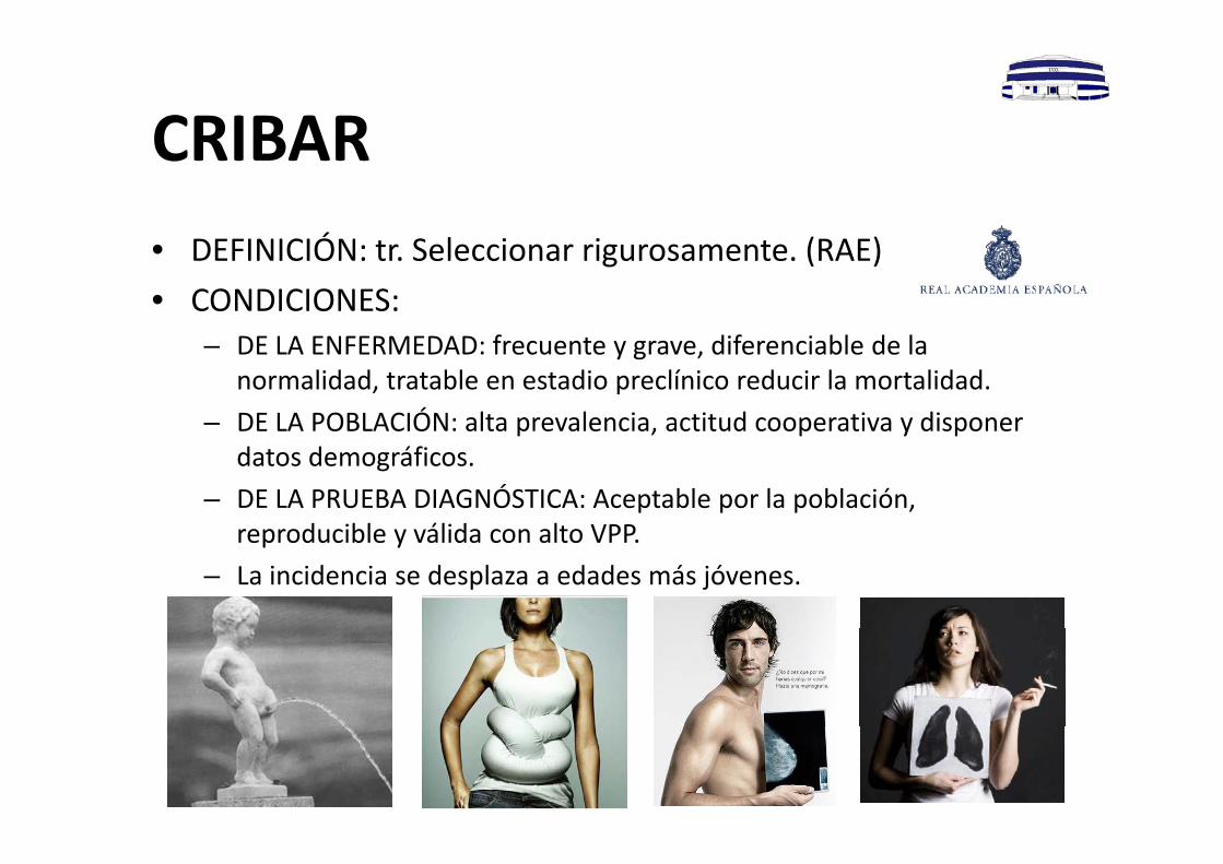
CRIBARCRIBARÓ• DEFINICIÓN: tr. Seleccionar rigurosamente. (RAE)
• CONDICIONES:– DE LA ENFERMEDAD: frecuente y grave, diferenciable de la
normalidad, tratable en estadio preclínico reducir la mortalidad.– DE LA POBLACIÓN: alta prevalencia, actitud cooperativa y disponer p , p y p
datos demográficos.– DE LA PRUEBA DIAGNÓSTICA: Aceptable por la población,
reproducible y válida con alto VPPreproducible y válida con alto VPP.– La incidencia se desplaza a edades más jóvenes.
CÁNCER DE PULMÓN. Un serio problema sociosanitario
– Primera causa de muerte de etiología oncológica– Solo 15% de los pacientes son resecables– Cuando presentan síntomas el 75% tienen metástasis o está localmente avanzado
– Mal pronóstico (12‐15% de supervivencia a los 5 años)– Estadio IV, supervivencia < 3% a 5 años– Aumento de la incidencia en la mujer– Cambio de patrón histológico. Adenocarcinoma
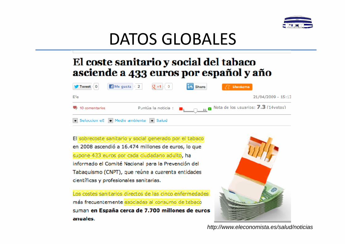
DATOS GLOBALESDATOS GLOBALES
http://www.eleconomista.es/salud/noticias
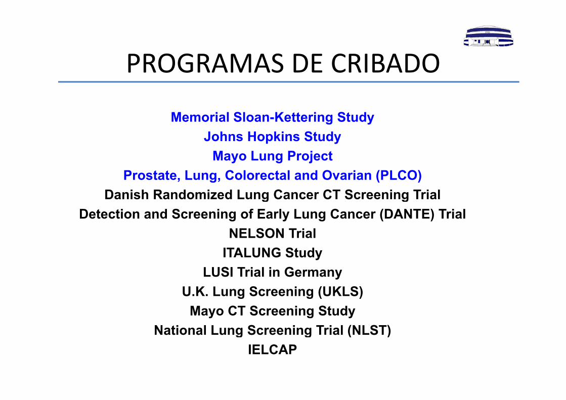
PROGRAMAS DE CRIBADOPROGRAMAS DE CRIBADO
Memorial Sloan-Kettering StudyJohns Hopkins Study
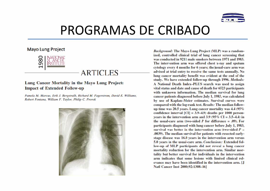
Mayo Lung ProjectMayo Lung ProjectProstate, Lung, Colorectal and Ovarian (PLCO)
Danish Randomized Lung Cancer CT Screening Trialg gDetection and Screening of Early Lung Cancer (DANTE) Trial
NELSON TrialITALUNG Study
LUSI Trial in GermanyU K Lung Screening (UKLS)U.K. Lung Screening (UKLS)
Mayo CT Screening StudyNational Lung Screening Trial (NLST)g g ( )
IELCAP
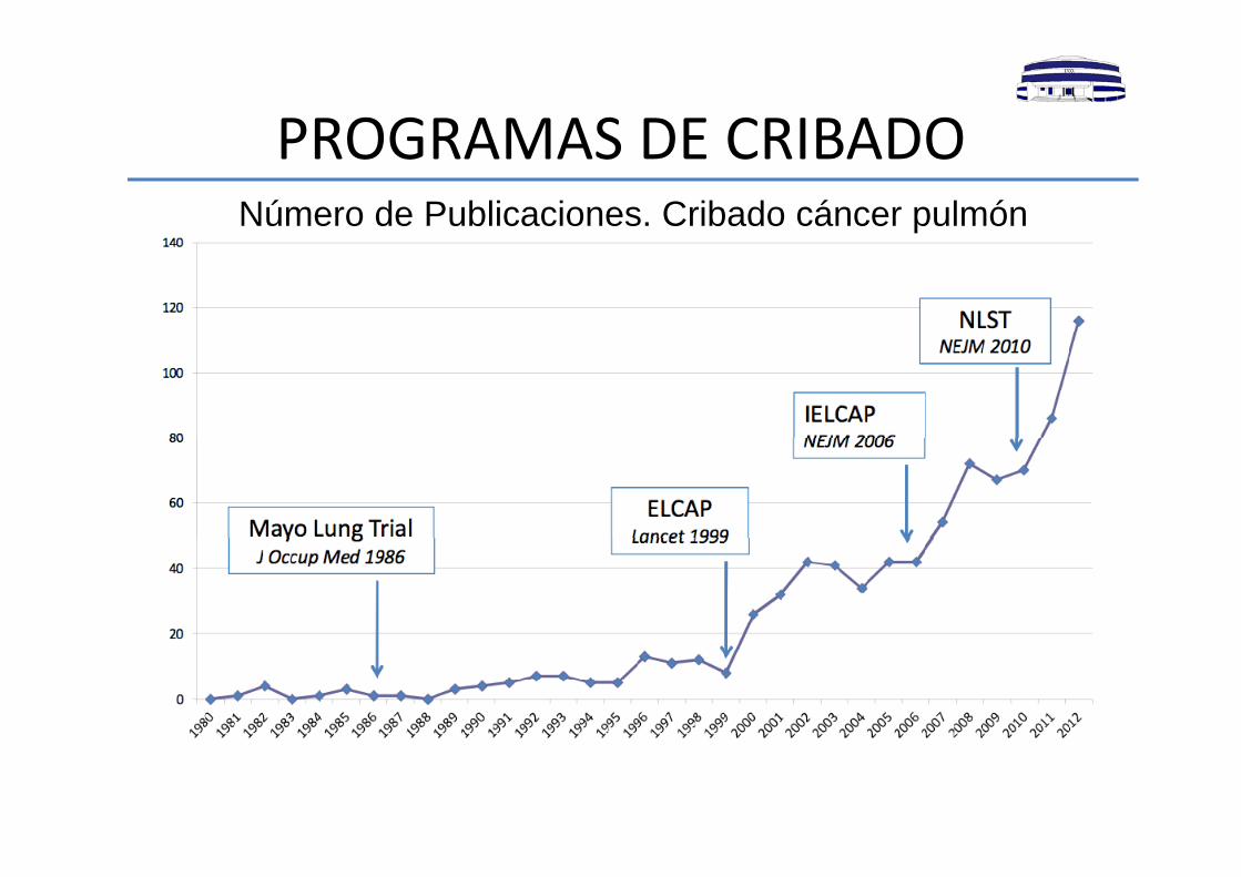
PROGRAMAS DE CRIBADOPROGRAMAS DE CRIBADONúmero de Publicaciones. Cribado cáncer pulmón
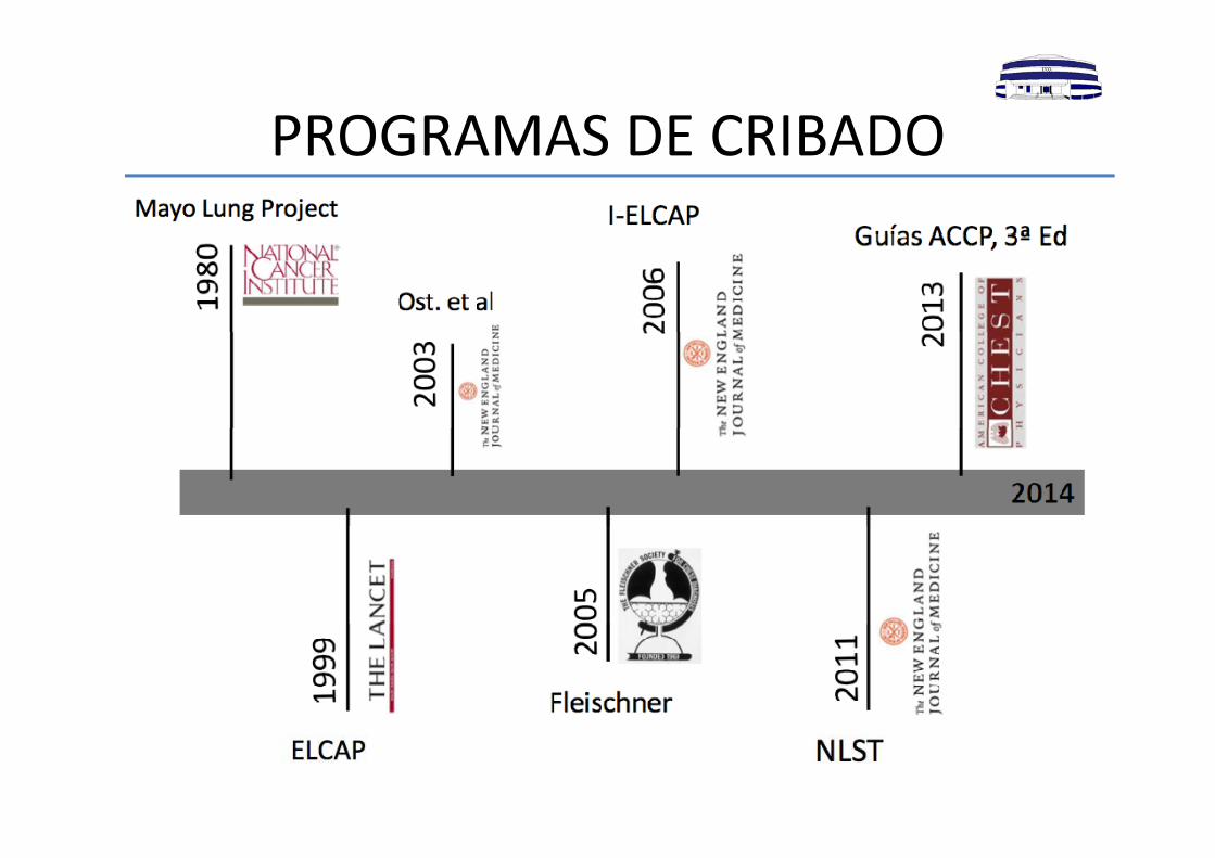
PROGRAMAS DE CRIBADOPROGRAMAS DE CRIBADO
PROGRAMAS DE CRIBADOPROGRAMAS DE CRIBADO
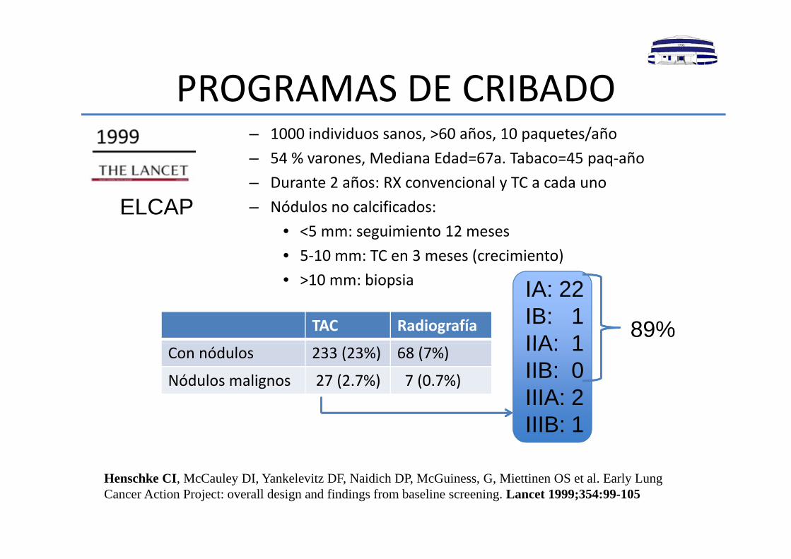
PROGRAMAS DE CRIBADOPROGRAMAS DE CRIBADO– 1000 individuos sanos, >60 años, 10 paquetes/año
54 % M di Ed d 67 T b 45 ñ– 54 % varones, Mediana Edad=67a. Tabaco=45 paq‐año – Durante 2 años: RX convencional y TC a cada uno – Nódulos no calcificados:ELCAP
• <5 mm: seguimiento 12 meses• 5‐10 mm: TC en 3 meses (crecimiento)• >10 mm: biopsia• >10 mm: biopsia
TAC Radiografía
IA: 22IB: 1IIA: 1 89%
Con nódulos 233 (23%) 68 (7%)
Nódulos malignos 27 (2.7%) 7 (0.7%)
IIA: 1IIB: 0IIIA: 2IIIB: 1
Henschke CI, McCauley DI, Yankelevitz DF, Naidich DP, McGuiness, G, Miettinen OS et al. Early Lung Cancer Action Project: overall design and findings from baseline screening. Lancet 1999;354:99-105
PROGRAMAS DE CRIBADOPROGRAMAS DE CRIBADO
PROGRAMAS DE CRIBADOPROGRAMAS DE CRIBADO
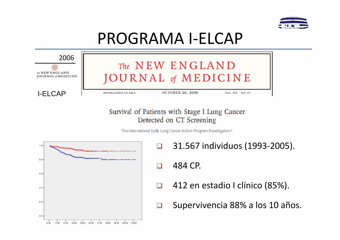
PROGRAMA I ELCAPPROGRAMA I‐ELCAP
I ELCAPI-ELCAP
31 567 individuos (1993‐2005) 31.567 individuos (1993 2005).
484 CP.
412 en estadio I clínico (85%).
l ñ Supervivencia 88% a los 10 años.
PROGRAMAS DE CRIBADOPROGRAMAS DE CRIBADO
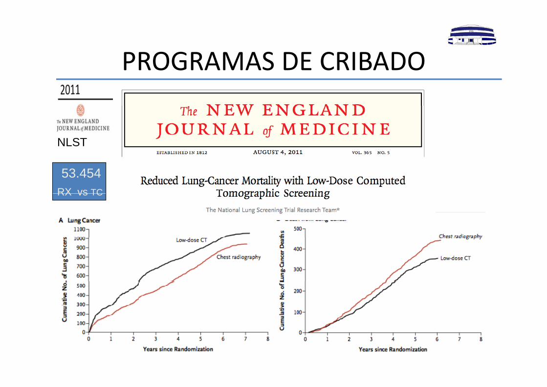
NLST
53.454RX vs TCRX vs TC

PROGRAMAS DE CRIBADOPROGRAMAS DE CRIBADO
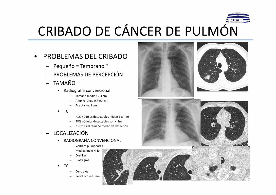
CRIBADO DE CÁNCER DE PULMÓNCRIBADO DE CÁNCER DE PULMÓNA= 0% B=20%• PROBLEMAS DEL CRIBADO
– Pequeño = Temprano ?– PROBLEMAS DE PERCEPCIÓN
A 0% B 20%
PROBLEMAS DE PERCEPCIÓN– TAMAÑO
• Radiografía convencional– Tamaño medio : 2,4 cm– Amplio rango 0,7‐9,4 cm– Aceptable: 1 cm
• TC– <1% nódulos detectables miden 1,5 mm C=30% D=50%D=50%– 48% nódulos detectables son < 3mm– 3 mm es el tamaño medio de detección
– LOCALIZACIÓN• RADIOGRAFÍA CONVENCIONALRADIOGRAFÍA CONVENCIONAL
– Vértices pulmonares– Mediastino e Hilio– Costillas– Diafragma
• TC– Centrales– Periféricos (< 3mm
CRIBADO DE CÁNCER DE PULMÓNCRIBADO DE CÁNCER DE PULMÓN
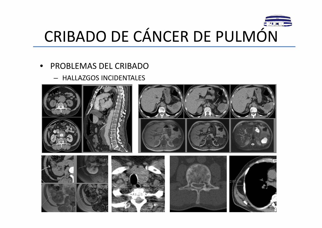
• PROBLEMAS DEL CRIBADO– HALLAZGOS INCIDENTALES
CRIBADO DE CÁNCER DE PULMÓNCRIBADO DE CÁNCER DE PULMÓN
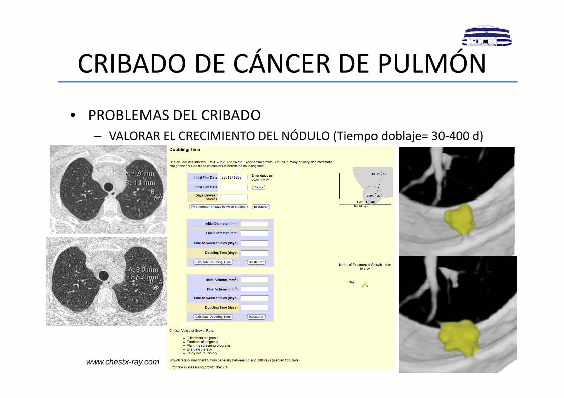
• PROBLEMAS DEL CRIBADO– VALORAR EL CRECIMIENTO DEL NÓDULO (Tiempo doblaje= 30‐400 d)
www.chestx-ray.com
CRIBADO DE CÁNCER DE PULMÓNCRIBADO DE CÁNCER DE PULMÓN
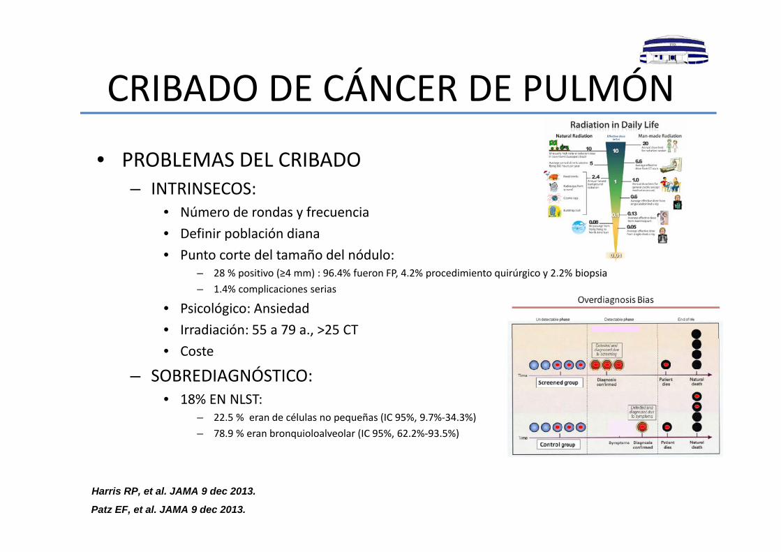
• PROBLEMAS DEL CRIBADO– INTRINSECOS:
• Número de rondas y frecuenciaNúmero de rondas y frecuencia• Definir población diana• Punto corte del tamaño del nódulo:
– 28 % positivo (≥4 mm) : 96.4% fueron FP, 4.2% procedimiento quirúrgico y 2.2% biopsia28 % positivo (≥4 mm) : 96.4% fueron FP, 4.2% procedimiento quirúrgico y 2.2% biopsia– 1.4% complicaciones serias
• Psicológico: Ansiedad• Irradiación: 55 a 79 a., >25 CT• Coste
– SOBREDIAGNÓSTICO:• 18% EN NLST:18% EN NLST:
– 22.5 % eran de células no pequeñas (IC 95%, 9.7%‐34.3%)– 78.9 % eran bronquioloalveolar (IC 95%, 62.2%‐93.5%)
Patz EF, et al. JAMA 9 dec 2013.
Harris RP, et al. JAMA 9 dec 2013.
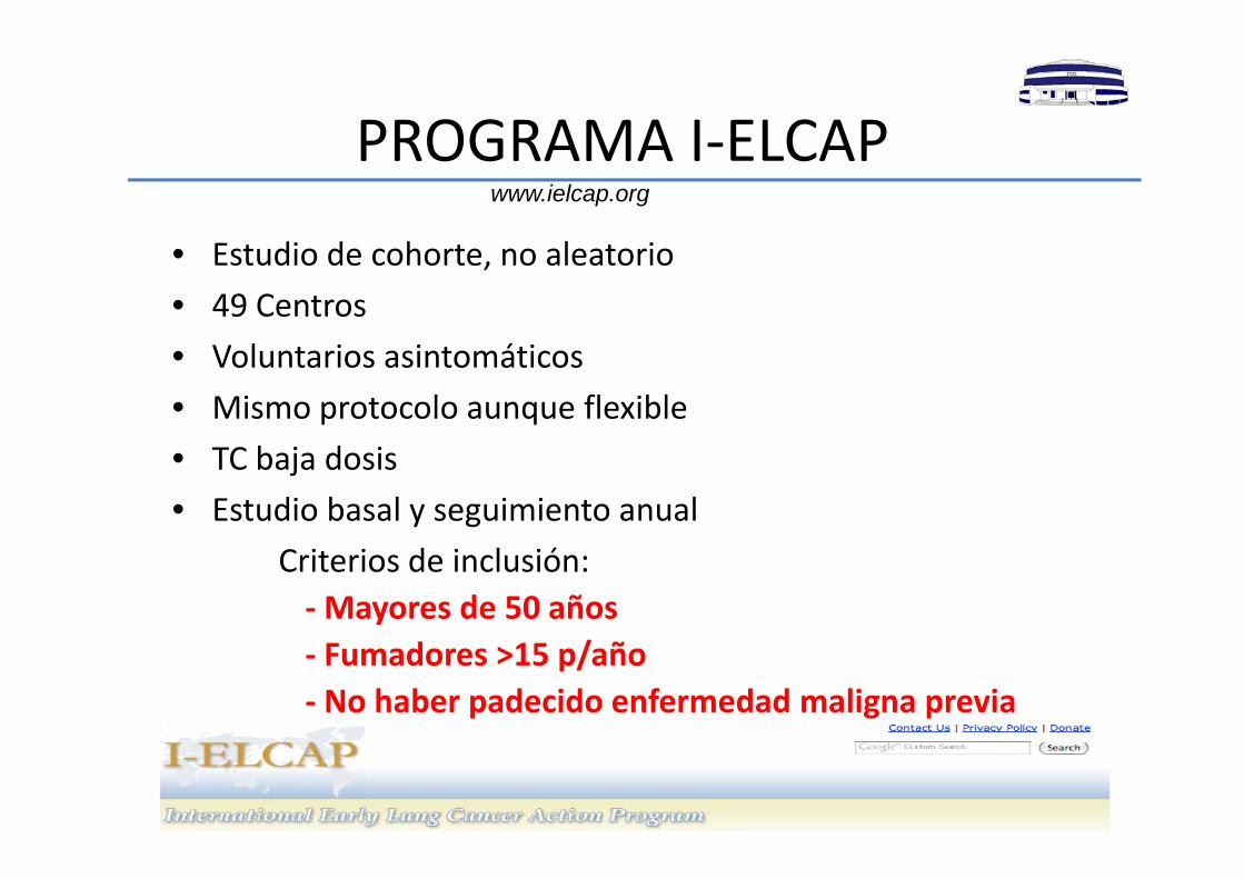
PROGRAMA I ELCAPPROGRAMA I‐ELCAPwww.ielcap.org
• Estudio de cohorte, no aleatorio• 49 Centros• Voluntarios asintomáticos• Mismo protocolo aunque flexible• TC baja dosis• Estudio basal y seguimiento anual
Criterios de inclusión: ‐Mayores de 50 años‐ Fumadores >15 p/año‐ No haber padecido enfermedad maligna previa
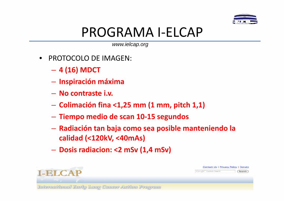
PROGRAMA I ELCAPPROGRAMA I‐ELCAPwww.ielcap.org
• PROTOCOLO DE IMAGEN:– 4 (16) MDCT – Inspiración máxima– No contraste i.v.– Colimación fina <1,25 mm (1 mm, pitch 1,1)– Tiempo medio de scan 10‐15 segundos– Radiación tan baja como sea posible manteniendo la calidad (<120kV, <40mAs)
– Dosis radiacion: <2 mSv (1,4 mSv)
PROGRAMA I ELCAPPROGRAMA I‐ELCAPwww.ielcap.org
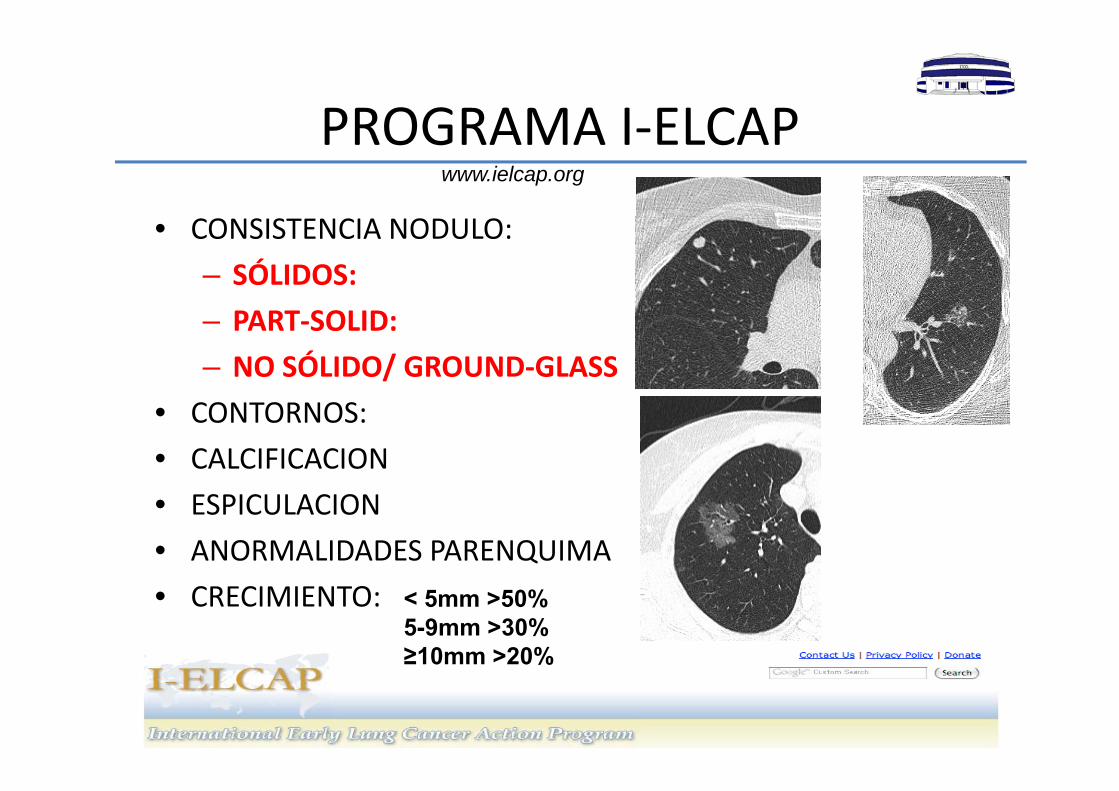
• CONSISTENCIA NODULO:– SÓLIDOS:– PART‐SOLID:– NO SÓLIDO/ GROUND‐GLASS
• CONTORNOS:• CALCIFICACION• ESPICULACION• ANORMALIDADES PARENQUIMA• CRECIMIENTO: < 5mm >50%
5-9mm >30%≥10mm >20%
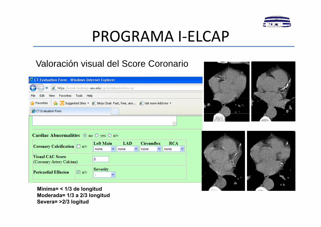
PROGRAMA I ELCAPPROGRAMA I‐ELCAPValoración visual del Score CoronarioValoración visual del Score Coronario
Mínima= < 1/3 de longitudgModerada= 1/3 a 2/3 longitudSevera= >2/3 logitud
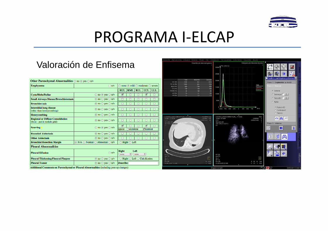
PROGRAMA I ELCAPPROGRAMA I‐ELCAPValoración de EnfisemaValoración de Enfisema
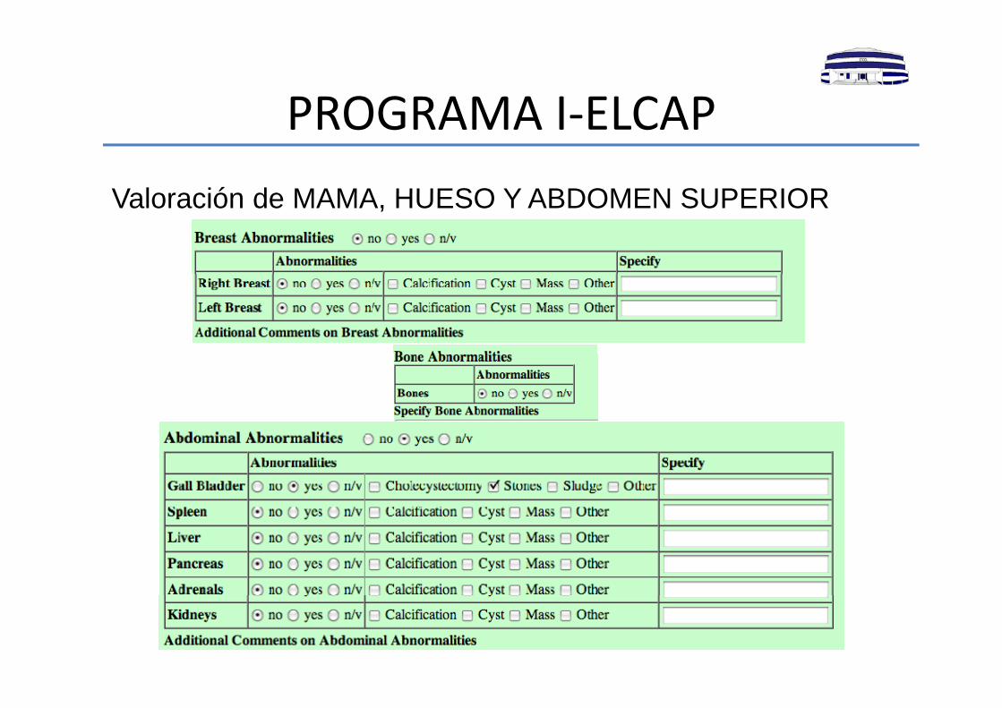
PROGRAMA I ELCAPPROGRAMA I‐ELCAPValoración de MAMA HUESO Y ABDOMEN SUPERIORValoración de MAMA, HUESO Y ABDOMEN SUPERIOR
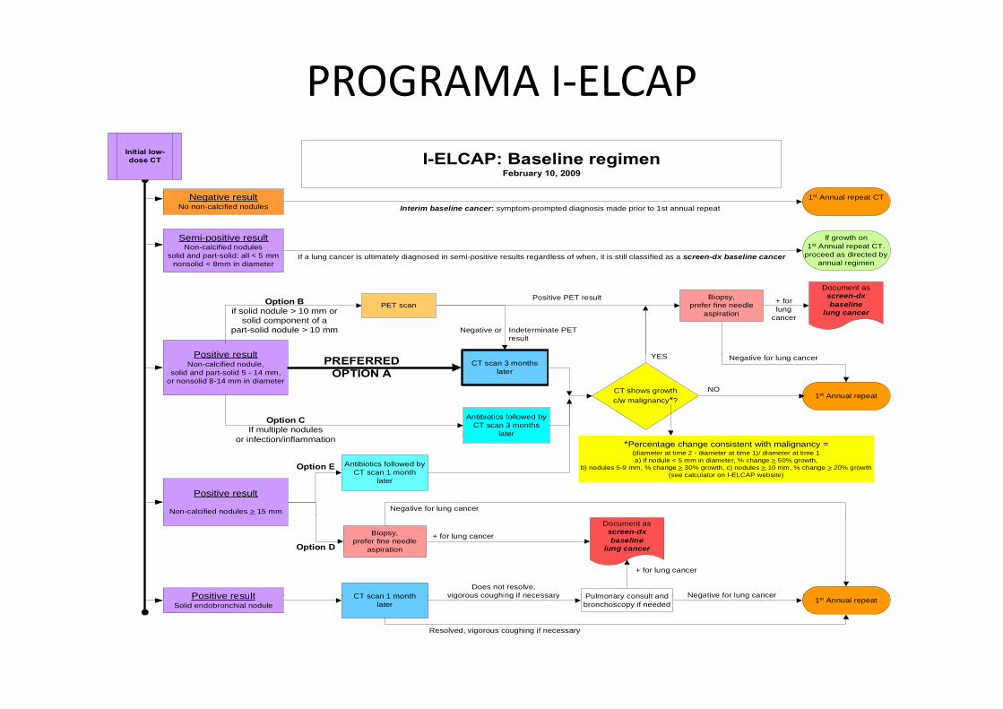
PROGRAMA I‐ELCAPI-ELCAP: Baseline regimen
February 10, 2009
Initial low-dose CT
Semi-positive resultNon-calcified nodules
solid and part-solid: all < 5 mmnonsolid < 8mm in diameter
Negative resultNo non-calcified nodules
1st Annual repeat CT
If growth on1st Annual repeat CT.
proceed as directed byannual regimen
Interim baseline cancer: symptom-prompted diagnosis made prior to 1st annual repeat
If a lung cancer is ultimately diagnosed in semi-positive results regardless of when, it is still classified as a screen-dx baseline cancer
YES
Negative or Indeterminate PETresult
Positive result
PET scanOption Bif solid nodule > 10 mm or
solid component of apart-solid nodule > 10 mm
Positive PET result Biopsy,prefer fine needle
aspiration
Document asscreen-dxbaseline
lung cancer+ forlung
cancer
PREFERREDOPTION A
CT shows growthc/w malignancy*? 1st Annual repeat
Option CIf multiple nodules
YESPositive resultNon-calcified nodule,
solid and part-solid 5 - 14 mm,or nonsolid 8-14 mm in diameter
CT scan 3 monthslater
Antibiotics followed byCT scan 3 months
later
NO
Negative for lung cancer
Positive result
Non-calcified nodules > 15 mm
Option E
or infection/inflammationlater
Antibiotics followed byCT scan 1 month
later
Negative for lung cancer
*Percentage change consistent with malignancy =(diameter at time 2 - diameter at time 1)/ diameter at time 1a) if nodule < 5 mm in diameter, % change > 50% growth,
b) nodules 5-9 mm, % change > 30% growth, c) nodules > 10 mm, % change > 20% growth(see calculator on I-ELCAP website)
Positive result
Option D
Pulmonary consult and
Biopsy,prefer fine needle
aspiration
Does not resolve,vigorous coughing if necessaryCT scan 1 month
Document asscreen-dxbaseline
lung cancer
+ for lung cancer
+ for lung cancer
Negative for lung cancerPositive resultSolid endobronchial nodule
Pulmonary consult andbronchoscopy if needed
Resolved, vigorous coughing if necessary
vigorous coughing if necessaryCT scan 1 monthlater 1st Annual repeat
Negative for lung cancer
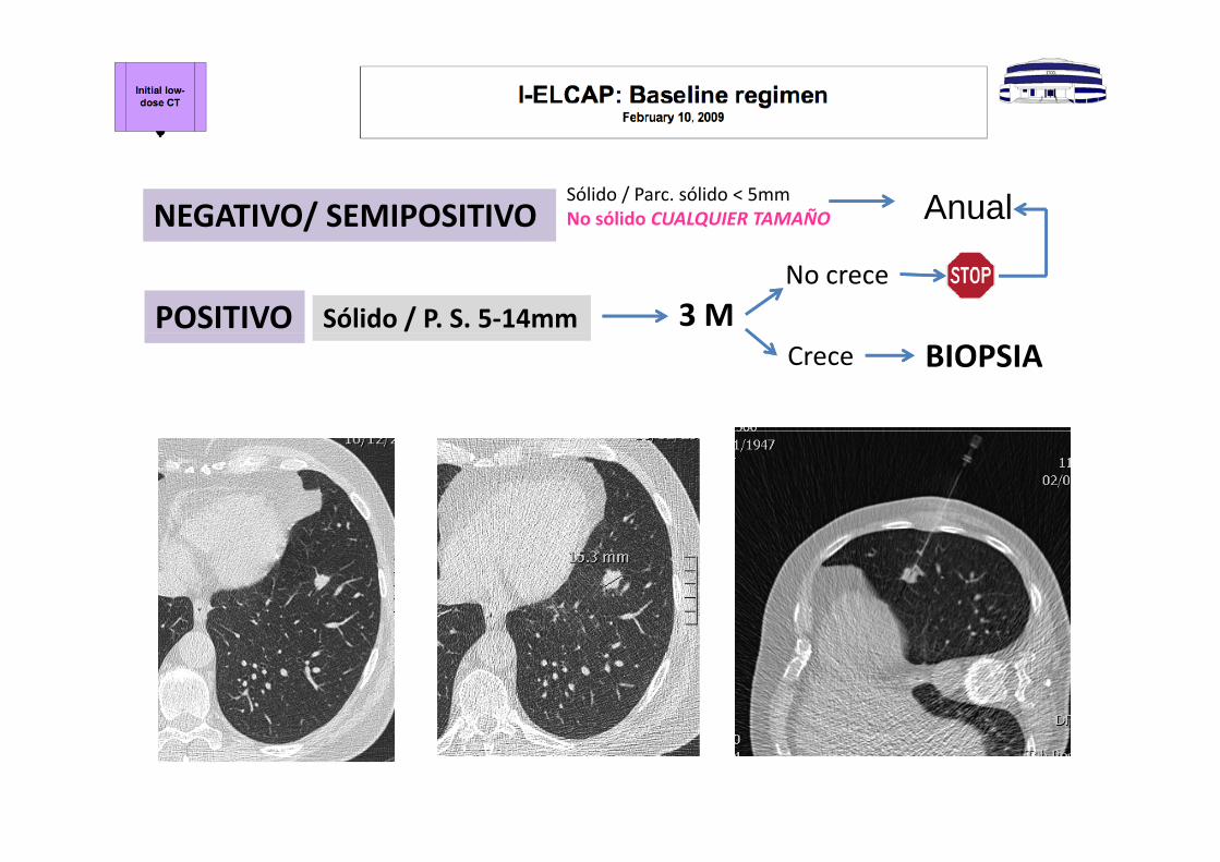
Sólido / Parc. sólido < 5mmNo sólido CUALQUIER TAMAÑONEGATIVO/ SEMIPOSITIVO Anual
POSITIVO Sólido / P. S. 5‐14mm 3 MNo crece
Crece BIOPSIA
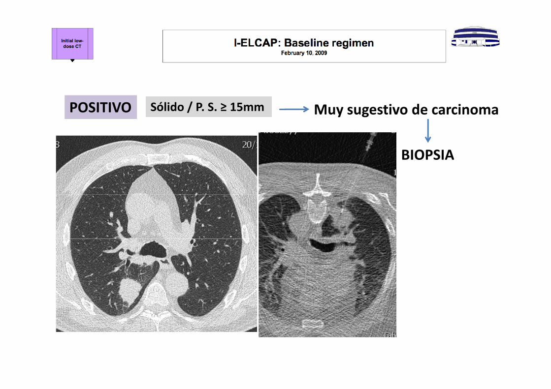
POSITIVO Sólido / P S ≥ 15mm M ti d iPOSITIVO Sólido / P. S. ≥ 15mm Muy sugestivo de carcinoma
BIOPSIABIOPSIA
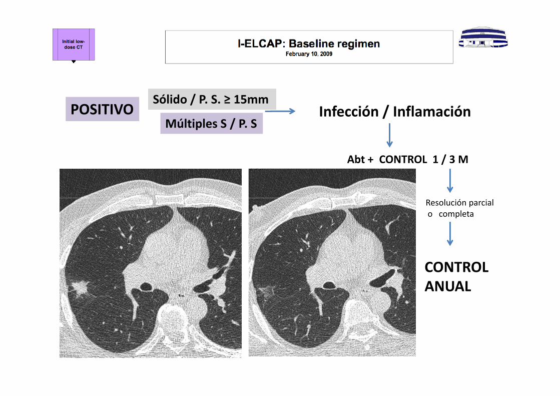
POSITIVO I f ió / I fl ióSólido / P. S. ≥ 15mm
POSITIVO Infección / InflamaciónMúltiples S / P. S
Abt + CONTROL 1 / 3 M
Resolución parcialo completa
CONTROL ANUALANUAL
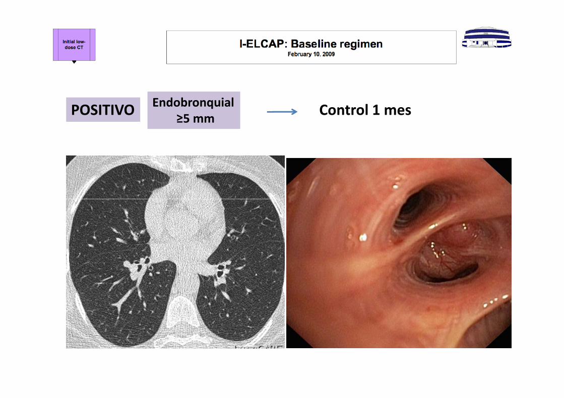
POSITIVO Control 1 mesEndobronquialPOSITIVO Control 1 mesq≥5 mm
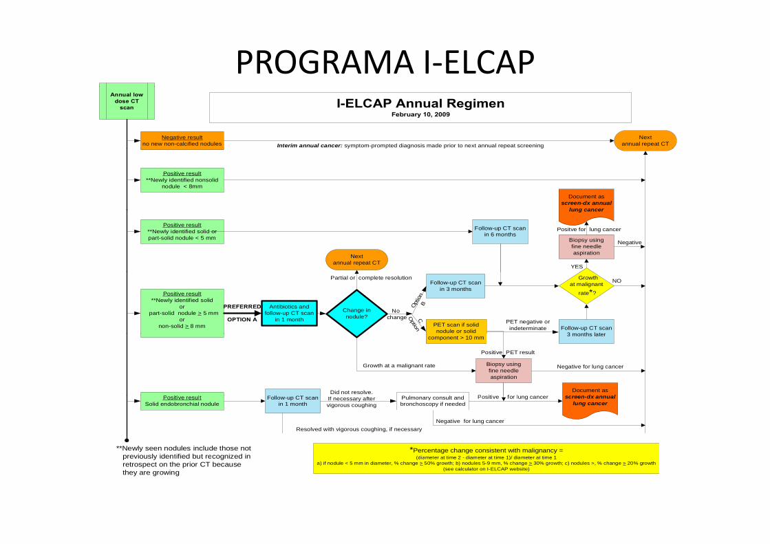
PROGRAMA I‐ELCAPI-ELCAP Annual Regimen
February 10, 2009
Annual lowdose CT
scan
Negative resultno new non-calcified nodules
Nextannual repeat CTInterim annual cancer: symptom-prompted diagnosis made prior to next annual repeat screening
Positive result**Newly identified nonsolid
nodule < 8mm
Document asscreen-dx annual
lung cancer
Positive result**Newly identified solid orpart-solid nodule < 5 mm
Follow-up CT scanin 6 months
YES
Nextannual repeat CT
Biopsy usingfine needleaspiration
lung cancer
Positve for lung cancer
Negative
Positive result**Newly identified solid
or part-solid nodule > 5 mm
ornon-solid > 8 mm
Change innodule?
PREFERRED
OPTION A COption
Opt
ion
BAntibiotics andfollow-up CT scan
in 1 month
Partial or complete resolution
No change
Follow-up CT scanin 3 months
PET scan if solidnodule or solid
Growthat malignant
rate*?
Follow-up CT scan3 months later
PET negative orindeterminate
NO
P i i l
Growth at a malignant rate
component > 10 mm 3 months later
Biopsy usingfine needleaspiration
Did not resolve.
Positive PET result
Document asd lP iti f l
Negative for lung cancer
Positive resultSolid endobronchial nodule
Follow-up CT scanin 1 month
Resolved with vigorous coughing, if necessary
If necessary aftervigorous coughing
Pulmonary consult andbronchoscopy if needed
*Percentage change consistent with malignancy =(diameter at time 2 diameter at time 1)/ diameter at time 1
**Newly seen nodules include those notpreviously identified but recognized in
Negative for lung cancer
screen-dx annuallung cancer
Positive for lung cancer
(diameter at time 2 - diameter at time 1)/ diameter at time 1a) if nodule < 5 mm in diameter, % change > 50% growth; b) nodules 5-9 mm, % change > 30% growth; c) nodules >, % change > 20% growth
(see calculator on I-ELCAP website)
previously identified but recognized in retrospect on the prior CT because they are growing
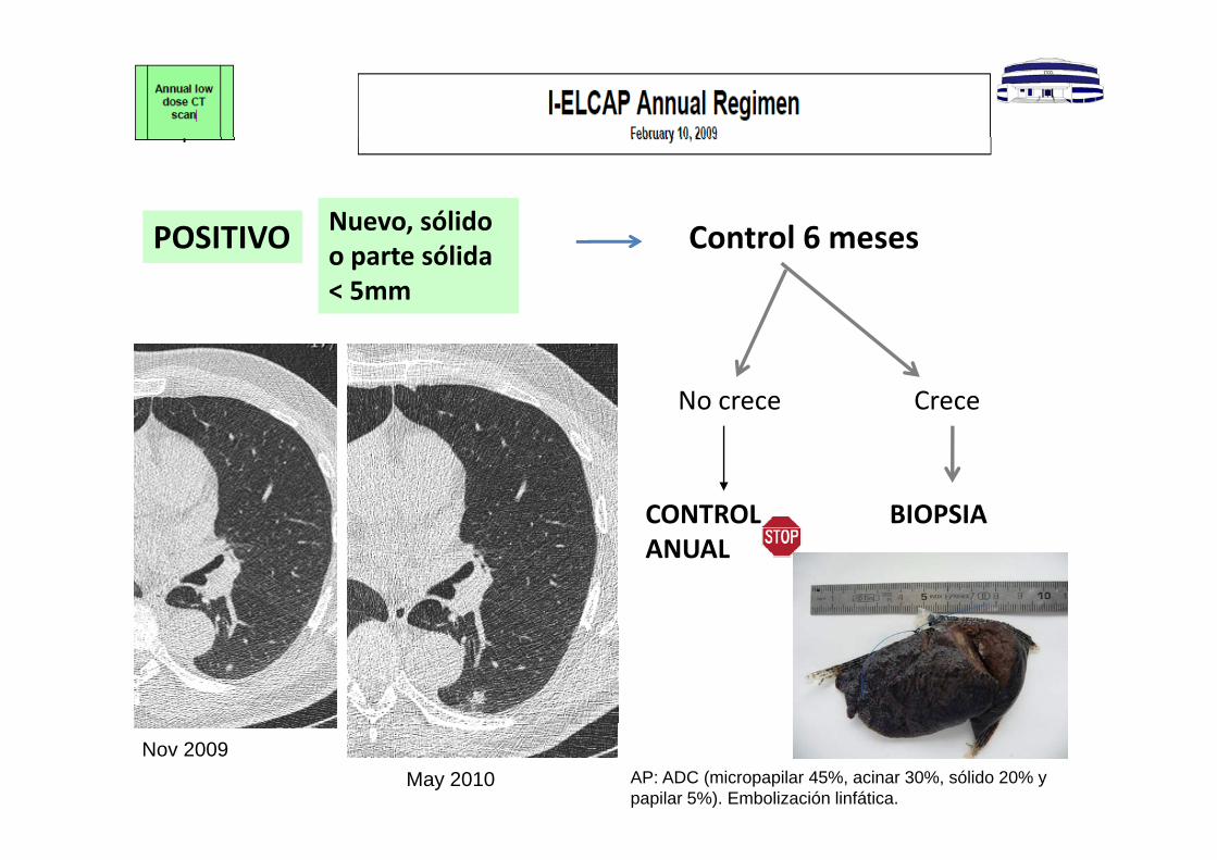
POSITIVO Nuevo, sólido Control 6 mesesPOSITIVO o parte sólida < 5mm
Control 6 meses
No crece Crece
CONTROL BIOPSIAANUAL
Nov 2009May 2010 AP: ADC (micropapilar 45%, acinar 30%, sólido 20% y
papilar 5%). Embolización linfática.
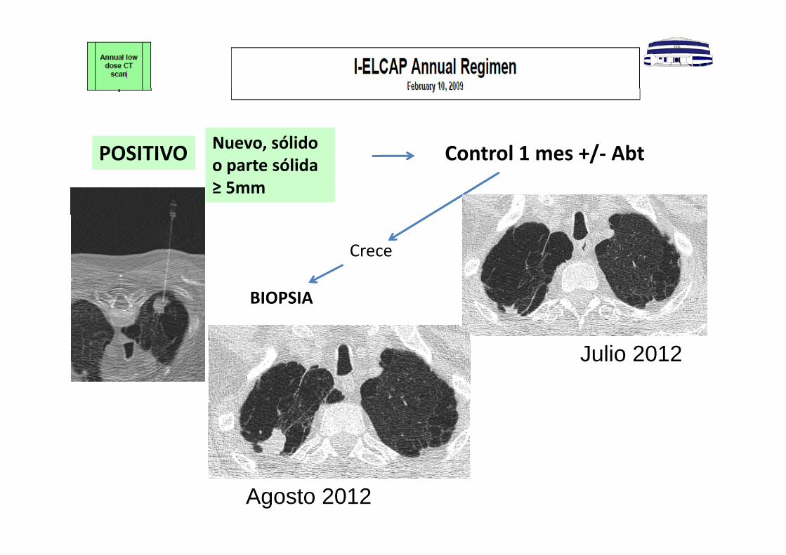
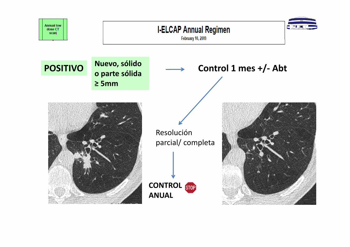
POSITIVO Nuevo, sólido Control 1 mes +/ AbtPOSITIVO o parte sólida ≥ 5mm
Control 1 mes +/‐ Abt
Crece
BIOPSIA
Julio 2012
Agosto 2012
POSITIVO Nuevo, sólido Control 1 mes +/ AbtPOSITIVO o parte sólida ≥ 5mm
Control 1 mes +/‐ Abt
Resolución parcial/ completa
CONTROL ANUAL
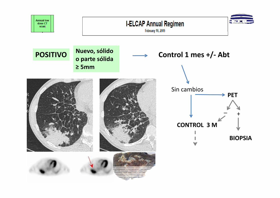
POSITIVO Nuevo, sólido Control 1 mes +/ AbtPOSITIVO o parte sólida ≥ 5mm
Control 1 mes +/‐ Abt
PETSin cambios
+_
CONTROL 3 M
BIOPSIA
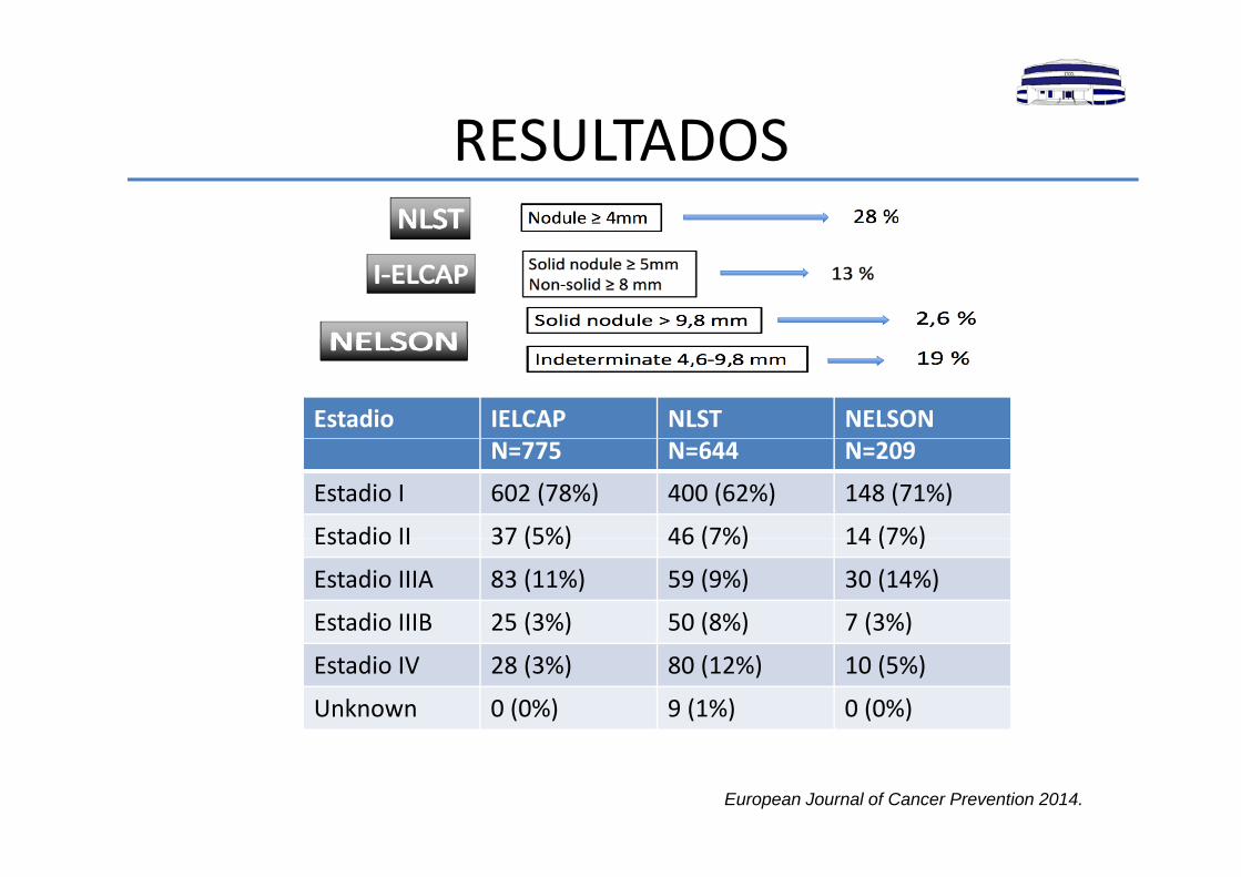
RESULTADOSRESULTADOS
Estadio IELCAP NLST NELSONN=775 N=644 N=209
Estadio I 602 (78%) 400 (62%) 148 (71%)
Estadio II 37 (5%) 46 (7%) 14 (7%)Estadio II 37 (5%) 46 (7%) 14 (7%)
Estadio IIIA 83 (11%) 59 (9%) 30 (14%)
Estadio IIIB 25 (3%) 50 (8%) 7 (3%)
Estadio IV 28 (3%) 80 (12%) 10 (5%)
Unknown 0 (0%) 9 (1%) 0 (0%)
European Journal of Cancer Prevention 2014.
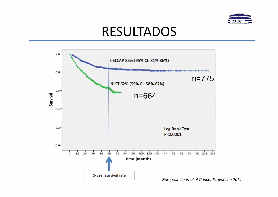
RESULTADOSRESULTADOS
775n=775
n=66466
European Journal of Cancer Prevention 2014.
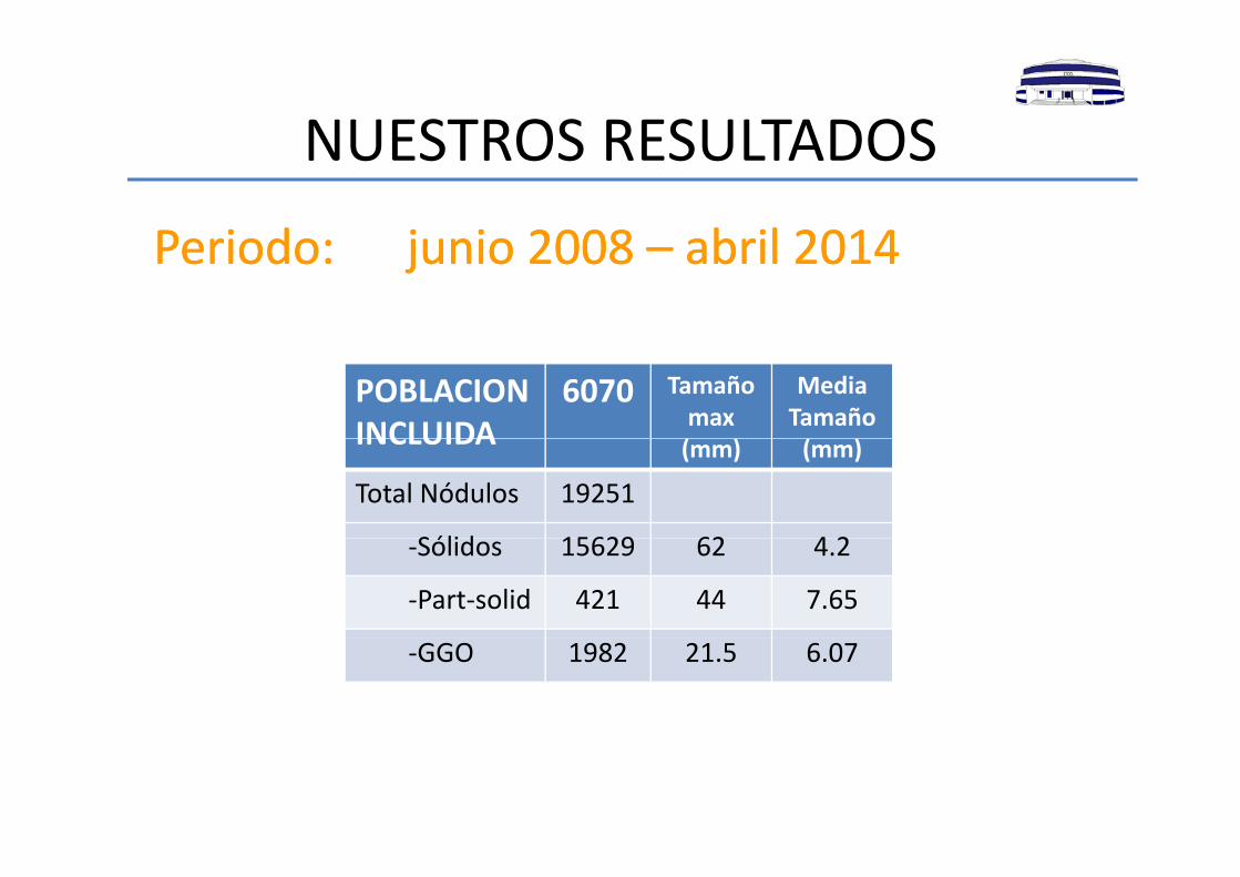
NUESTROS RESULTADOSNUESTROS RESULTADOS
Periodo junio 2008Periodo junio 2008 abril 2014abril 2014Periodo: junio 2008 Periodo: junio 2008 –– abril 2014abril 2014
POBLACION INCLUIDA
6070 Tamaño max
Media Tamaño INCLUIDA (mm) (mm)
Total Nódulos 19251
Sólid 15629 62 4 2‐Sólidos 15629 62 4.2
‐Part‐solid 421 44 7.65
‐GGO 1982 21.5 6.07
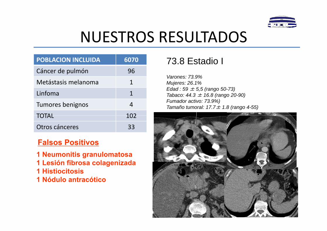
NUESTROS RESULTADOSNUESTROS RESULTADOSPOBLACION INCLUIDA 6070 73 8 Estadio ICáncer de pulmón 96
Metástasis melanoma 1
73.8 Estadio IVarones: 73.9%Mujeres: 26.1%Edad : 59 ± 5 5 (rango 50-73)
Linfoma 1
Tumores benignos 4
TOTAL 102
Edad : 59 ± 5,5 (rango 50-73)Tabaco: 44.3 ± 16.8 (rango 20-90)Fumador activo: 73.9%)Tamaño tumoral: 17.7± 1.8 (rango 4-55)
TOTAL 102
Otros cánceres 33
Falsos Positivos1 Neumonitis granulomatosa1 Lesión fibrosa colagenizada
Falsos Positivos
1 Histiocitosis1 Nódulo antracótico
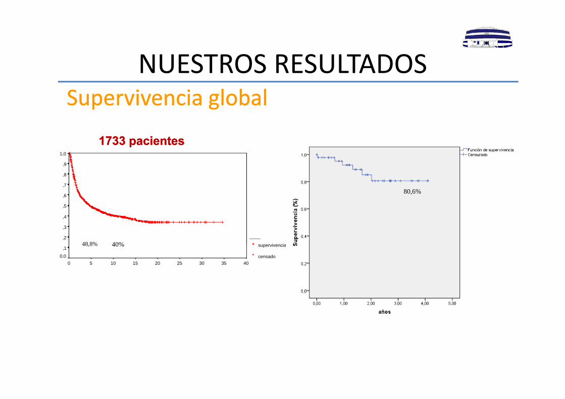
NUESTROS RESULTADOSNUESTROS RESULTADOSSupervivencia globalSupervivencia global
1 0
p gp g
1733 pacientes1733 pacientes1,0
,9
,8
,7
,680,6%
supervivencia
,5
,4
,3
,240%48 8% supervivencia
censado4035302520151050
,1
0,0
40%48,8%
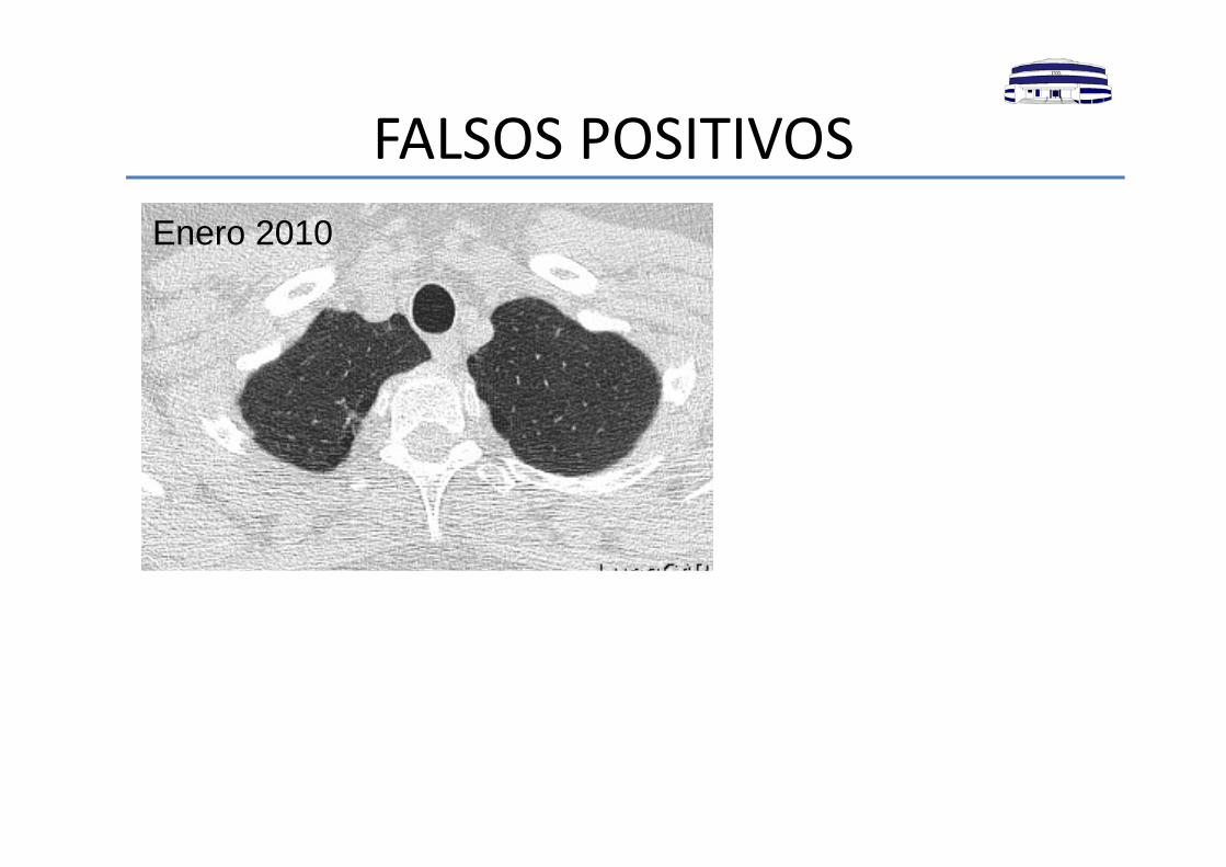
FALSOS POSITIVOSFALSOS POSITIVOSEnero 2010Enero 2010
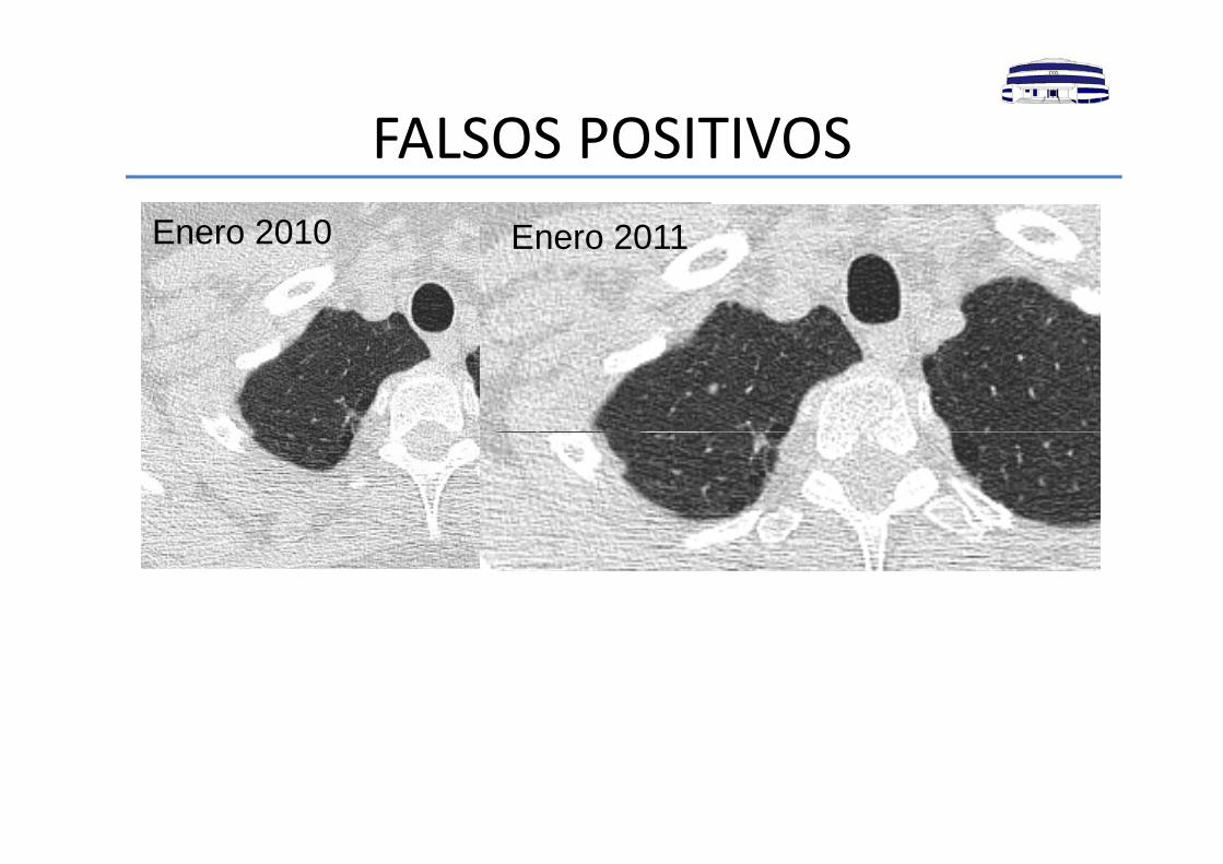
FALSOS POSITIVOSFALSOS POSITIVOSEnero 2010 Enero 2011Enero 2010 Enero 2011
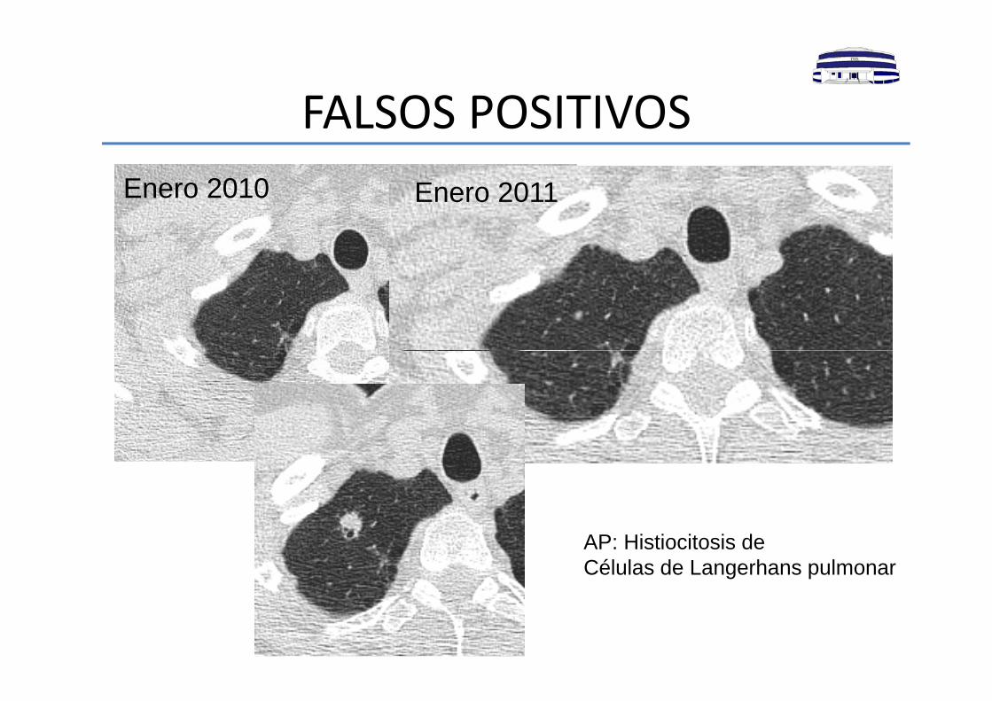
FALSOS POSITIVOSFALSOS POSITIVOSEnero 2010 Enero 2011Enero 2010 Enero 2011
AP: Histiocitosis deCélulas de Langerhans pulmonar
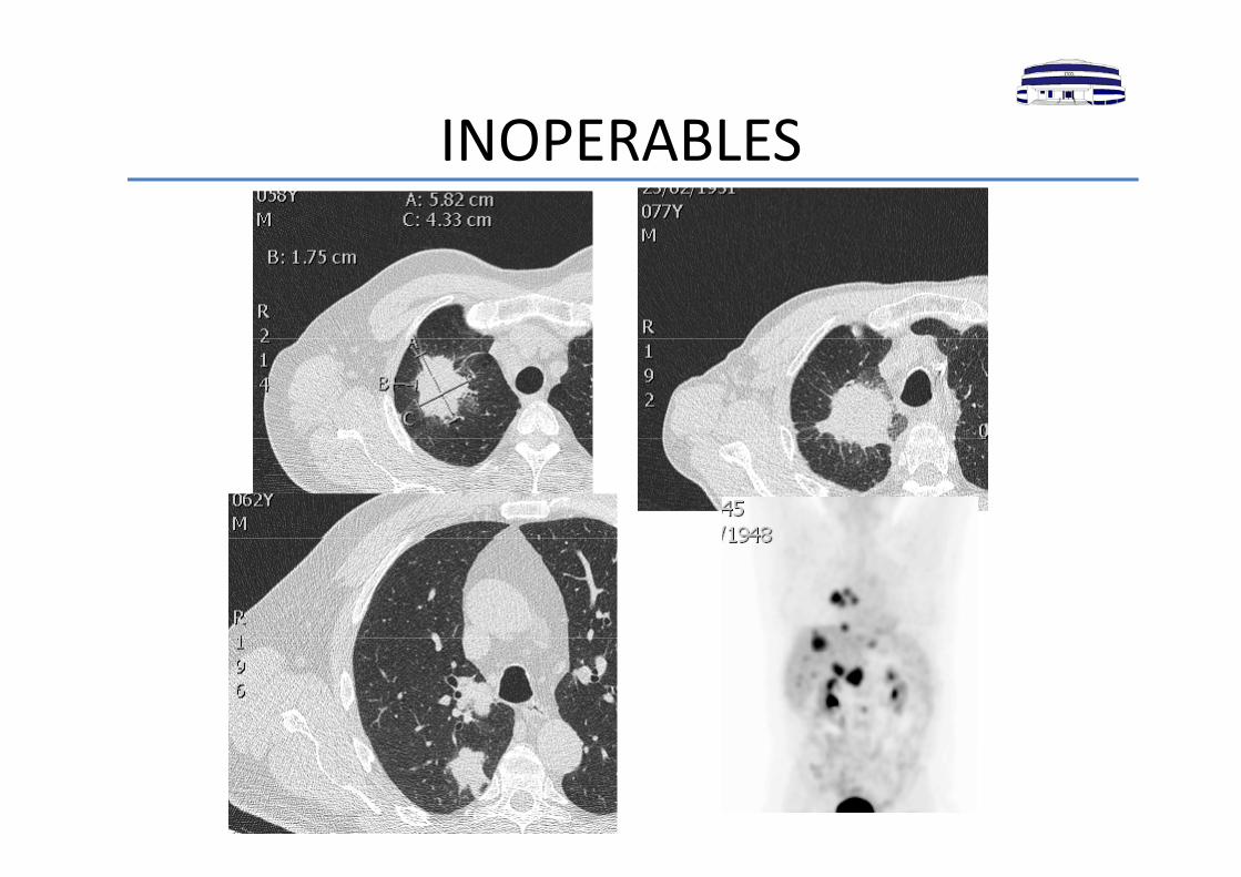
INOPERABLESINOPERABLES
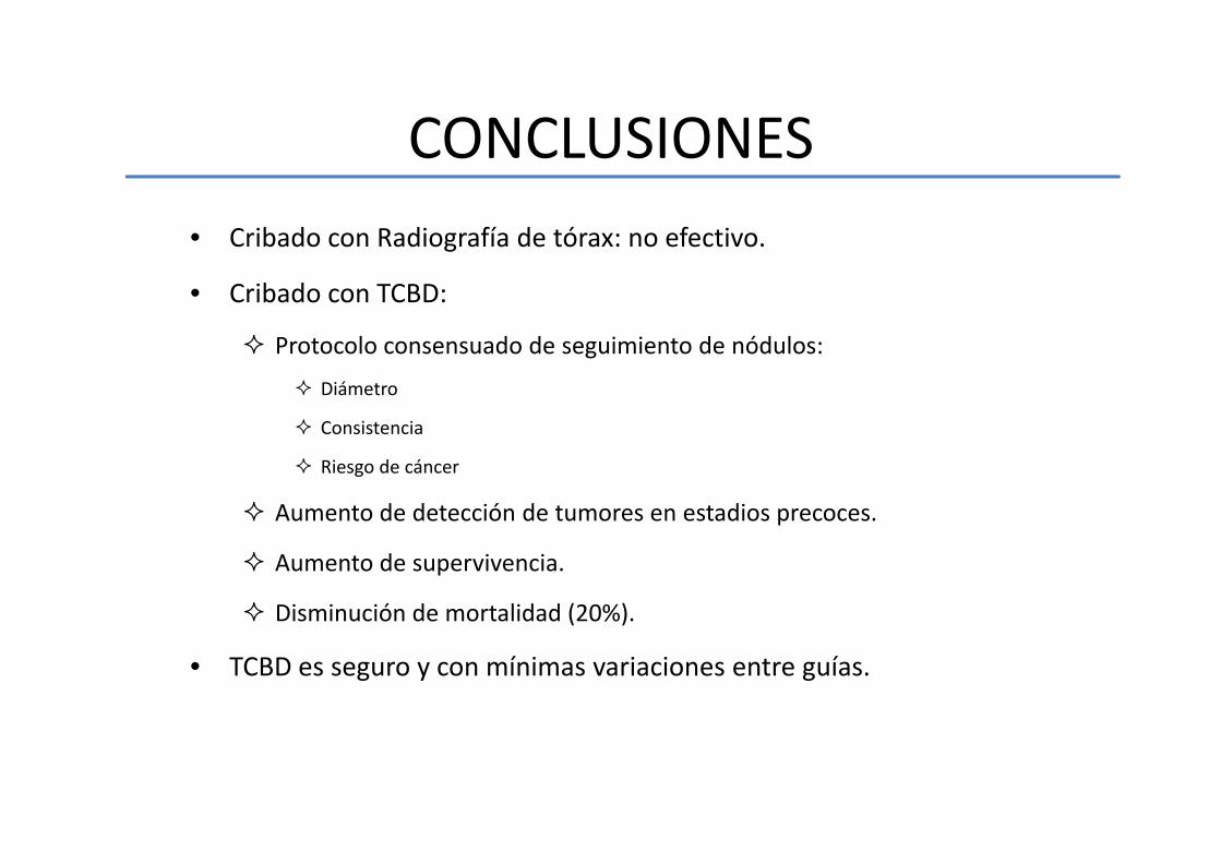
CONCLUSIONESCONCLUSIONES• Cribado con Radiografía de tórax: no efectivo• Cribado con Radiografía de tórax: no efectivo.
• Cribado con TCBD:
Protocolo consensuado de seguimiento de nódulos:
Diámetro
Consistencia Consistencia
Riesgo de cáncer
Aumento de detección de tumores en estadios precoces.
Aumento de supervivencia.
Disminución de mortalidad (20%).
• TCBD es seguro y con mínimas variaciones entre guías.
CRIBADO DE CÁNCER DE PULMÓNCRIBADO DE CÁNCER DE PULMÓNRecomendaciones de cribado
• American Association for Thoracic Surgery (AATS)• American College of Chest Physicians (ACCP)• American Society of Clinical Oncology (ASCO)American Society of Clinical Oncology (ASCO)• American Cancer Society• International Association for the Study of Lung Cancer
(IASLC)(IASLC)• National Comprehensive Cancer Network (NCCN)
JAMA, March 20,2013‐vol 309,No.11
MUCHAS GRACIAS
Recommended


![Símbolos del catálogo Catalogue symbolsA] [02] Manguera...Símbolos del catálogo Catalogue symbols Diámetro interno / Internal diameter Diámetro exterior / External diameter Diámetro](https://img.pdfslide.es/doc/110x75/5eaa6ff049f5fa538c64e509/smbolos-del-catlogo-catalogue-symbols-a-02-manguera-smbolos-del-catlogo.jpg)