DEFINICION
Neoplasia = Nuevo Crecimiento
• Una masa anormal de tejido que prolifera en forma excesiva y sin
coordinación con el tejido normal y persiste en su crecimiento aún
después de cesar el estímulo que provocó el cambio, compitiendo
con células y tejidos normales respecto a sus necesidades
metabólicas
• Su nutrición depende del huésped • Autónomo • Clonalidad
TUMOR: Aumento de volumen de un tejido producido por
edema, hemorragia, granuloma, neoplasia, etc.
CÁNCER: Crecimiento tisular producido por la proliferación continua de células anormales con capacidad de invasión y destruc- ción de otros tejidos.
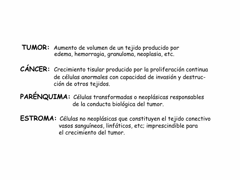
PARÉNQUIMA: Células transformadas o neoplásicas responsables de la conducta biológica del tumor.
ESTROMA: Células no neoplásicas que constituyen el tejido conectivo vasos sanguíneos, linfáticos, etc; imprescindible para el crecimiento del tumor.
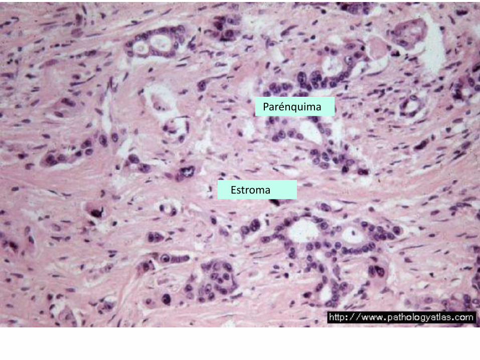
Parénquima
Estroma
CLASIFICACIÓN:
Epitelial: Escamoso, glandular, transicional, etc.
Mesenquimal: Músculo liso, músculo estriado, fibroso, hueso, etc.
Nervioso: Gliales, neuronas, nervios, etc.
Linfoproliferativo: Linfocitos, células plasmáticas, etc. Endotelial, Embrionarios, etc.
BENIGNOS / MALIGNOS
NOMENCLATURA:
BENIGNOS: “oma” - Adenoma, papiloma, cistoadenoma. Pólipo. - Fibroma, osteoma, leiomioma, condroma, etc. - Schwannoma, neurofibroma, etc. MALIGNOS: “carcinoma” / “sarcoma” - Adenocarcinoma, carcinoma escamoso, carcinoma de células transicionales. - Fibrosarcoma, osteosarcoma, leiomiosarcoma. Schwannoma maligno, meningioma maligno, etc. Linfoma, seminoma, hepatoma, astrocitoma, melanoma, etc.
TUMORES BENIGNOS:
- Solo crecen hasta un determinado tamaño - Normalmente no crecen muy rápido - No destruyen células normales - No se propagan al tejido que los rodea - Normalmente no producen efectos
secundarios graves - Por lo general crecen de forma ordenada
TUMORES MALIGNOS:
- LA MAYORÍA CRECE RAPIDAMENTE - CRECEN ILIMITADAMENTE - PUEDEN INVADIR Y DESTRUIR ESTRUCTURAS
ADYACENTES - PUEDEN PROPAGARSE A SITIOS DISTANTES
(METÁSTASIS) - PRESENTAN UN AMPLIO MARGEN DE
DIFERENCIACIÓN - PUEDEN PROVOCAR LA MUERTE
TUMORES BENIGNOS
NOMBRE DE CÉLULA DE PROCEDENCIA+“OMA”
• FIBROBLASTOS FIBROMA
• PATRÓN GLANDULAR ADENOMA
• MASAS QUÍSTICAS CISTOADENOMA
• MASA QUISTICA CON PAPILAS EN SU INTERIOR CISTOADENOMA PAPILAR
• VERRUCOSA, DIGITIFORME SOBRE EPITELIO PAPILOMA
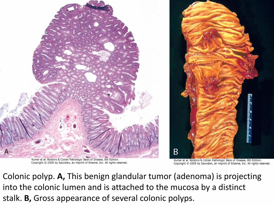
Colonic polyp. A, This benign glandular tumor (adenoma) is projecting into the colonic lumen and is attached to the mucosa by a distinct stalk. B, Gross appearance of several colonic polyps.
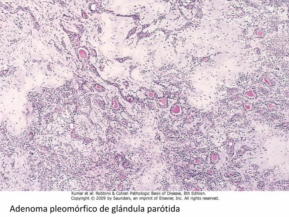
Adenoma pleomórfico de glándula parótida
TUMORES MALIGNOS
Formados a partir de una capa embrionaria • Origen: Tejidos mesenquimales Sarcomas • Tejido epitelial (cualquiera de las 3 capas
embrionarias) Carcinomas patrón glandular Adenocarcinomas epitelial Carcinoma escamoso Formados por varios células totipotenciales (mas de
una capa embrionaria) • Teratoma
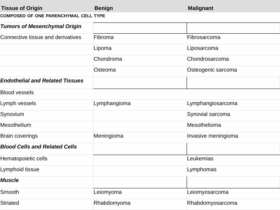
Tissue of Origin Benign Malignant
COMPOSED OF ONE PARENCHYMAL CELL TYPE
Tumors of Mesenchymal Origin
Connective tissue and derivatives Fibroma Fibrosarcoma
Lipoma Liposarcoma
Chondroma Chondrosarcoma
Osteoma Osteogenic sarcoma
Endothelial and Related Tissues
Blood vessels
Lymph vessels Lymphangioma Lymphangiosarcoma
Synovium Synovial sarcoma
Mesothelium Mesothelioma
Brain coverings Meningioma Invasive meningioma
Blood Cells and Related Cells
Hematopoietic cells Leukemias
Lymphoid tissue Lymphomas
Muscle
Smooth Leiomyoma Leiomyosarcoma
Striated Rhabdomyoma Rhabdomyosarcoma
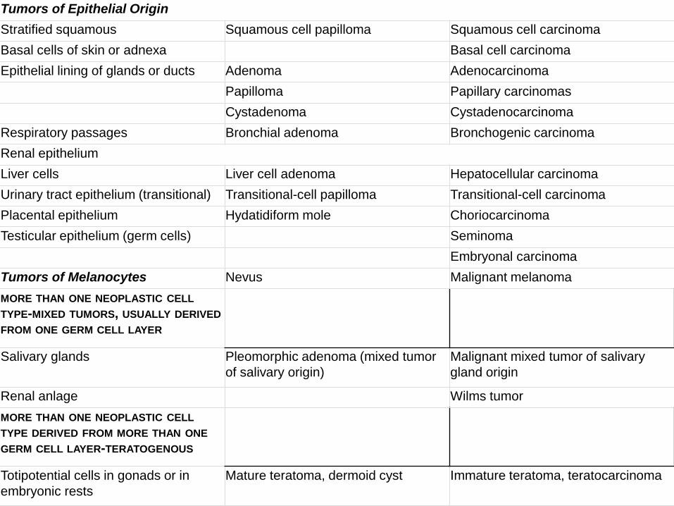
Tumors of Epithelial Origin
Stratified squamous Squamous cell papilloma Squamous cell carcinoma
Basal cells of skin or adnexa Basal cell carcinoma
Epithelial lining of glands or ducts Adenoma Adenocarcinoma
Papilloma Papillary carcinomas
Cystadenoma Cystadenocarcinoma
Respiratory passages Bronchial adenoma Bronchogenic carcinoma
Renal epithelium
Liver cells Liver cell adenoma Hepatocellular carcinoma
Urinary tract epithelium (transitional) Transitional-cell papilloma Transitional-cell carcinoma
Placental epithelium Hydatidiform mole Choriocarcinoma
Testicular epithelium (germ cells) Seminoma
Embryonal carcinoma
Tumors of Melanocytes Nevus Malignant melanoma
MORE THAN ONE NEOPLASTIC CELL
TYPE-MIXED TUMORS, USUALLY DERIVED
FROM ONE GERM CELL LAYER
Salivary glands Pleomorphic adenoma (mixed tumor
of salivary origin)
Malignant mixed tumor of salivary
gland origin
Renal anlage Wilms tumor
MORE THAN ONE NEOPLASTIC CELL
TYPE DERIVED FROM MORE THAN ONE
GERM CELL LAYER-TERATOGENOUS
Totipotential cells in gonads or in
embryonic rests
Mature teratoma, dermoid cyst Immature teratoma, teratocarcinoma
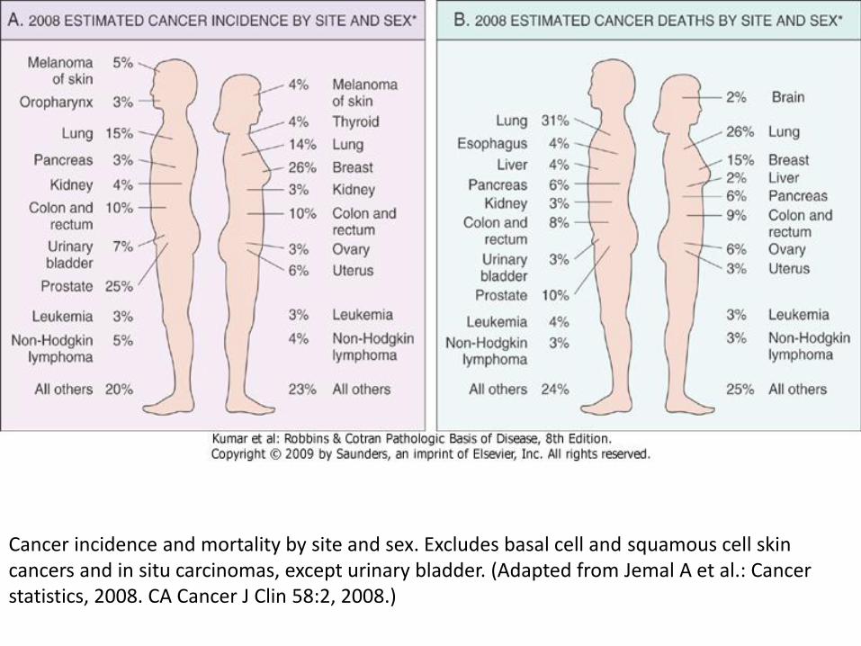
Cancer incidence and mortality by site and sex. Excludes basal cell and squamous cell skin cancers and in situ carcinomas, except urinary bladder. (Adapted from Jemal A et al.: Cancer statistics, 2008. CA Cancer J Clin 58:2, 2008.)
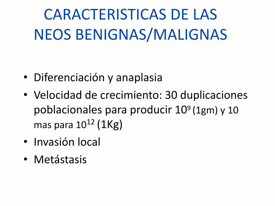
CARACTERISTICAS DE LAS NEOS BENIGNAS/MALIGNAS
• Diferenciación y anaplasia
• Velocidad de crecimiento: 30 duplicaciones poblacionales para producir 109 (1gm) y 10
mas para 1012 (1Kg)
• Invasión local
• Metástasis

CARACTERISTICAS DE LAS NEOPLASIAS MALIGNAS
• ANAPLASIA: CARENCIA DE DIAFERENCIACION, CELULAS GIGANTES
• PLEOMORFISMO: VARIACION EN TAMAÑO Y FORMA
• MORFOLOGIA NUCLEAR ANORMAL: HIPERCROMATISMO
• MITOSIS
• PERDIDA DE POLARIDAD
• NECROSIS
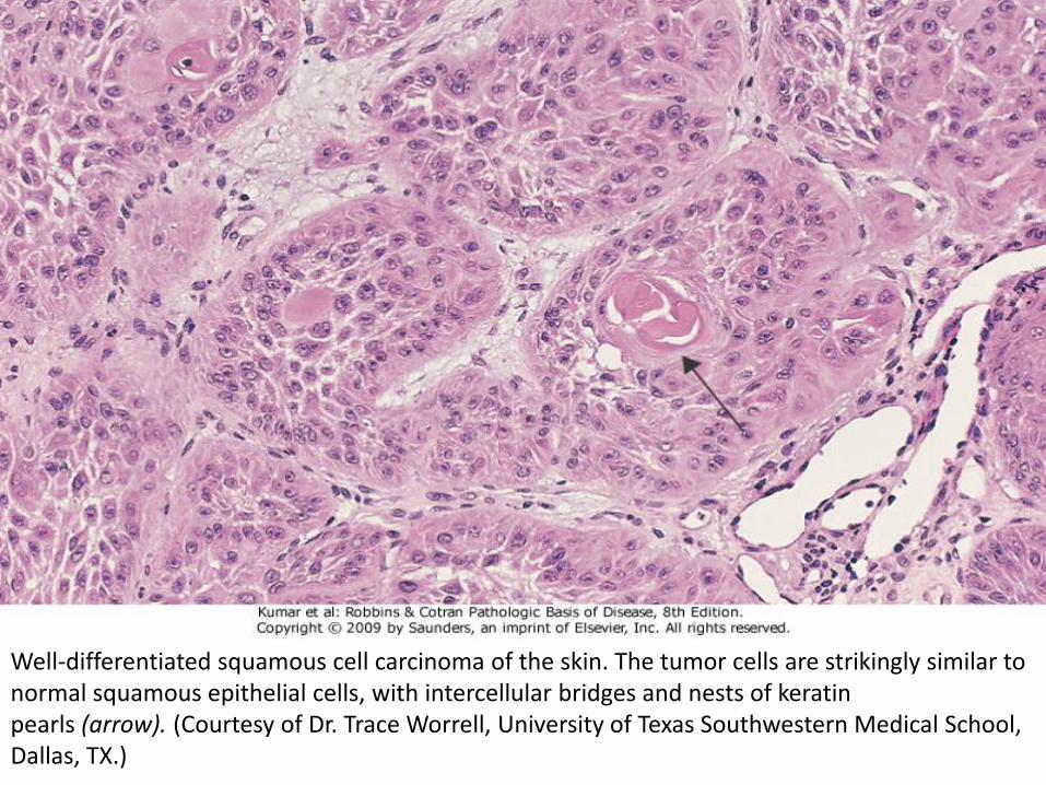
Well-differentiated squamous cell carcinoma of the skin. The tumor cells are strikingly similar to normal squamous epithelial cells, with intercellular bridges and nests of keratin pearls (arrow). (Courtesy of Dr. Trace Worrell, University of Texas Southwestern Medical School, Dallas, TX.)
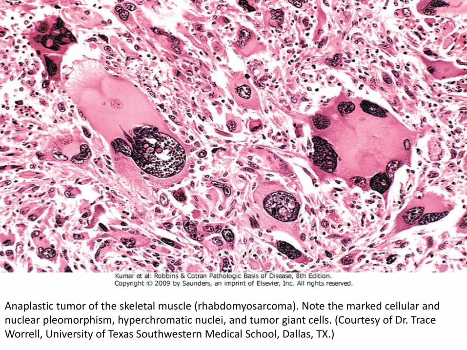
Anaplastic tumor of the skeletal muscle (rhabdomyosarcoma). Note the marked cellular and nuclear pleomorphism, hyperchromatic nuclei, and tumor giant cells. (Courtesy of Dr. Trace Worrell, University of Texas Southwestern Medical School, Dallas, TX.)
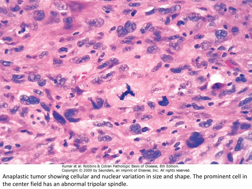
Anaplastic tumor showing cellular and nuclear variation in size and shape. The prominent cell in the center field has an abnormal tripolar spindle.
GRADOS Y ESTADIAJE
• LOS METODOS PARA CUANTIFICAR LA AGRESIVIDAD DE UNA NEOPLASIA DETERMINADA, SU APARENTE EXTENSION Y DISEMINACION EN EL PACIENTE SON NECESARIAS PARA DETERMINAR EL PRONÓSTICO Y PARA COMPARAR LOS RESULTADOS FINALES DE PROTOCOLOS ESTABLECIDOS.
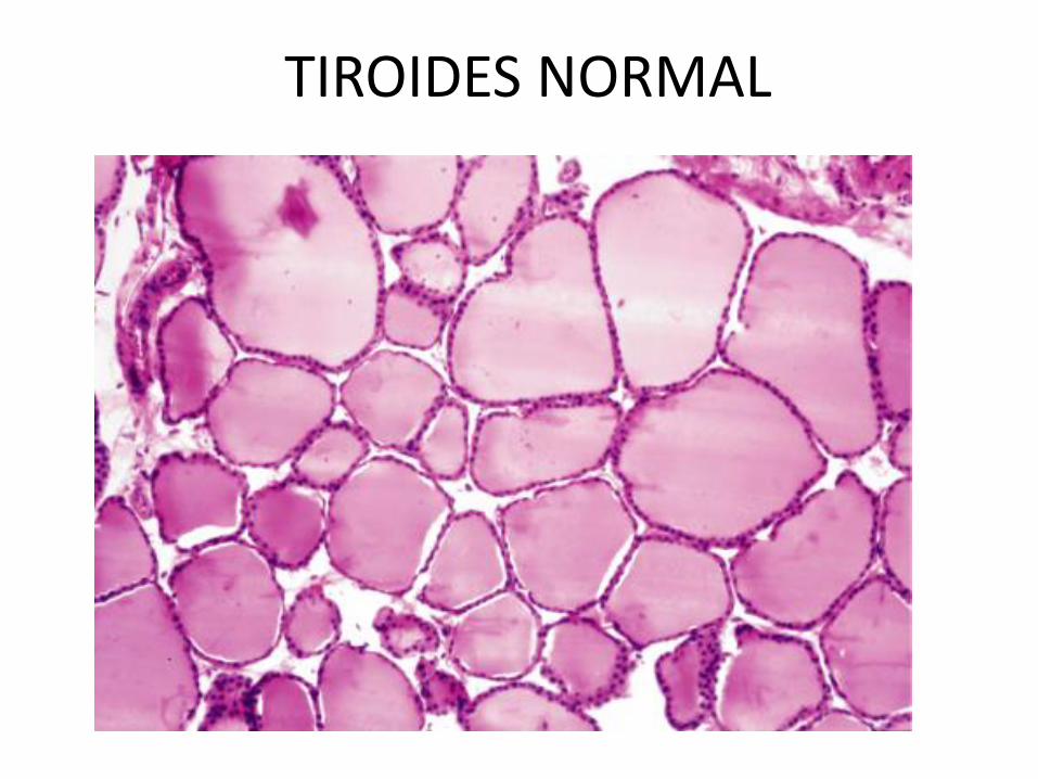
TIROIDES NORMAL
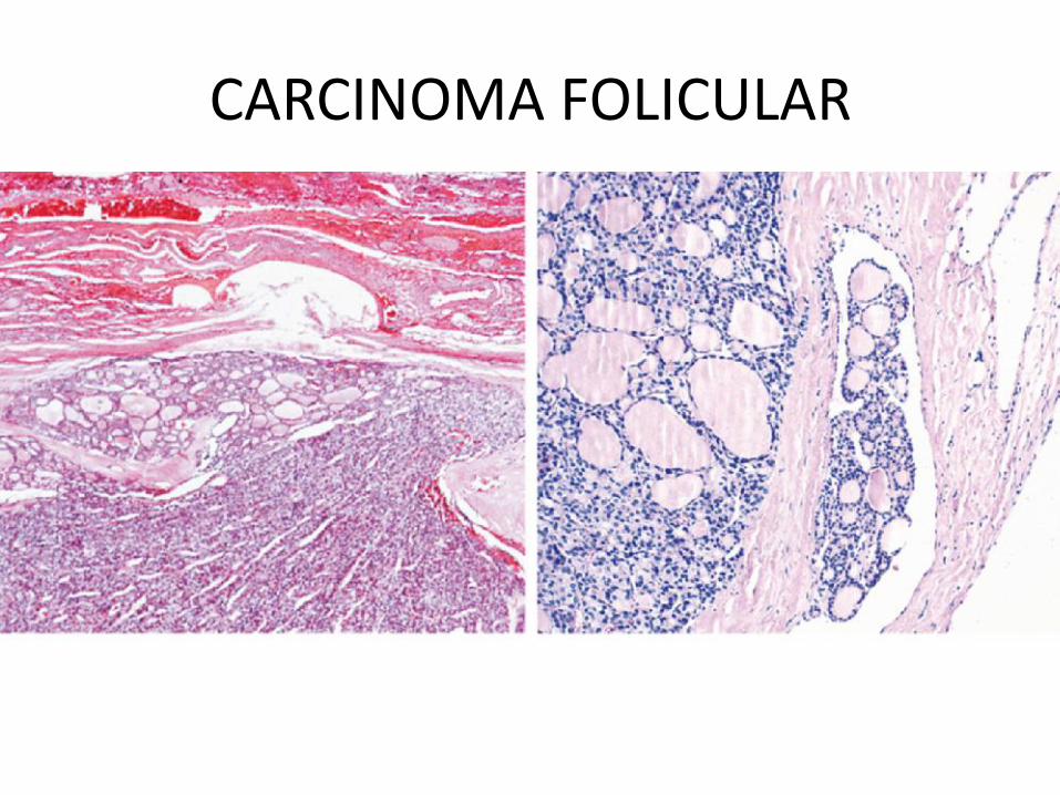
CARCINOMA FOLICULAR
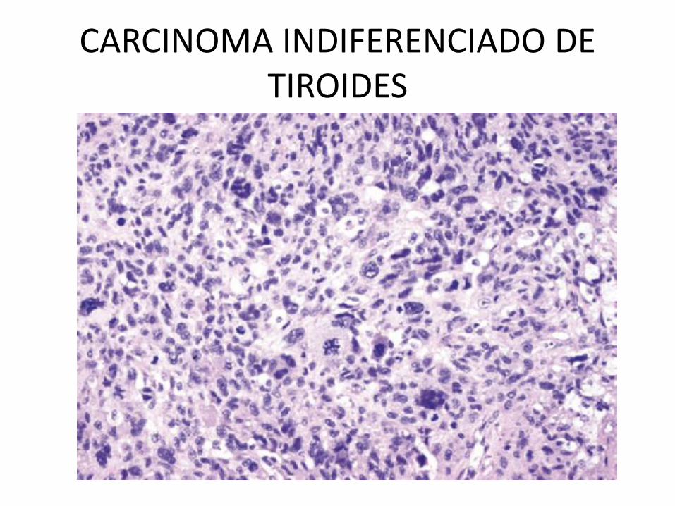
CARCINOMA INDIFERENCIADO DE TIROIDES
GRADO DE UNA NEOPLASIA MALIGNA
• ES BASADO EN EL GRADO DE DIFERNCIACION DE LAS CELULAS TUMORALES.
• EN ALGUNOS CASOS EN EL NUMERO DE MITOSIS Y CARACTERISTICAS ARQUITECTURALES.
• GENERALMENTE VAN DE DOS CATEGORIAS A CUATRO.
• TIENE MENOR RELEVANCIA CLINICA QUE EL ESTADIAJE.
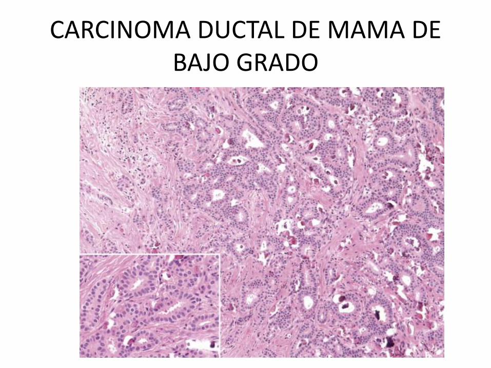
CARCINOMA DUCTAL DE MAMA DE BAJO GRADO
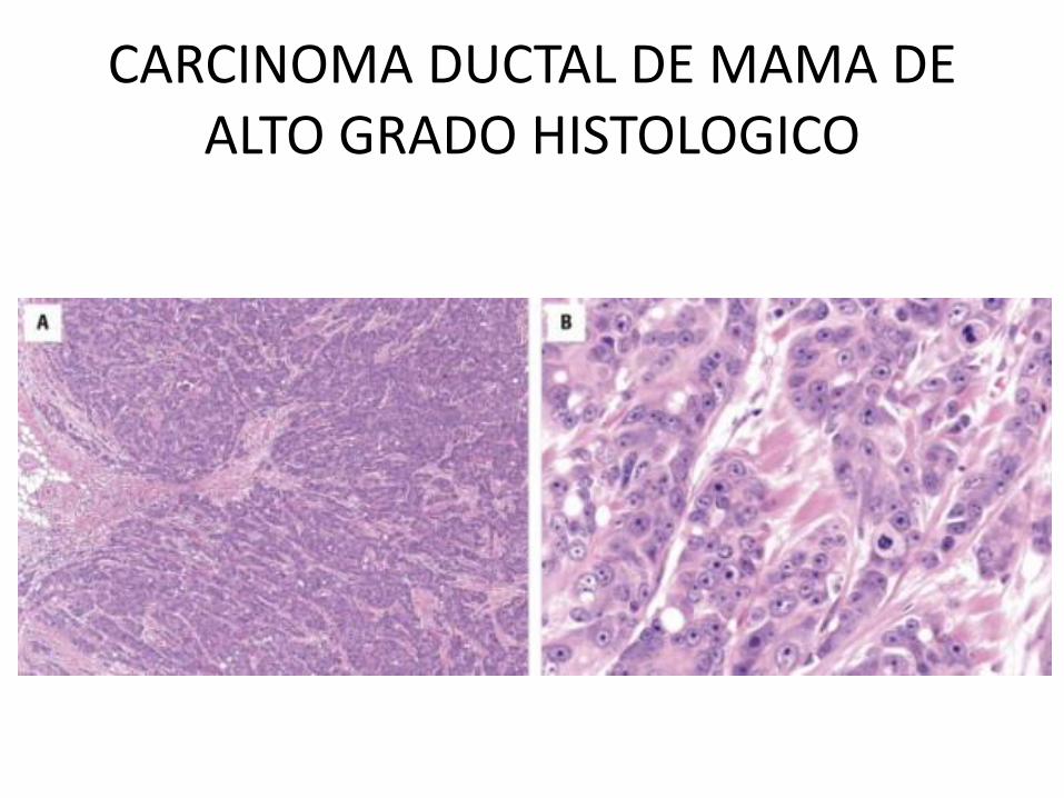
CARCINOMA DUCTAL DE MAMA DE ALTO GRADO HISTOLOGICO
ESTADIAJE
EL ESTADIAJE DEL CANCER ES BASADO EN:
• EL TAMAÑO DE LA LESION PRIMARIA,
• LA EXTENSION DE LA DISEMINACION A GANGLIOS LINFATICOS REGIONALES, Y
• LA PRESENCIA O AUSENCIA DE METASTASIS.
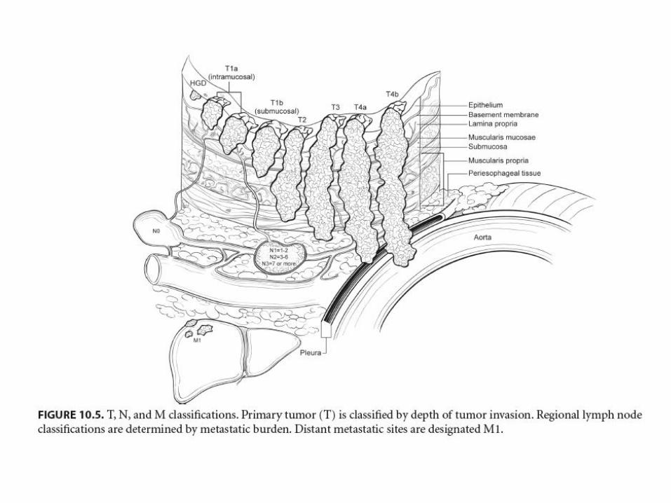
LA MAS UTILIZADA ACTUALMENTE ES LA DE LA AMERICAN JOINT COMMITTEE ON CANCER STAGING: TNM
ASPECTOS CLINICOS DEL CANCER
• AUNQUE LOS TUMORES MALIGNOS AMENAZAN MAS LA VIDA, CUALQUIER TUMOR PUEDE CAUSAR MORBILIDAD Y MORTALIDAD.
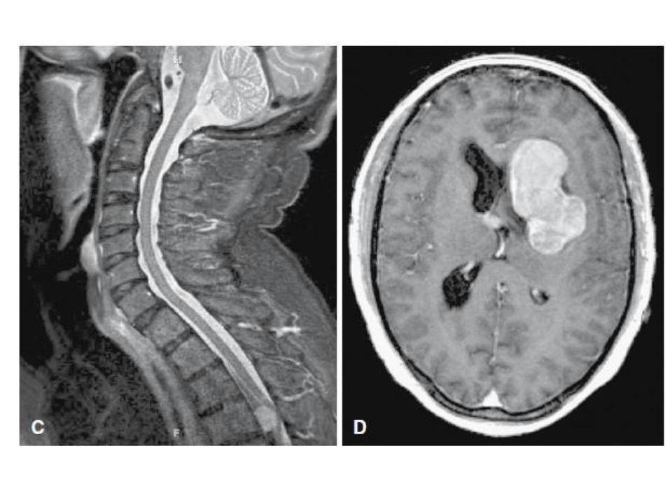
• TANTO LOS TUMORES MALIGNOS COMO BENIGNOS PUEDEN CAUSAR PROBLEMAS POR:
A) LOCALIZACION Y COMPROMISO DE
ESTRUCTURAS ADYACENTES.
• ACTIVIDAD FUNCIONAL TAL COMO SINTESIS DE HORMONAS O EL DESARROLLO DE SINDROMES PARANEOPLASICOS.
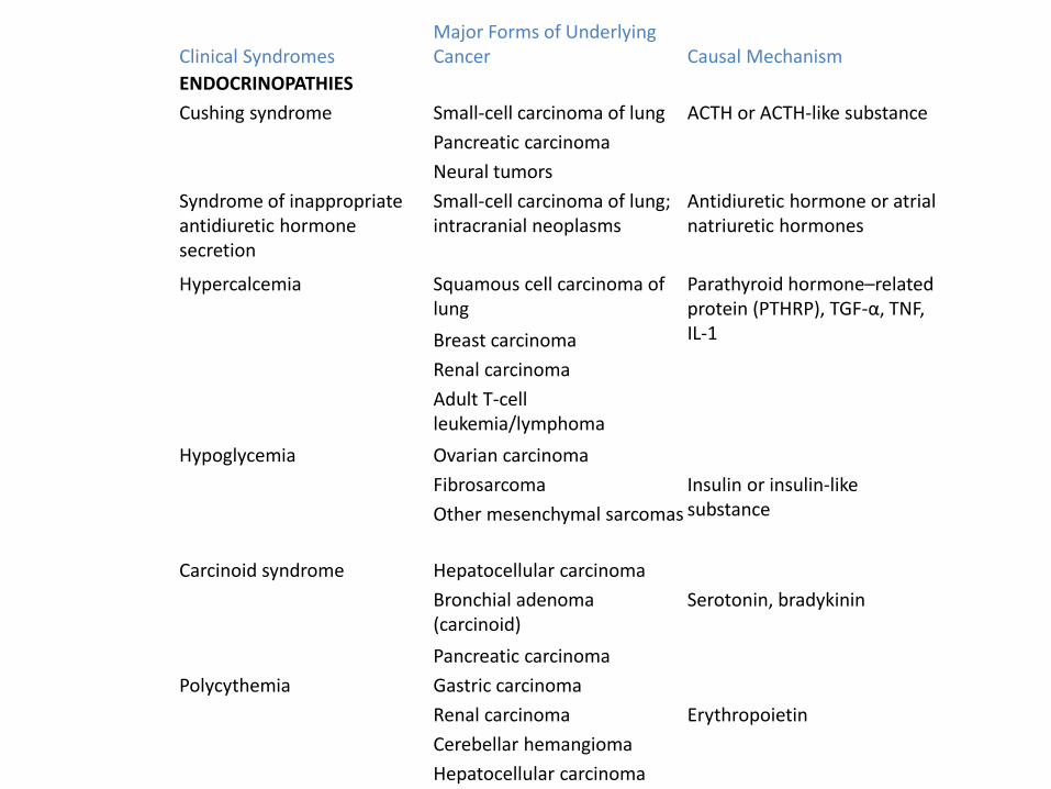
Clinical Syndromes Major Forms of Underlying Cancer Causal Mechanism
ENDOCRINOPATHIES
Cushing syndrome Small-cell carcinoma of lung ACTH or ACTH-like substance
Pancreatic carcinoma
Neural tumors
Syndrome of inappropriate antidiuretic hormone secretion
Small-cell carcinoma of lung; intracranial neoplasms
Antidiuretic hormone or atrial natriuretic hormones
Hypercalcemia Squamous cell carcinoma of lung
Parathyroid hormone–related protein (PTHRP), TGF-α, TNF, IL-1 Breast carcinoma
Renal carcinoma
Adult T-cell leukemia/lymphoma
Hypoglycemia Ovarian carcinoma
Fibrosarcoma Insulin or insulin-like substance Other mesenchymal sarcomas
Carcinoid syndrome Hepatocellular carcinoma
Bronchial adenoma (carcinoid)
Serotonin, bradykinin
Pancreatic carcinoma
Polycythemia Gastric carcinoma
Renal carcinoma Erythropoietin
Cerebellar hemangioma
Hepatocellular carcinoma
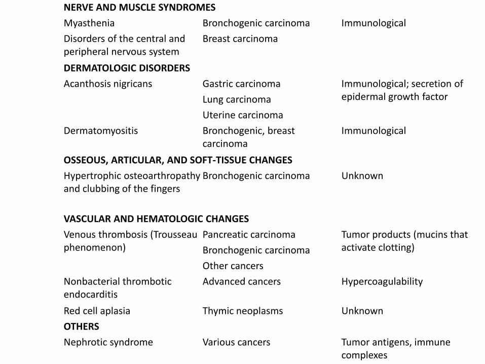
NERVE AND MUSCLE SYNDROMES
Myasthenia Bronchogenic carcinoma Immunological
Disorders of the central and peripheral nervous system
Breast carcinoma
DERMATOLOGIC DISORDERS
Acanthosis nigricans Gastric carcinoma Immunological; secretion of epidermal growth factor Lung carcinoma
Uterine carcinoma
Dermatomyositis Bronchogenic, breast carcinoma
Immunological
OSSEOUS, ARTICULAR, AND SOFT-TISSUE CHANGES
Hypertrophic osteoarthropathy and clubbing of the fingers
Bronchogenic carcinoma Unknown
VASCULAR AND HEMATOLOGIC CHANGES
Venous thrombosis (Trousseau phenomenon)
Pancreatic carcinoma Tumor products (mucins that activate clotting) Bronchogenic carcinoma
Other cancers
Nonbacterial thrombotic endocarditis
Advanced cancers Hypercoagulability
Red cell aplasia Thymic neoplasms Unknown
OTHERS
Nephrotic syndrome Various cancers Tumor antigens, immune complexes
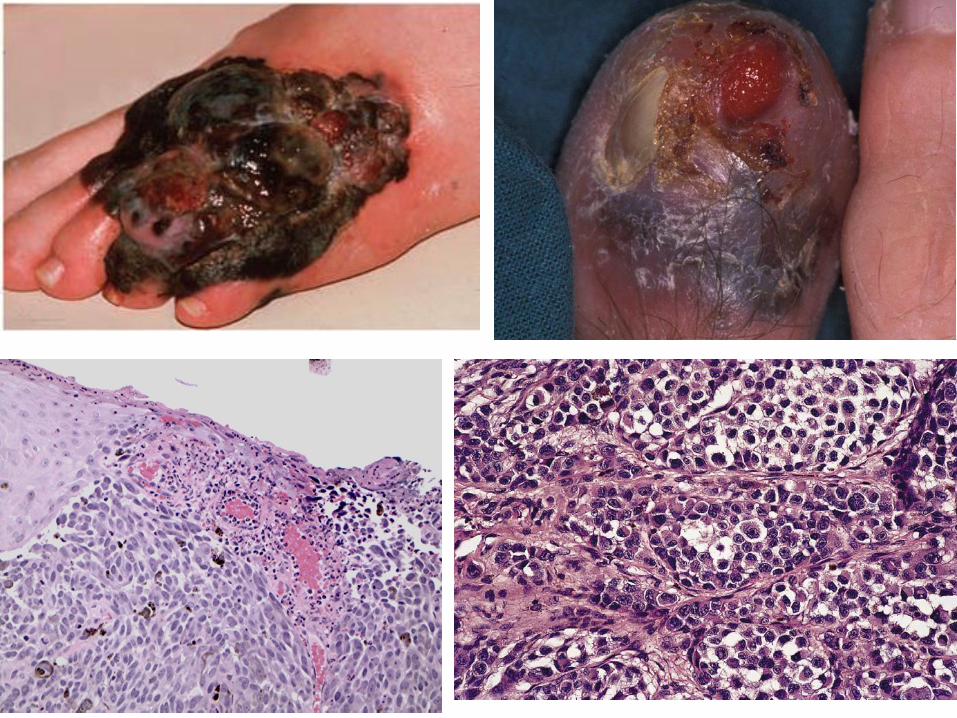
• SANGRADO E INFECCIONES CUANDO EL TUMOR SE ULCERA POR LA SUPERFICIE ADYACENTE
• SINTOMAS QUE RESULTAN DE RUPTURA E INFARTO.
• SINDROME CONSUNTIVO Y CAQUEXIA.
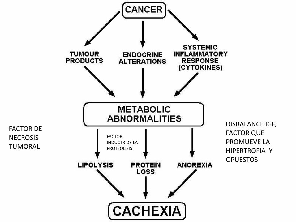
FACTOR DE NECROSIS TUMORAL
DISBALANCE IGF, FACTOR QUE PROMUEVE LA HIPERTROFIA Y OPUESTOS
FACTOR INDUCTR DE LA PROTEOLISIS
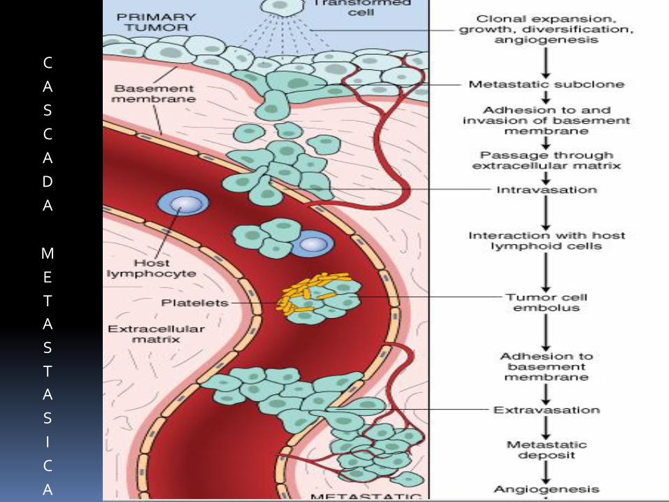
C
A
S
C
A
D
A
M
E
T
A
S
T
A
S
I
C
A

GRACIAS
Recommended