Embed Size (px)
Citation preview
Diagnòstic integrat en eritropatologia: Membranopaties
María del Mar Mañú PereiraVall d’Hebron campus – Rare anaemia research area
ERN-EuroBloodNet Scientific [email protected]
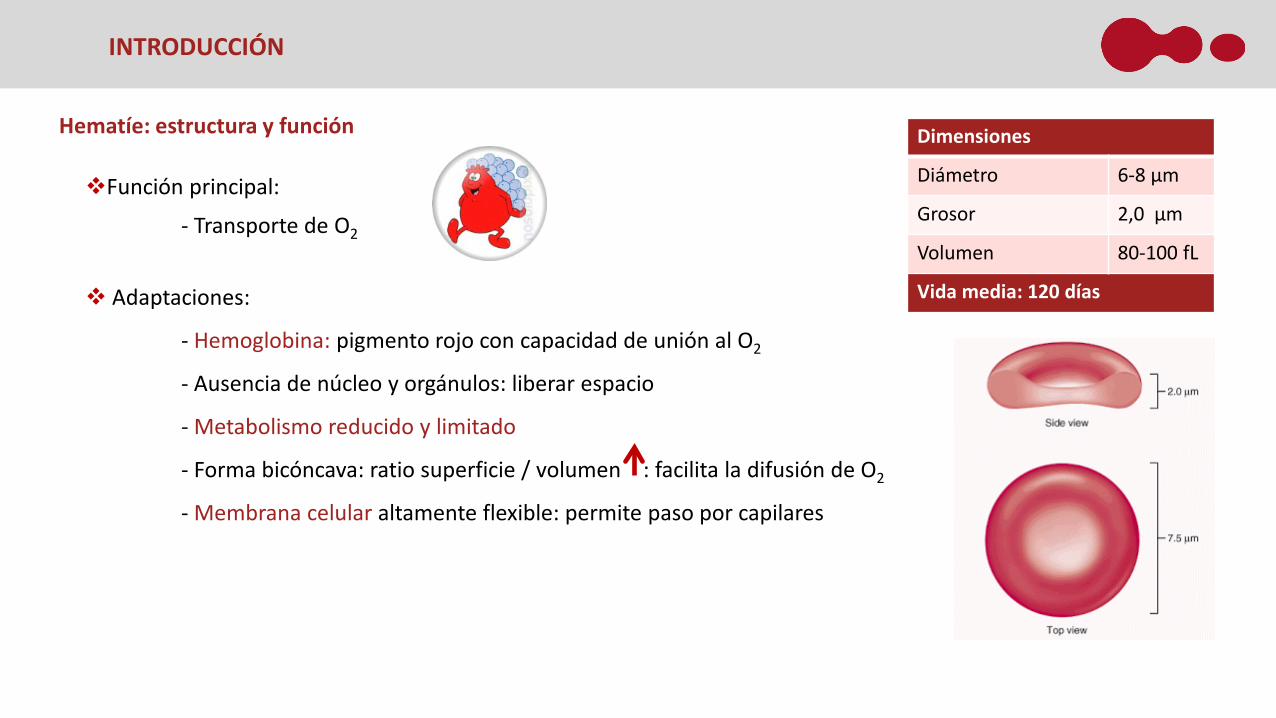
Hematíe: estructura y función
Función principal:
- Transporte de O2
Adaptaciones:
- Hemoglobina: pigmento rojo con capacidad de unión al O2
- Ausencia de núcleo y orgánulos: liberar espacio
- Metabolismo reducido y limitado
- Forma bicóncava: ratio superficie / volumen : facilita la difusión de O2
- Membrana celular altamente flexible: permite paso por capilares
Dimensiones
Diámetro 6-8 µm
Grosor 2,0 µm
Volumen 80-100 fL
Vida media: 120 días
INTRODUCCIÓN
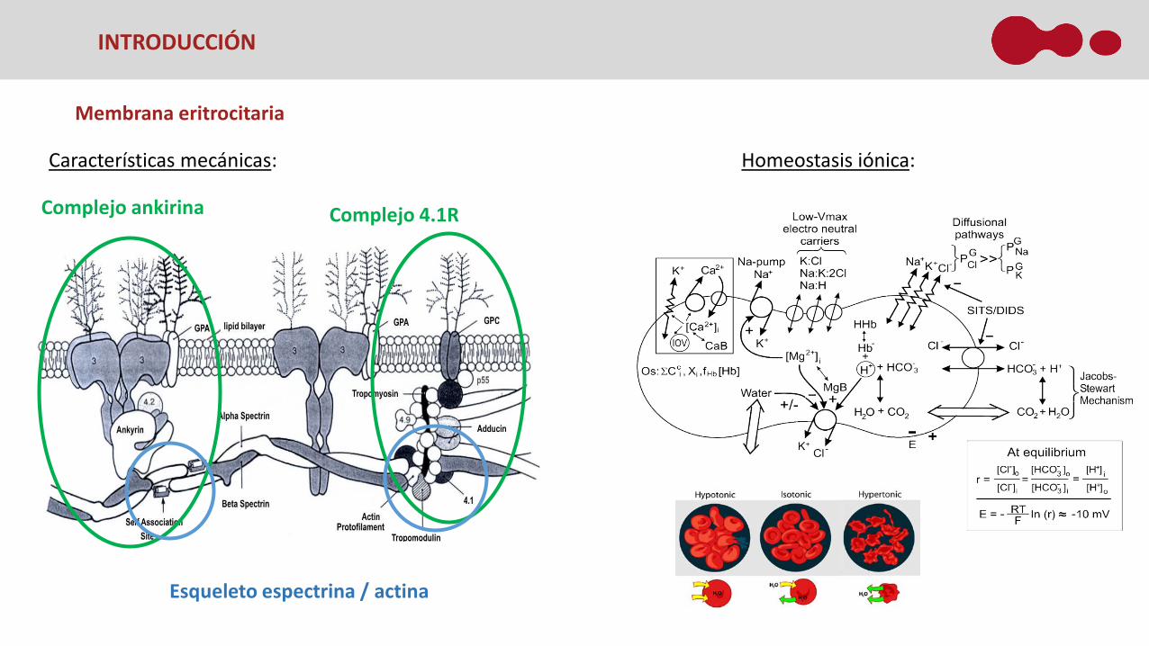
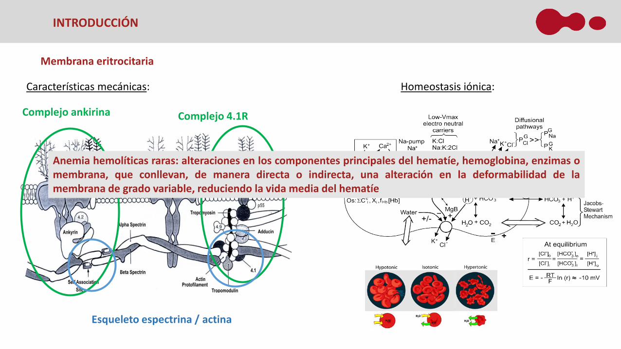
Membrana eritrocitaria
Características mecánicas: Homeostasis iónica:
INTRODUCCIÓN
Complejo ankirina Complejo 4.1R
Esqueleto espectrina / actina
Membrana eritrocitaria
Características mecánicas: Homeostasis iónica:
INTRODUCCIÓN
Complejo ankirina Complejo 4.1R
Esqueleto espectrina / actina
Anemia hemolíticas raras: alteraciones en los componentes principales del hematíe, hemoglobina, enzimas omembrana, que conllevan, de manera directa o indirecta, una alteración en la deformabilidad de lamembrana de grado variable, reduciendo la vida media del hematíe
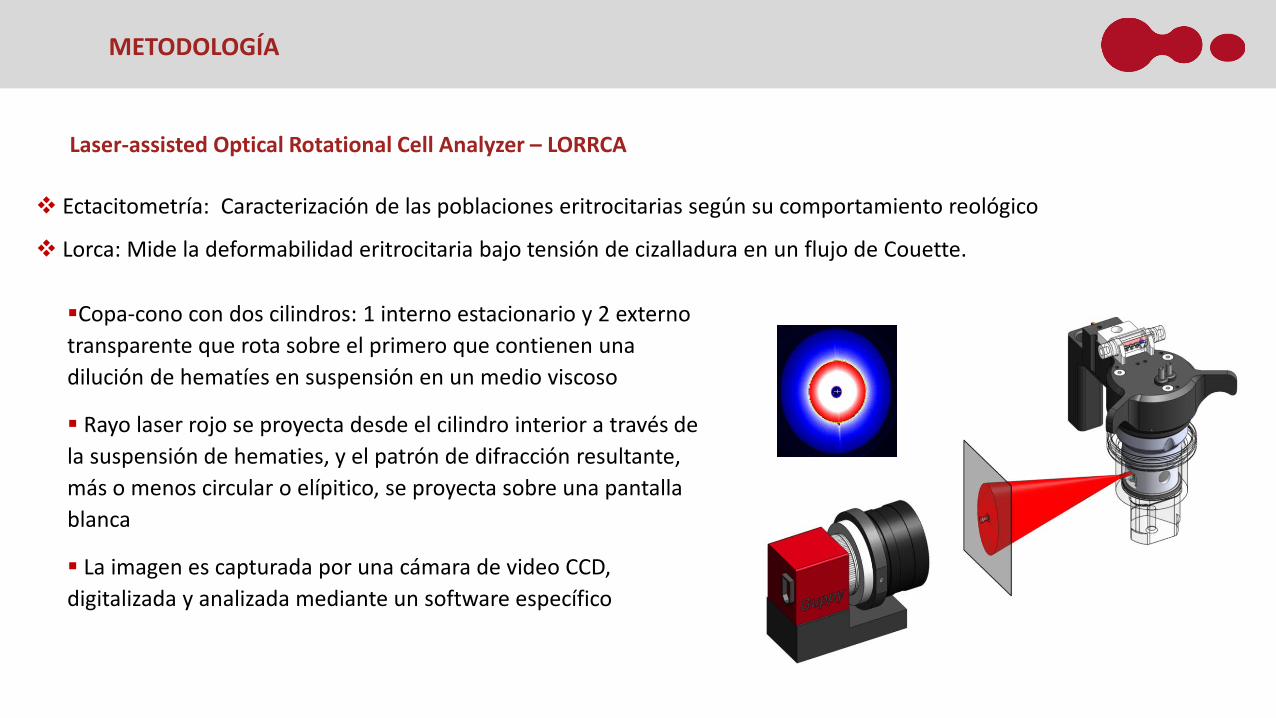
Laser-assisted Optical Rotational Cell Analyzer – LORRCA
METODOLOGÍA
Copa-cono con dos cilindros: 1 interno estacionario y 2 externo transparente que rota sobre el primero que contienen una dilución de hematíes en suspensión en un medio viscoso
Rayo laser rojo se proyecta desde el cilindro interior a través de la suspensión de hematies, y el patrón de difracción resultante, más o menos circular o elípitico, se proyecta sobre una pantalla blanca
La imagen es capturada por una cámara de video CCD, digitalizada y analizada mediante un software específico
Ectacitometría: Caracterización de las poblaciones eritrocitarias según su comportamiento reológico
Lorca: Mide la deformabilidad eritrocitaria bajo tensión de cizalladura en un flujo de Couette.
Copyright © 2014 RR Mechatronics
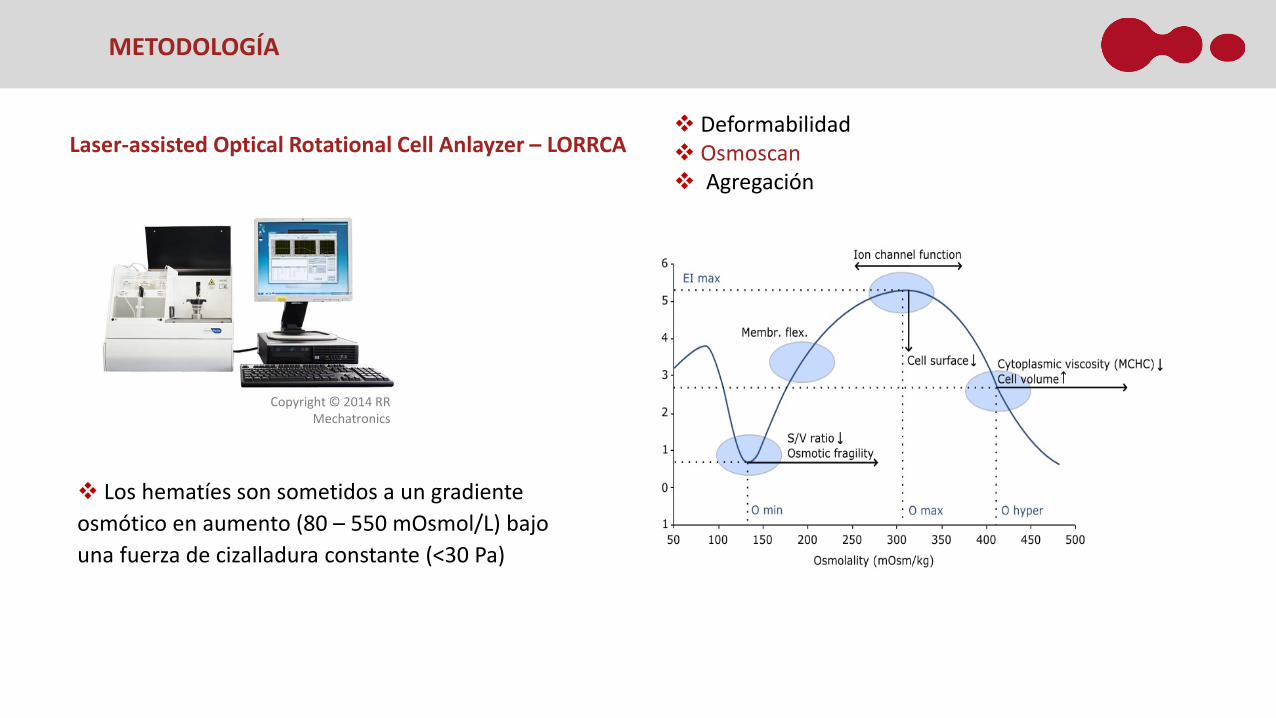
Laser-assisted Optical Rotational Cell Anlayzer – LORRCA
METODOLOGÍA
Deformabilidad Osmoscan Agregación
Los hematíes son sometidos a un gradiente osmótico en aumento (80 – 550 mOsmol/L) bajo una fuerza de cizalladura constante (<30 Pa)
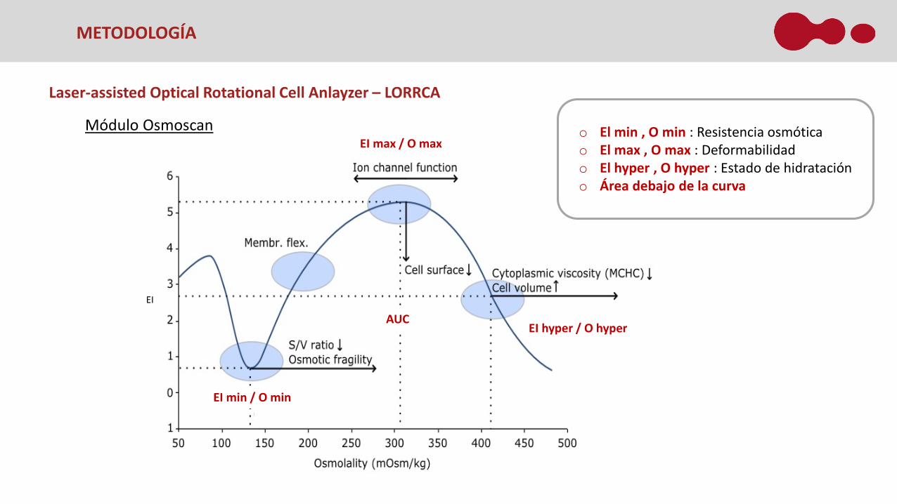
Laser-assisted Optical Rotational Cell Anlayzer – LORRCA
Módulo Osmoscan
Copyright © 2014 RR Mechatronics
EI
EI max
EI max EI max EI maxEI min / O min
EI max / O max
EI hyper / O hyperAUC
o El min , O min : Resistencia osmóticao El max , O max : Deformabilidado El hyper , O hyper : Estado de hidratacióno Área debajo de la curva
METODOLOGÍA
Aplicación del LoRRCa
Diagnóstico de anemias hemolíticas debidas alteraciones de la membrana y/o homeostasis iónica
APLICACIONES
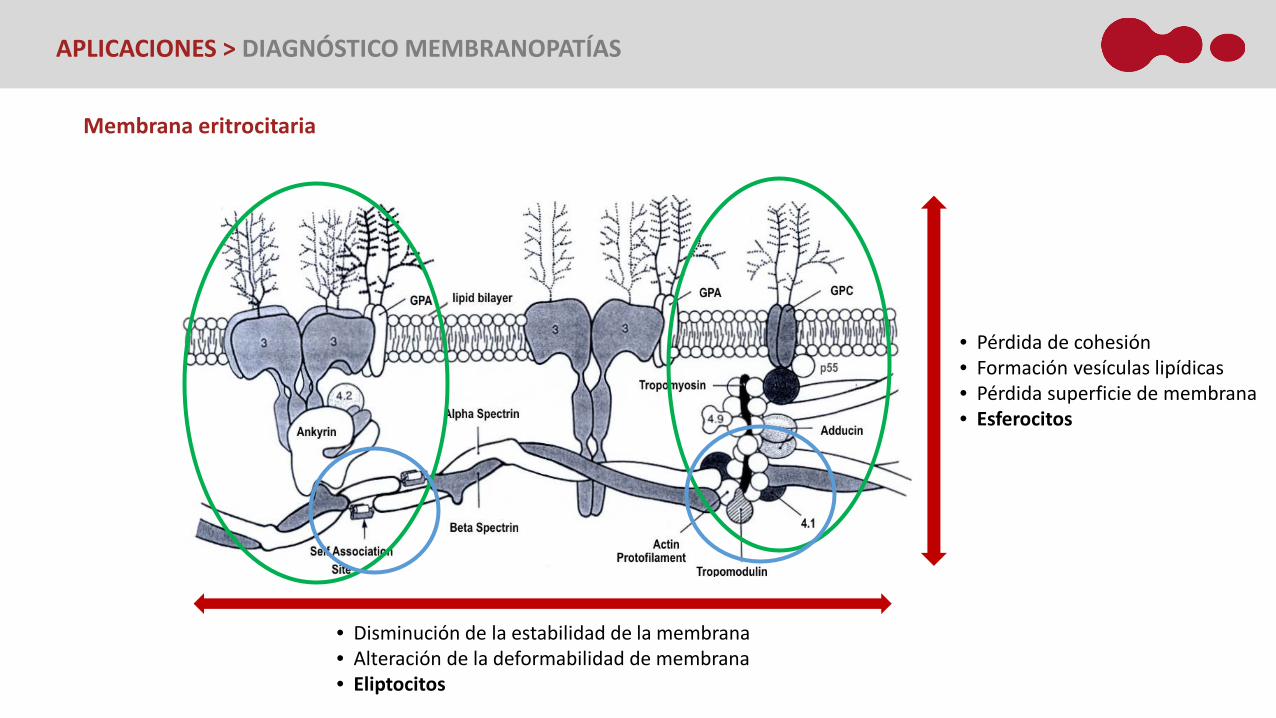
• Pérdida de cohesión• Formación vesículas lipídicas• Pérdida superficie de membrana• Esferocitos
• Disminución de la estabilidad de la membrana• Alteración de la deformabilidad de membrana• Eliptocitos
APLICACIONES > DIAGNÓSTICO MEMBRANOPATÍAS
Membrana eritrocitaria
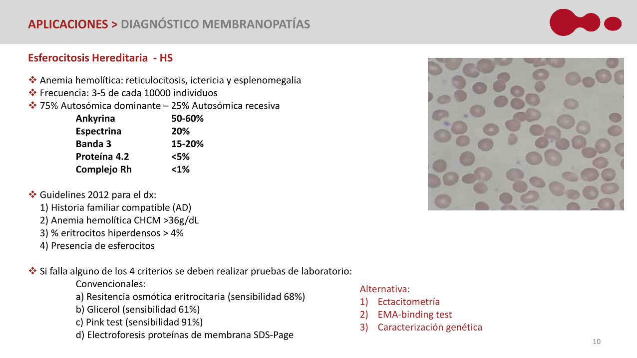
Anemia hemolítica: reticulocitosis, ictericia y esplenomegalia Frecuencia: 3-5 de cada 10000 individuos 75% Autosómica dominante – 25% Autosómica recesiva
Ankyrina 50-60%Espectrina 20%Banda 3 15-20%Proteína 4.2 <5%Complejo Rh <1%
Guidelines 2012 para el dx:1) Historia familiar compatible (AD)2) Anemia hemolítica CHCM >36g/dL3) % eritrocitos hiperdensos > 4%4) Presencia de esferocitos
Si falla alguno de los 4 criterios se deben realizar pruebas de laboratorio: Convencionales:a) Resitencia osmótica eritrocitaria (sensibilidad 68%)b) Glicerol (sensibilidad 61%)c) Pink test (sensibilidad 91%)d) Electroforesis proteínas de membrana SDS-Page
10
Esferocitosis Hereditaria - HS
APLICACIONES > DIAGNÓSTICO MEMBRANOPATÍAS
Alternativa: 1) Ectacitometría2) EMA-binding test3) Caracterización genética
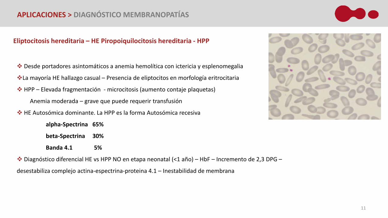
Desde portadores asintomáticos a anemia hemolítica con ictericia y esplenomegalia
La mayoría HE hallazgo casual – Presencia de eliptocitos en morfología eritrocitaria
HPP – Elevada fragmentación - microcitosis (aumento contaje plaquetas)
Anemia moderada – grave que puede requerir transfusión
HE Autosómica dominante. La HPP es la forma Autosómica recesiva
alpha-Spectrina 65%
beta-Spectrina 30%
Banda 4.1 5%
Diagnóstico diferencial HE vs HPP NO en etapa neonatal (<1 año) – HbF – Incremento de 2,3 DPG –
desestabiliza complejo actina-espectrina-proteina 4.1 – Inestabilidad de membrana
11
Eliptocitosis hereditaria – HE Piropoiquilocitosis hereditaria - HPP
APLICACIONES > DIAGNÓSTICO MEMBRANOPATÍAS
12
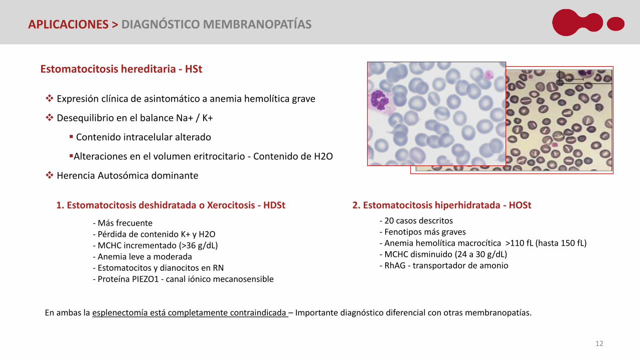
Expresión clínica de asintomático a anemia hemolítica grave
Desequilibrio en el balance Na+ / K+
Contenido intracelular alterado
Alteraciones en el volumen eritrocitario - Contenido de H2O
Herencia Autosómica dominante
- Más frecuente- Pérdida de contenido K+ y H2O- MCHC incrementado (>36 g/dL)- Anemia leve a moderada- Estomatocitos y dianocitos en RN- Proteína PIEZO1 - canal iónico mecanosensible
En ambas la esplenectomía está completamente contraindicada – Importante diagnóstico diferencial con otras membranopatías.
- 20 casos descritos- Fenotipos más graves- Anemia hemolítica macrocítica >110 fL (hasta 150 fL)- MCHC disminuido (24 a 30 g/dL)- RhAG - transportador de amonio
APLICACIONES > DIAGNÓSTICO MEMBRANOPATÍAS
Estomatocitosis hereditaria - HSt
1. Estomatocitosis deshidratada o Xerocitosis - HDSt 2. Estomatocitosis hiperhidratada - HOSt
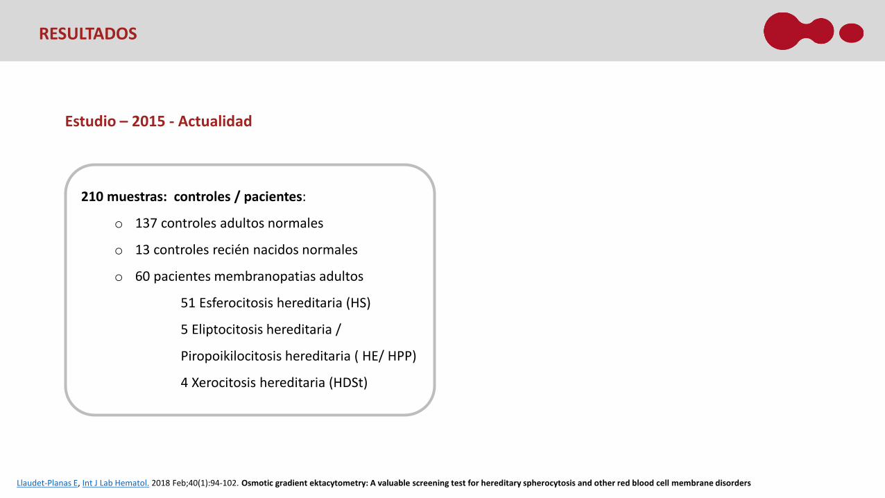
Estudio – 2015 - Actualidad
210 muestras: controles / pacientes:
o 137 controles adultos normales
o 13 controles recién nacidos normales
o 60 pacientes membranopatias adultos
51 Esferocitosis hereditaria (HS)
5 Eliptocitosis hereditaria /
Piropoikilocitosis hereditaria ( HE/ HPP)
4 Xerocitosis hereditaria (HDSt)
RESULTADOS
Llaudet-Planas E, Int J Lab Hematol. 2018 Feb;40(1):94-102. Osmotic gradient ektacytometry: A valuable screening test for hereditary spherocytosis and other red blood cell membrane disorders
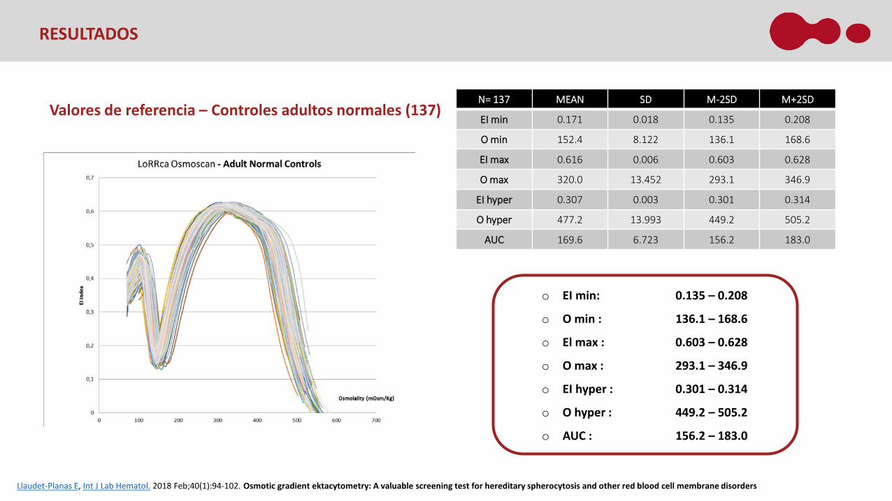
Valores de referencia – Controles adultos normales (137)
o EI min: 0.135 – 0.208
o O min : 136.1 – 168.6
o El max : 0.603 – 0.628
o O max : 293.1 – 346.9
o EI hyper : 0.301 – 0.314
o O hyper : 449.2 – 505.2
o AUC : 156.2 – 183.0
N= 137 MEAN SD M-2SD M+2SD
EI min 0.171 0.018 0.135 0.208
O min 152.4 8.122 136.1 168.6
EI max 0.616 0.006 0.603 0.628
O max 320.0 13.452 293.1 346.9
EI hyper 0.307 0.003 0.301 0.314
O hyper 477.2 13.993 449.2 505.2
AUC 169.6 6.723 156.2 183.0
RESULTADOS
Llaudet-Planas E, Int J Lab Hematol. 2018 Feb;40(1):94-102. Osmotic gradient ektacytometry: A valuable screening test for hereditary spherocytosis and other red blood cell membrane disorders
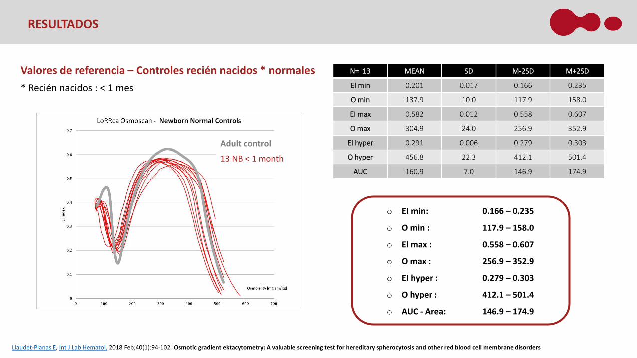
o EI min: 0.166 – 0.235
o O min : 117.9 – 158.0
o El max : 0.558 – 0.607
o O max : 256.9 – 352.9
o EI hyper : 0.279 – 0.303
o O hyper : 412.1 – 501.4
o AUC - Area: 146.9 – 174.9
N= 13 MEAN SD M-2SD M+2SD
EI min 0.201 0.017 0.166 0.235
O min 137.9 10.0 117.9 158.0
EI max 0.582 0.012 0.558 0.607
O max 304.9 24.0 256.9 352.9
EI hyper 0.291 0.006 0.279 0.303
O hyper 456.8 22.3 412.1 501.4
AUC 160.9 7.0 146.9 174.9
Adult control
13 NB < 1 month
Valores de referencia – Controles recién nacidos * normales* Recién nacidos : < 1 mes
RESULTADOS
Llaudet-Planas E, Int J Lab Hematol. 2018 Feb;40(1):94-102. Osmotic gradient ektacytometry: A valuable screening test for hereditary spherocytosis and other red blood cell membrane disorders
Llaudet-Planas E, Int J Lab Hematol. 2018 Feb;40(1):94-102. Osmotic gradient ektacytometry: A valuable screening test for hereditary spherocytosis and other red blood cell membrane disorders
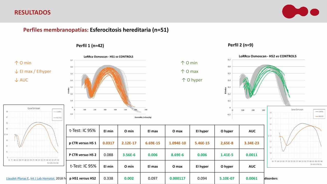
Perfiles membranopatías: Esferocitosis hereditaria (n=51)
RESULTADOS
↑ O min
↓ EI max / EIhyper
↓ AUC
↑ O min
↑ O max
↑ O hyper
EI min O min El max O max EI hyper O hyper AUC
p CTR versus HS 1 0.0317 2.12E-17 6.69E-15 1.094E-10 5.46E-15 2,65E-8 3.34E-23
P CTR versus HS 2 0.088 3.56E-6 0.006 8.69E-6 0.006 1.41E-5 0.0011
t-Test: IC 95%
Perfil 1 (n=42) Perfil 2 (n=9)
EI min O min El max O max EI hyper O hyper AUC
p HS1 versus HS2 0.338 0.002 0.097 0.000117 0.094 5.10E-07 0.0061
t-Test: IC 95%

Llaudet-Planas E, Int J Lab Hematol. 2018 Feb;40(1):94-102. Osmotic gradient ektacytometry: A valuable screening test for hereditary spherocytosis and other red blood cell membrane disorders
Perfiles membranopatías
Eliptocitosis hereditaria/ Piropoiquilocitosis hereditaria (n=5)
RESULTADOS
Perfil forma trapezoidal
↓ EI max / EI hyper
EI min: 0.137 – 0,174
O min : 130.8 – 163.2
El max : 0.463 – 0.621
O max : 228.1 – 332.3
EI hyper : 0.232 – 0.310
O hyper : 465.2 – 478.0
AUC : 120.0 – 174.3
EI min O min El max O max EI hyper O hyper AUC
p CTR versus HE 0.0178 0.2574 0.0144 0.0277 0.0143 0.328 0.0211
t-Test: IC 95%
Llaudet-Planas E, Int J Lab Hematol. 2018 Feb;40(1):94-102. Osmotic gradient ektacytometry: A valuable screening test for hereditary spherocytosis and other red blood cell membrane disorders
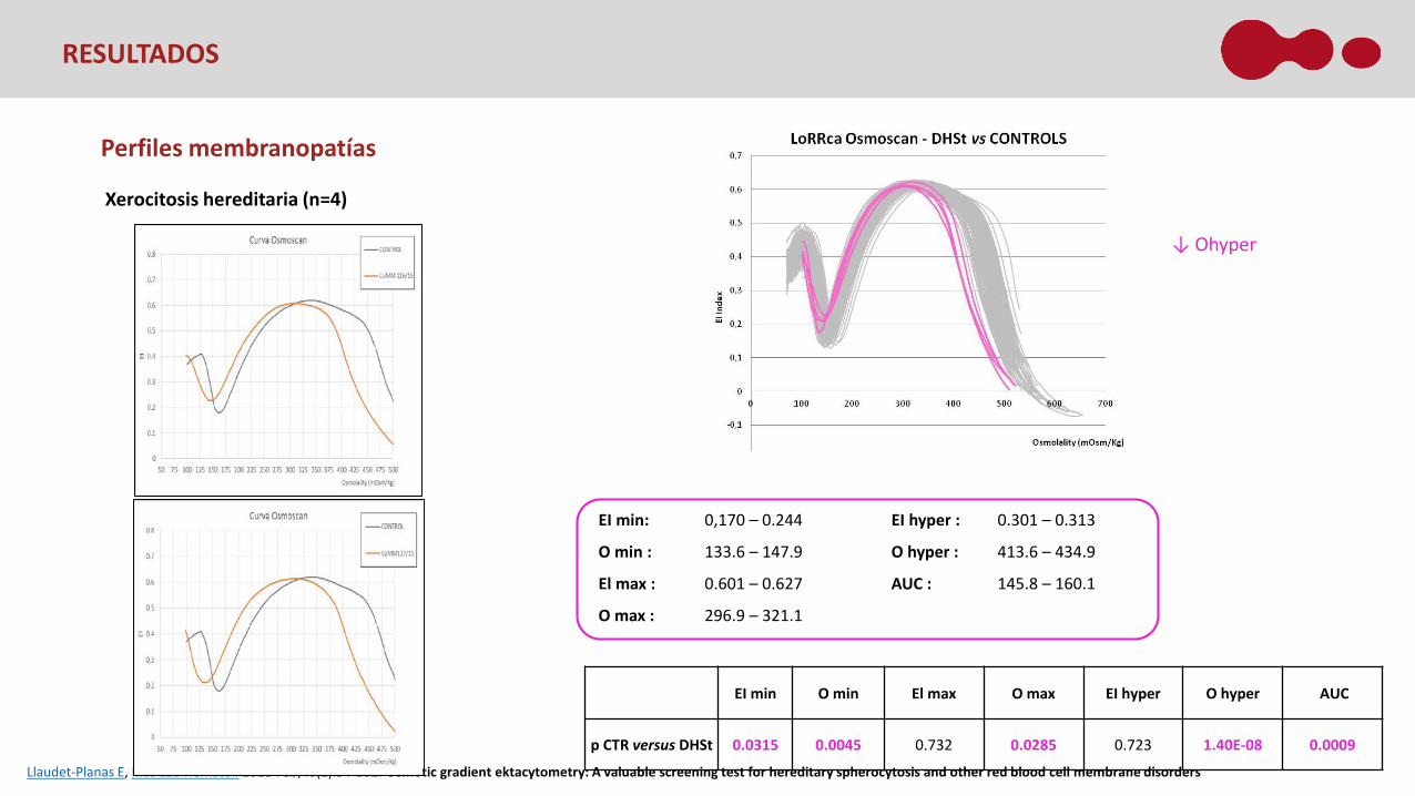
Perfiles membranopatías
Xerocitosis hereditaria (n=4)
RESULTADOS
↓ Ohyper
EI min O min El max O max EI hyper O hyper AUC
p CTR versus DHSt 0.0315 0.0045 0.732 0.0285 0.723 1.40E-08 0.0009
EI min: 0,170 – 0.244
O min : 133.6 – 147.9
El max : 0.601 – 0.627
O max : 296.9 – 321.1
EI hyper : 0.301 – 0.313
O hyper : 413.6 – 434.9
AUC : 145.8 – 160.1
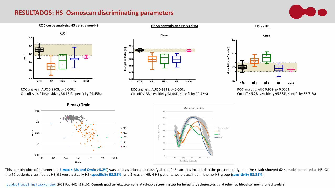
ROC curve analysis: HS versus non-HS
ROC analysis: AUC 0.9903; p<0.0001Cut-off <-14.9%(sensitivity 86.15%, specificity 99.45%)
ROC analysis: AUC 0.9998, p<0.0001Cut-off < -3%(sensitivity 98.46%, specificity 99.42%)
ROC analysis: AUC 0.959, p<0.0001Cut-off > 5.2%(sensitivity 95.38%, specificity 85.71%)
HS vs controls and HS vs dHSt HS vs HE
This combination of parameters (EImax <-3% and Omin >5.2%) was used as criteria to classify all the 246 samples included in the present study, and the result showed 62 samples detected as HS. Ofthe 62 patients classified as HS, 61 were actually HS (specificity 98.38%) and 1 was an HE. 4 HS patients were classified in the no-HS group (sensitivity 93.85%)
Llaudet-Planas E, Int J Lab Hematol. 2018 Feb;40(1):94-102. Osmotic gradient ektacytometry: A valuable screening test for hereditary spherocytosis and other red blood cell membrane disorders
RESULTADOS: HS Osmoscan discriminating parameters
INTRODUCCIÓN METODOLOGÍA RESULTADOS CONCLUSIONES
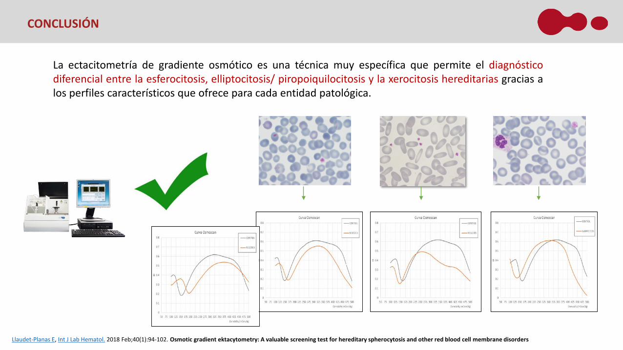
La ectacitometría de gradiente osmótico es una técnica muy específica que permite el diagnósticodiferencial entre la esferocitosis, elliptocitosis/ piropoiquilocitosis y la xerocitosis hereditarias gracias alos perfiles característicos que ofrece para cada entidad patológica.
CONCLUSIÓN
Llaudet-Planas E, Int J Lab Hematol. 2018 Feb;40(1):94-102. Osmotic gradient ektacytometry: A valuable screening test for hereditary spherocytosis and other red blood cell membrane disorders
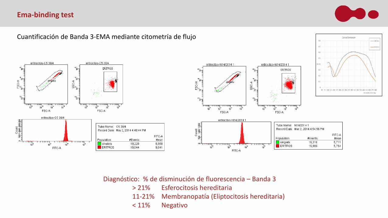
Diagnóstico: % de disminución de fluorescencia – Banda 3> 21% Esferocitosis hereditaria11-21% Membranopatía (Eliptocitosis hereditaria)< 11% Negativo
Ema-binding test
Cuantificación de Banda 3-EMA mediante citometría de flujo
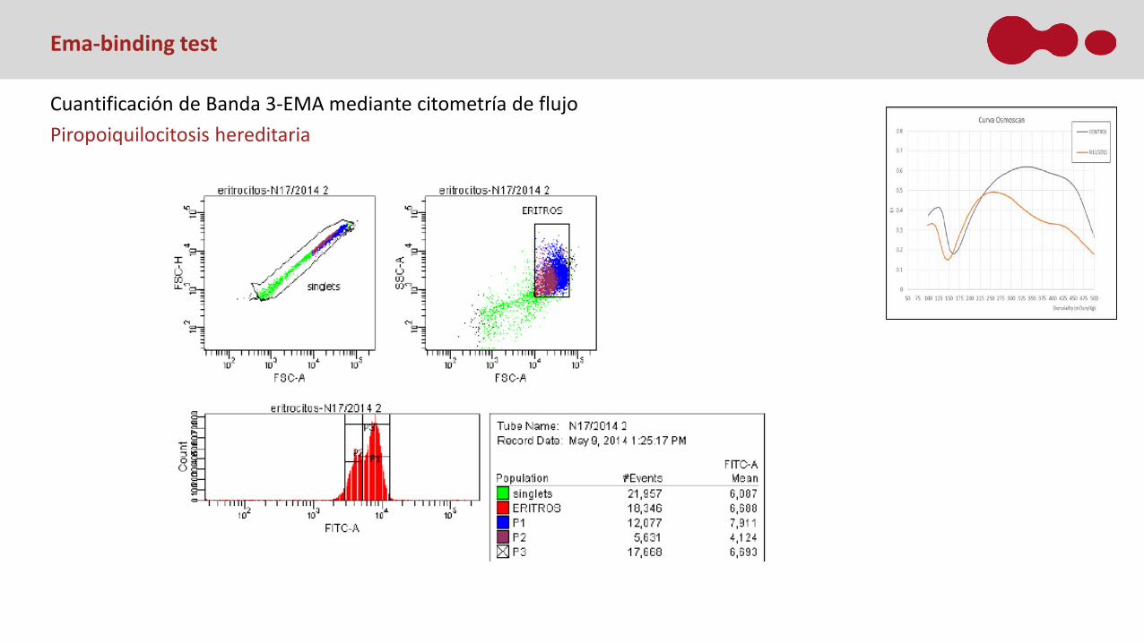
Cuantificación de Banda 3-EMA mediante citometría de flujoPiropoiquilocitosis hereditaria
Ema-binding test
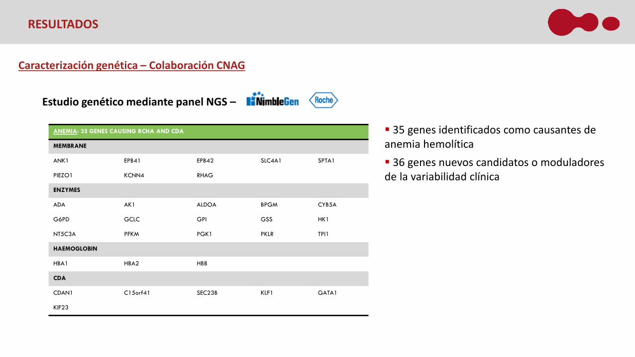
Caracterización genética – Colaboración CNAG
Estudio genético mediante panel NGS –
INTRODUCCIÓN METODOLOGÍA RESULTADOS CONCLUSIONESRESULTADOS
ANEMIA: 35 GENES CAUSING RCHA AND CDA
MEMBRANE
ANK1 EPB41 EPB42 SLC4A1 SPTA1
PIEZO1 KCNN4 RHAG
ENZYMES
ADA AK1 ALDOA BPGM CYB5A
G6PD GCLC GPI GSS HK1
NT5C3A PFKM PGK1 PKLR TPI1
HAEMOGLOBIN
HBA1 HBA2 HBB
CDA
CDAN1 C15orf41 SEC23B KLF1 GATA1
KIF23
35 genes identificados como causantes de anemia hemolítica 36 genes nuevos candidatos o moduladores de la variabilidad clínica
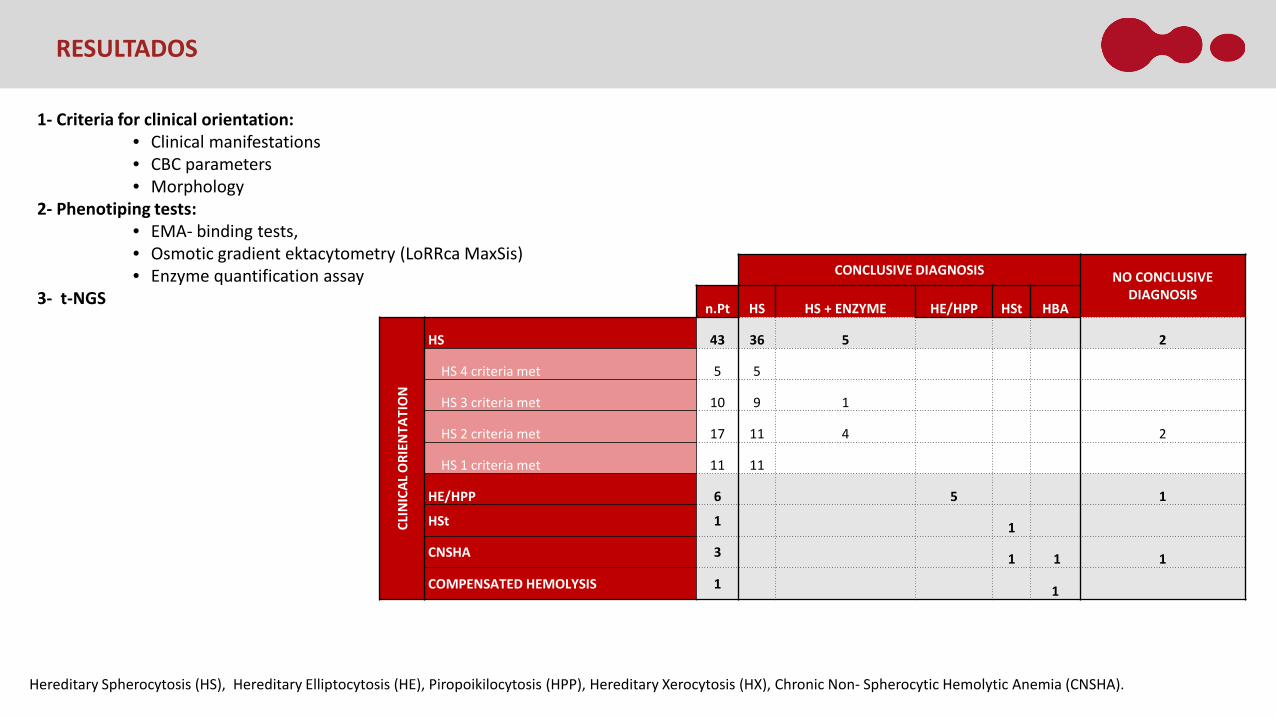
Hereditary Spherocytosis (HS), Hereditary Elliptocytosis (HE), Piropoikilocytosis (HPP), Hereditary Xerocytosis (HX), Chronic Non- Spherocytic Hemolytic Anemia (CNSHA).
CONCLUSIVE DIAGNOSIS NO CONCLUSIVEDIAGNOSIS
n.Pt HS HS + ENZYME HE/HPP HSt HBA
CLIN
ICAL
ORI
ENTA
TIO
N
HS 43 36 5 2
HS 4 criteria met 5 5
HS 3 criteria met 10 9 1
HS 2 criteria met 17 11 4 2
HS 1 criteria met 11 11
HE/HPP 6 5 1
HSt 1 1
CNSHA 3 1 1 1
COMPENSATED HEMOLYSIS 1 1
1- Criteria for clinical orientation:• Clinical manifestations• CBC parameters• Morphology
2- Phenotiping tests: • EMA- binding tests, • Osmotic gradient ektacytometry (LoRRca MaxSis)• Enzyme quantification assay
3- t-NGS
RESULTADOS
RESULTADOS
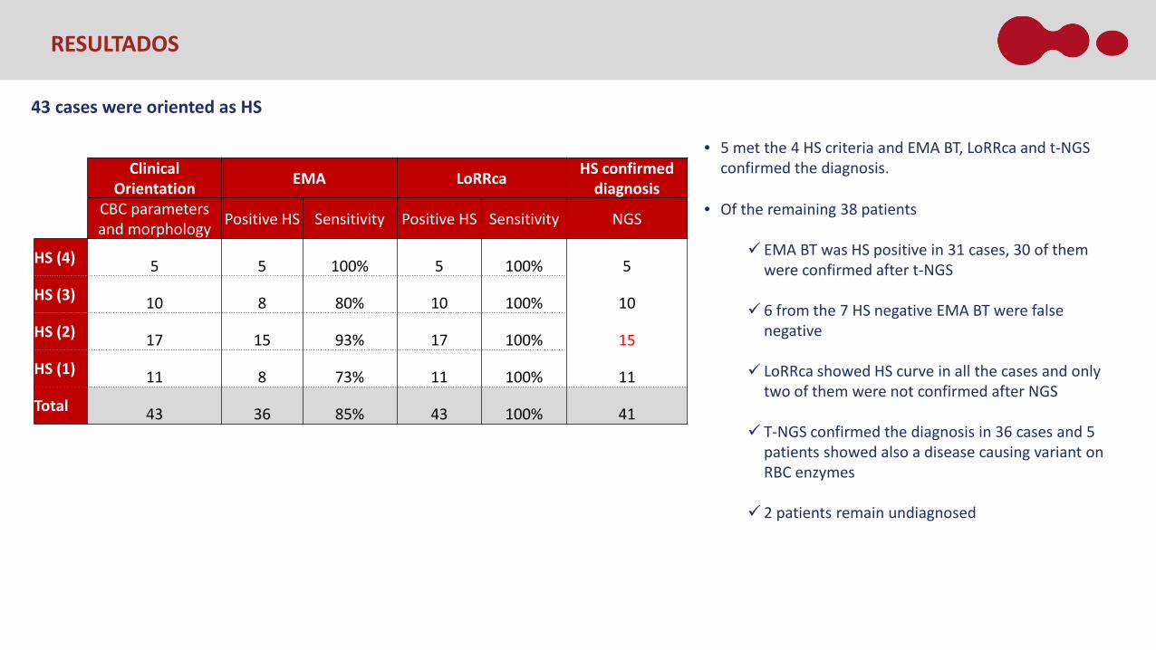
ClinicalOrientation EMA LoRRca HS confirmed
diagnosisCBC parametersand morphology Positive HS Sensitivity Positive HS Sensitivity NGS
HS (4) 5 5 100% 5 100% 5
HS (3) 10 8 80% 10 100% 10
HS (2) 17 15 93% 17 100% 15
HS (1) 11 8 73% 11 100% 11
Total 43 36 85% 43 100% 41
• Of the remaining 38 patients
EMA BT was HS positive in 31 cases, 30 of them were confirmed after t-NGS
6 from the 7 HS negative EMA BT were false negative
LoRRca showed HS curve in all the cases and only two of them were not confirmed after NGS
T-NGS confirmed the diagnosis in 36 cases and 5 patients showed also a disease causing variant on RBC enzymes
2 patients remain undiagnosed
43 cases were oriented as HS
• 5 met the 4 HS criteria and EMA BT, LoRRca and t-NGS confirmed the diagnosis.
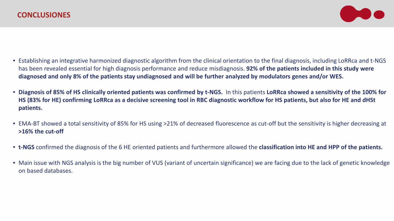
• Establishing an integrative harmonized diagnostic algorithm from the clinical orientation to the final diagnosis, including LoRRca and t-NGS has been revealed essential for high diagnosis performance and reduce misdiagnosis. 92% of the patients included in this study were diagnosed and only 8% of the patients stay undiagnosed and will be further analyzed by modulators genes and/or WES.
• Diagnosis of 85% of HS clinically oriented patients was confirmed by t-NGS. In this patients LoRRca showed a sensitivity of the 100% for HS (83% for HE) confirming LoRRca as a decisive screening tool in RBC diagnostic workflow for HS patients, but also for HE and dHStpatients.
• EMA-BT showed a total sensitivity of 85% for HS using >21% of decreased fluorescence as cut-off but the sensitivity is higher decreasing at >16% the cut-off
• t-NGS confirmed the diagnosis of the 6 HE oriented patients and furthermore allowed the classification into HE and HPP of the patients.
• Main issue with NGS analysis is the big number of VUS (variant of uncertain significance) we are facing due to the lack of genetic knowledge on based databases.
CONCLUSIONES
APLICACIONES > PRONÓSTICO ENFERMEDAD DE CÉLULAS FALCIFORME
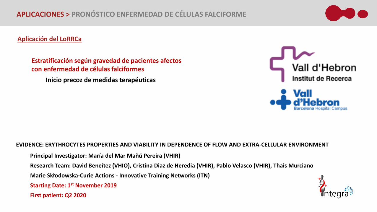
Aplicación del LoRRCa
Estratificación según gravedad de pacientes afectos con enfermedad de células falciformes
Inicio precoz de medidas terapéuticas
EVIDENCE: ERYTHROCYTES PROPERTIES AND VIABILITY IN DEPENDENCE OF FLOW AND EXTRA-CELLULAR ENVIRONMENT
Principal Investigator: María del Mar Mañú Pereira (VHIR)Research Team: David Beneítez (VHIO), Cristina Diaz de Heredia (VHIR), Pablo Velasco (VHIR), Thais MurcianoMarie Skłodowska-Curie Actions - Innovative Training Networks (ITN)Starting Date: 1st November 2019First patient: Q2 2020
APLICACIONES > PRONÓSTICO ENFERMEDAD DE CÉLULAS FALCIFORME
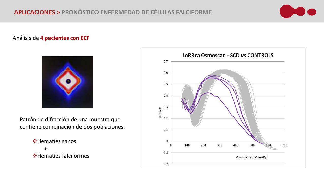
Análisis de 4 pacientes con ECF
Patrón de difracción de una muestra que contiene combinación de dos poblaciones:
Hematíes sanos +
Hematíes falciformes
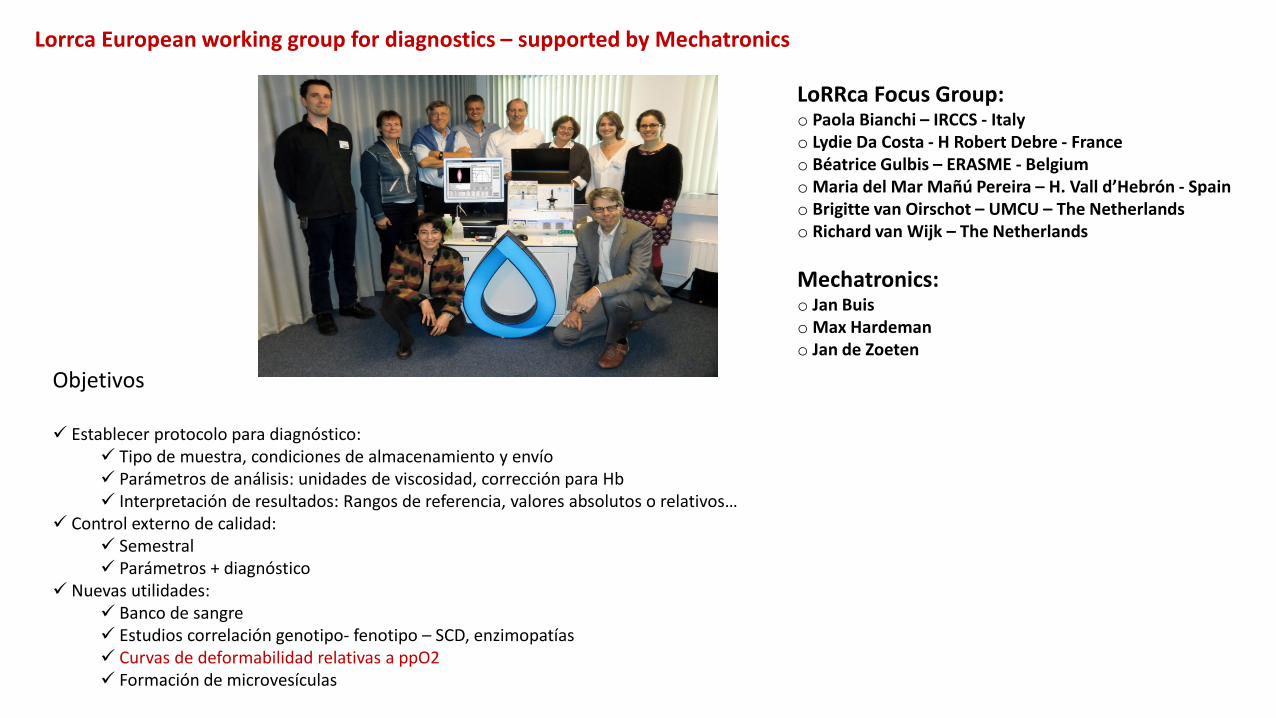
Lorrca European working group for diagnostics – supported by Mechatronics
LoRRca Focus Group:o Paola Bianchi – IRCCS - Italyo Lydie Da Costa - H Robert Debre - Franceo Béatrice Gulbis – ERASME - Belgiumo Maria del Mar Mañú Pereira – H. Vall d’Hebrón - Spaino Brigitte van Oirschot – UMCU – The Netherlandso Richard van Wijk – The Netherlands
Mechatronics:o Jan Buiso Max Hardemano Jan de Zoeten
Objetivos
Establecer protocolo para diagnóstico: Tipo de muestra, condiciones de almacenamiento y envío Parámetros de análisis: unidades de viscosidad, corrección para Hb Interpretación de resultados: Rangos de referencia, valores absolutos o relativos…
Control externo de calidad: Semestral Parámetros + diagnóstico
Nuevas utilidades: Banco de sangre Estudios correlación genotipo- fenotipo – SCD, enzimopatías Curvas de deformabilidad relativas a ppO2 Formación de microvesículas
Copyright © 2014 RR Mechatronics
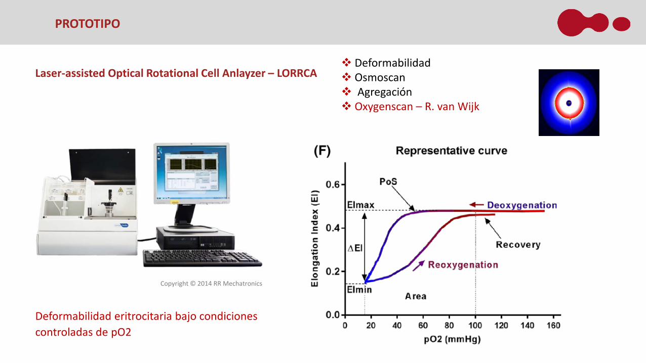
Laser-assisted Optical Rotational Cell Anlayzer – LORRCA
PROTOTIPO
Deformabilidad Osmoscan Agregación Oxygenscan – R. van Wijk
Deformabilidad eritrocitaria bajo condiciones controladas de pO2
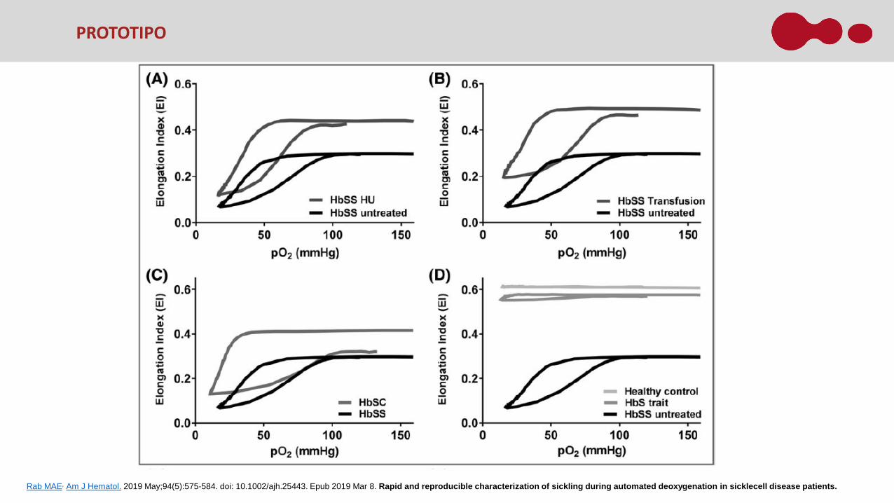
Rab MAE, Am J Hematol. 2019 May;94(5):575-584. doi: 10.1002/ajh.25443. Epub 2019 Mar 8. Rapid and reproducible characterization of sickling during automated deoxygenation in sicklecell disease patients.
PROTOTIPO
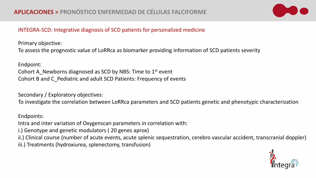
Primary objective: To assess the prognostic value of LoRRca as biomarker providing information of SCD patients severity
Endpoint:Cohort A_Newborns diagnosed as SCD by NBS: Time to 1st eventCohort B and C_Pediatric and adult SCD Patients: Frequency of events
Secondary / Exploratory objectives:To investigate the correlation between LoRRca parameters and SCD patients genetic and phenotypic characterization
Endpoints:Intra and inter variation of Oxygenscan parameters in correlation with: i.) Genotype and genetic modulators ( 20 genes aprox)ii.) Clinical course (number of acute events, acute splenic sequestration, cerebro vascular accident, transcranial doppler)iii.) Treatments (hydroxiurea, splenectomy, transfusion)
APLICACIONES > PRONÓSTICO ENFERMEDAD DE CÉLULAS FALCIFORME
INTEGRA-SCD: Integrative diagnosis of SCD patients for personalized medicine
Vall d’Hebrón Campus
HOSPITAL UNIVERSITARI VALL D’HEBRÓN (HUVH)
SERVEI D’HEMATOLOGIA CLÍNICA
UNITAT D’ERITROPATOLOGIA I DIAGNÒSTIC INTEGRAT
• David Beneítez (VHIO)• Ana Ortuño• Adoración Blanco• Barbara Tazón (VHIO)
SERVEI D’ONCOLOGIA I HEMATOLOGIA PEDIÀTRIQUES
• Cristina Diaz de Heredia (VHIR)• Pablo Velasco (VHIR)• Thais Murciano• Mari Luz Uría• Maribel Benítez• Laura Murillo• Laura Alonso
INSTITUT D’INVESTIGACIÓ VALL D’HEBRÓN (VHIR)
RECERCA TRANSLACIONAL EN CÀNCER EN LA INFÀNCIA I L'ADOLESCÈNCIA
RECERCA EN ANÈMIES MINORITÀRIES
• María del Mar Mañú Pereira
• Victoria Gutiérrez Valle
• Valeria Rizzuto