Embed Size (px)
Citation preview
Tip Revista Especializada en Ciencias
Químico-Biológicas
ISSN: 1405-888X
Universidad Nacional Autónoma de
México
México
Mendoza-Meza, Dary Luz; España-Puccini, Pierine
Cytotoxic and genotoxic activity of phenolic fractions from Ulomoides dermestoides
Fairmaire, 1893 (Coleoptera, Tenebrionidae), in HaCat Cells
Tip Revista Especializada en Ciencias Químico-Biológicas, vol. 19, núm. 2, agosto, 2016,
pp. 83-91
Universidad Nacional Autónoma de México
Distrito Federal, México
Available in: http://www.redalyc.org/articulo.oa?id=43246005001
How to cite
Complete issue
More information about this article
Journal's homepage in redalyc.org
Scientific Information System
Network of Scientific Journals from Latin America, the Caribbean, Spain and Portugal
Non-profit academic project, developed under the open access initiative

D.R. © TIP Revista Especializada en Ciencias Químico-Biológicas, 19(2):83-91, 2016
CytotoxiC and genotoxiC aCtivity of phenoliC fraCtions from Ulomoides dermestoides fairmaire, 1893 (Coleoptera,
tenebrionidae), in haCat Cells
Dary Luz Mendoza-Meza* and Pierine España-PucciniGrupo de Investigación en Productos Naturales y Bioquímica de Macromoléculas. Programa de
Química, Facultad de Ciencias Básicas, Universidad del Atlántico. Km 7 antigua vía a Puerto Colombia, Departamento del Atlántico, Colombia. E-mail: *[email protected].
Nota: Artículo recibido el 03 de marzo de 2016 y aceptado el 04 de junio de 2016.
ARTÍCULO ORIGINAL
abstraCt Ulomoides dermestoides (Fairmaire, 1893) is a beetle used in alternative medicine treatments in some South American countries. The objective of this study was to evaluate the cytotoxic and genotoxic effects of phenolic fractions (PF) from U. dermestoides. The PF were separated from crude extracts in acetone (PFAc) and ethanol (PFEtOH). The total phenolic content (TPC) was determined by Folin-Ciocalteu test. Volatile and semi-volatile compounds presents in crude extracts were identified by GC-MS; moreover, phenolic fractions were analyzed by HPLC-MS. The cellular viability, after exposition to phenolic fractions, was determined by Trypan blue exclusion test and MTT reduction assay on immortalized human keratinocyte cell line (HaCat); the degree of DNA damage was detected by alkaline comet-DNA assay. The TPC in PFAc and PFEtOH were: 11.34±0.88 mgGAE/g and 6.52±1.28 mgGAE/g, respectively (mean.dif: 4.951; p value = 0.0000). In both samples, HPLC-MS showed a pseudo-molecular ion [M-H]− at m/z 153, tentatively identified as protocatechuic acid. The results of cytotoxic assays suggest that the viability of HaCat cells depends on the concentration and exposure time of each treatment. Furthermore, the comet assay revealed moderate genotoxic effect after 48 hours of exposure to PFAc (40 to 160 µg.mL-1); cytotoxic/genotoxic activity of this fraction could be related to the higher phenol contents. Key Words: beetles, cell viability, cytotoxicity, genotoxicity, phenolic fractions, Ulomoides dermestoides.
Actividad citotóxica y genotóxica de fracciones fenólicas de Ulomoides dermestoides Fairmaire, 1893 (Coleoptera, Tenebrionidae), en células HaCat
resumenUlomoides dermestoides (Fairmaire, 1893), es un escarabajo usado en medicina alternativa en algunos países de Sudamérica. El objetivo de este estudio fue evaluar el efecto citotóxico y genotóxico de fracciones fenólicas (FF) de extractos de U. dermestoides. Las FF se separaron desde extractos acetónicos (FFAc) y etanólicos (FFEtOH). El contenido de fenoles totales (CFT) se determinó mediante ensayo de Folin-Ciocalteu. Compuestos volátiles y semi-volátiles, presentes en los extractos crudos, se identificaron mediante CG-EM; por otra parte, las FF se analizaron por HPLC-EM. La viabilidad celular, después de exposición a las FF se determinó mediante la prueba de exclusión con azul de tripano y el ensayo de reducción con MTT, usando la línea celular de queratinocitos humanos inmortalizados (HaCat); el grado de daño en el ADN se detectó mediante el ensayo de ADN-cometa alcalino. CFT en FFAc y FFEtOH fueron: 11,34±0,88 mgAGE/g y 6,52±1,28 mgAGE/g, respectivamente (dif.media: 4,951; p value = 0.0000). En ambas muestras, HPLC-EM mostró un ion pseudo-molecular [M−H]− a 153m/z, identificado tentativamente como ácido protocateuico. Los resultados de los ensayos de citotoxicidad sugieren que la viabilidad de células HaCat depende de la concentración y el tiempo de exposición a cada tratamiento. Además, el ensayo cometa reveló efecto genotóxico moderado después de 48 h de exposición a FFAc (40 a 160 µg.mL-1). La actividad citotóxica/genotóxica de esta fracción podría estar relacionada con el contenido más alto de fenoles.Palabras Clave: escarabajos, viabilidad celular, citotoxicidad, genotoxicidad, fracciones fenólicas, Ulomoides dermestoides.
TIP Rev.Esp.Cienc.Quím.Biol.84 Vol. 19, No. 2
round the world, many cultures use insects and their products as nutraceutical1. The Ulomoides dermestoides Fairmaire, 1893 (Coleoptera, Tenebrionidae, Diaperinae) (synonymy: Martianers
introduCtion
Adermestoides; Palembus dermestoides) is a darkling beetle of Asian origin, known in oriental culture for its aphrodisiac and therapeutic properties2; in South America, this beetle is eaten alive as an alternative therapy for bronchial asthma, psoriasis, vitiligo, chronic skin diseases, rheumatoid arthritis, hemorrhoids, diabetes mellitus, inflammation and different types of cancer3-4.
During the last decade, the interest to research the therapeutic effects attributed to the U. dermestoides has risen. Santos et al., 2010 showed the anti-inflammatory properties of polar extracts of this beetle, using the carrageenan-induced edema test in mice5; Crespo et al., 2011 described cytotoxic and genotoxic effects of dichloromethane extracts from whole body of U. dermestoides and the main quinone in their defense secretion (1,4 -benzoquinone), on cellular line of lung cancer (A549)6.
Tobón et al., reported depressive activity on the central nervous system (CNS) of albino rats (Mus musculus Linnaeus, 1758) because of administration of oil extracted from U. dermestoides (3 mg.kg-1, orally)7.
Additionally, presence of secondary metabolites with antioxidant activity8 and antioxidant enzymes such as superoxide dismutase9-10 on whole body U. dermestoides extracts has been described. This is proof of existence of complex systems anti-free radical in the beetles, that could be explored in the search of novel therapeutic agents for treatment of human disease associated with oxidative stress. However, it is also necessary to know the effect of U. dermestoides extracts or their fractions, on viability of healthy human cells. Skin is an important barrier in protecting the body from external chemicals and HaCat cell are an in vitro model to investigate cytotoxic effects on epidermal tissues11-13. In addition, HaCat cells exhibited a remarkably stable genetic balance over extended culture periods, without shifting to the tumorigenic phenotype14, for these reasons HaCat cell in vitro were selected for this research.
The objective of this study was to investigate cytotoxic and/or genotoxic effects of phenolic fractions from U. dermestoides whole body extracts, on a line of immortalized human keratinocytes (HaCat cells).
materials and methodsSample. A culture of U. dermestoides was established from a broodstock whose taxonomic identity was validated in the entomology department of the Natural Science Institute of the Universidad Nacional, Bogotá, Colombia (collection code ICN-45905). The beetles were bred under controlled temperature conditions (27 ± 2°C) and relative humidity (70-75%) and were fed exclusively with wheat bread and wheat bran. After
90 days of culture, adult individuals were separated from the substratum and were stored at -70ºC.
Materials. Chemicals and solvents were obtained from Merck (Darmstadt, Germany) unless stated otherwise. Analytical grade chemicals were used. Solvents used in HPLC-MS, extraction and sample preparation were of HPLC grade.
Extracts. The frozen beetles were ground by friction with a mortar under a stream of liquid nitrogen until obtaining a pulverized material. Four grams of this material were mixed with 40 mL of extraction solvent, the mixture was submitted to constant agitation at 4°C, during 48h. The extraction solvents used were: absolute ethanol (EtOH), butanol (BuOH), acetone (Ac) and ethylic acetate (EtAc). Homogenates were then clarified using a fritted funnel and centrifuged at 3,500 g for 20 min at 4°C in a Beckman Refrigerated Benchtop equipment (model GPR), the supernatants were stored in amber glass vials at -20°C.
Phenolic fractions (PF). Separation of the PF was done with column chromatography (40 x 2 cm), using 20-60 mesh Amberlita® XAD®- 2 resin (Supelco, 10357), with a solvent flow rate of 10 mL.min-1. It was added to a 1 mL column of the total extract, and then two washes were done with 10 mL of distilled water; compounds were eluted from the column with 10 mL of methanol (MeOH)15. Eluates were lyophilized at - 40ºC and 1.33 mBar, in a FreeZone 1-liter Benchtop Freeze Dry System (Labconco, Kansas City, MO).
Total phenolic content (TPC). The TPC was determined with Folin-Ciocalteu method described by Ozgen et al., 200816. Samples were prepared by dissolving the lyophilized phenolic fractions to a concentration of 0.2 mg.mL-1. A volume of 2 mL of this solution was mixed with 2.5 mL of the Folin-Ciocalteu reagent (Panreac Quimica S.L.U., Barcelona, Espana), diluted at 10% v/v in distilled water. The mixture was incubated at room temperature for 2 min, then 2 mL of 7.5% w/v sodium carbonate were added, and the mixture was then incubated at 50ºC for 15 min. Absorbance of the sample was read in a Thermo Scientific Genesys 6 spectrophotometer (Madison, WI, USA) at a 765 nm wavelength, the results were interpolated in a gallic acid (Sigma, G7384) standard calibration curve (0.1 – 0.00312 mg.mL-1) and were expressed as milligram of Gallic Acid Equivalent (GAE) by gram of dry weight (mg GEA/g). The calculation was done using the following equation:
mg GEA/g = (C x V)/M.
Where:
C = Concentration, determined by the gallic acid standard curve (mg.mL-1).
V = Volume of sample used in the test (mL).M = Mass of the dry weight (g).
Mendoza-Meza, D.L. & España-Puccini, P.: Cytotoxic activity of Ulomoides dermestoides 85agosto, 2016
GC-MS. This procedure was performed at the Chromatography and Mass Spectrometry Laboratory of the Universidad Industrial de Santander. Preparation of the sample (total extracts of U. dermestoides) was carried out by direct injection of the samples into the chromatography equipment. The analysis was done in an AT 6890 Plus Series (Agilent Technologies, Palo Alto, California, USA.) gas chromatographer, coupled to a selective mass detector (Agilent Technologies, MSD-5975) operated in full scan mode. The columns that were employed in the analysis were DB-5MS (J & W Scientific, Folsom, CA, USA.) [5%phenyl-poly (dimethylsiloxane), 60 m x 0.25 mm x 0.25 μm]. The injection was carried out in Split mode (30:1), Voliny = 2 μL. Tentative identification of registered compounds in the samples was established based on their mass spectrum (EI, 70Ev), using Adams databases, Wiley 138 and NIST 2005 (W*/N05)17.
HPLC-MS. The analysis was performed at the Mass Spectrometry Laboratory at the Universidad de Antioquia (University Research-SIU). Phenolic fractions from U. dermestoides extracts, were analyzed on an Agilent 1100 series LC-MS (Agilent Technologies, Waldbronn, Germany) system equipped with a selective detector with single quadrupole mass (G1956A LC/MSD Quad VL System), a vacuum degassing module (G1379B), a binary pumping module (G1312A), a high performance autosampler (G1367B), a column thermostat (G1316A), and a diode-array detector (G1315A). Data were processed using Agilent LC-MSD ChemStation B.04.03 Data Analysis Software.
An Agilent Zorbax Eclipse Plus Rapid Resolution Stable Bond Poroshell XDB-C18 column 100 mm x 4.6 mm id and 3.5 μm particle size was used for the separation. The analysis was performed as described previously 18 with some modifications. Composition of the mobile phase was: Phase A consisting of 10% (v/v) CH3CN in H2O and 0.05% (v/v) of CH3-COOH; and phase B consisting of 90% (v/v) CH3CN in H2O and 0.05% (v/v) of CH3-COOH. The column elution was carried out with the following linear gradient, 0.0 min: 100% phase A with a flow rate of 0.5 mL min-1; 39.0 min: 100% phase B with a flow rate 0.5 mL min-1; 40.0 min: 100% phase B with a flow rate of 1.0 mL min-1; 41.0 min: 100% phase A with a flow rate of 1.0 mL min-1; 46.0 min: 100% phase A with a flow rate of 1.0 mL min-1; 47.0 min: 100% phase A with a constant flow rate of 0.5 mL min-1. The detector used was of diode arrangement, the temperature of the column at the beginning and end of the test was kept at 25°C and the pressure between 46.8 and 51.3 bar, respectively. The injection volume of the sample was 10 μL.
Mass analysis was performed by electrospray (ESI-API) using an ionization source of negative polarity. Equipment conditions were: 60 psi nebulizer pressure; drying gas flow of 13 L.min-1; quadrupole temperature at 100 ºC; gas temperature at 350°C;
source current of 8nA at the beginning and 31nA at the end. The ion scan was carried out in full scan mode.
Cytotoxicity and genotoxicity tests. These were performed on the HaCaT cell line (human keratinocytes), established at the Laboratory of Genetic Toxicology, Universidad de Antioquía, Colombia. Cytotoxic effects of the phenolic fractions (PFAc and PFEtOH) were evaluated by two methods, namely a MTT reduction test and trypan blue exclusion test. The genotoxic effect was assessed using the alkaline comet-DNA test.
Cell Cultures: HaCaT cells were cultured in Dulbecco’s modified Eagle’s medium (DMEM, Gibco BRL) supplemented with 5% of fetal bovine serum (FBS), penicillin (100 μg.mL-1), streptomycin (100 μg.mL-1) and incubated in a humidified atmosphere of 5% CO2 at 37 °C.
MTT reduction test: This is a colorimetric assay for assessing cell metabolic activity and consist in measure of metabolic reduction of 3(4,5-dimethylthiazol-2yl)2,5-diphenyltetrazolium bromide (MTT reagent). The reaction is catalyzed by mitochondrial enzyme succinate dehydrogenase, producing a blue colored compound (formazan). The test determines the mitochondrial functionality of the treated cells, since the amount of living cells is proportional to the amount of formazan produced19.
HaCaT cells were plated at density of 1x10⁴ cells/well in 96-well microplates and maintained under standard culture conditions. After allowing accession, the treatments were performed for each concentration and each sample; after 24 and 48 h of incubation 10 μL of MTT (Sigma M2128) at 5 mg.mL-1 (final concentration in the well: 0.5 mg.mL-1) were added and samples were incubated for 4h in the dark. Finally, to dissolve the formazan crystals, 100 µL of isopropanol - HCl (0.04 M) were added and absorbance at 570 nm (A570) was measured using a microplate reader Benchmark Plus (Bio-Rad Laboratories). The percentage of viability was calculated as the following equation.
A570 of untreated samplesViable cells (%) = –––––––––––––––––––––– x 100A570 of treated samples
The positive control of the assay was 1 mM H2O2 and the negative control was the sample solvent (EtOH). The results are shown as the mean ± SD (standard deviation) of three biological replicates per experimental group.
Trypan blue exclusion test: This test is based on the fact that living cells have intact cell membranes that exclude certain dyes such as trypan blue, while dead cells do not20. HaCat cell were plated (1 x 10⁴ cells/well) into 12-well microplates and were maintained under standard culture conditions. After allowing accession, treatments were performed for each sample and each concentration. After 24 and 48 h of exposure, the microplates were processed according to the protocol. For this purpose,
TIP Rev.Esp.Cienc.Quím.Biol.86 Vol. 19, No. 2
cells were detached with a trypsin-EDTA (0.1%) solution and equal volumes of cell suspension and trypan blue 0.4% w/v (Sigma, T6146) were mixed to quantify the cell viability using a hemocytometer. The percentage of living cells was determined by dividing the number total live cells by the total number of cells (both alive and dead cells). For this test, several trials were performed with the purpose of determining the range of concentrations at which cell viability was greater than 80% and thus, based on these resulting ranges, develop the genotoxicity studies. Results are shown as the mean ± SD of three biological replicates per experimental group.
DNA-Comet alkaline test: HaCat cell were plated into 12-well microplates at density of 1 x 10⁴ cells/well and were maintained under standard culture conditions. After allowing accession, treatments were performed for each sample and each concentration under evaluation; after 48 h exposure, cells were processed under guidelines of International Workshop on Genotoxicity Test Procedures21, adapted as follows: after treatment, aliquots of 2 x 10⁴ cells were re-suspended in 20 μL of simple medium (DMEM) and mixed with 80 μL of agarose low melting point (Sigma, A9414 molecular biology grade) at 37°C. Afterwards, the cell/agarose mix was layered on glass microscope slides, pre-coated with agarose of normal fusion point (Sigma, A9539 molecular biology grade). The coated slides were kept at 4°C for 15 min to allow solidification of the agarose. Subsequently, cells were then subjected to digestion for 1 hour in lysis buffer (2.5 M NaCl, 0.1 M Na2EDTA, 10 mM Tris base, 1% N-lauryl sarcosinate, 10% DMSO and 1% Triton X-100 (Sigma, T8787), adjusted at pH 10.0) and 30 min in denaturation buffer (50 mM NaOH/1mM EDTA, pH 13). After, slides were place in a horizontal submarine gel electrophoresis system (Bio-Rad Co., Hercules, CA, USA) containing Tris-Borate-EDTA buffer. The electrophoresis conditions were 25 volts and 300 mA. Then, plates were washed three times for 5 min with neutralizing buffer pH 7.4 (0.4M Tris). Finally, cells were dyed with 40 μL of ethidium bromide (20 μg.mL-1) (Sigma, E7637) and plates were read in a fluorescent microscope (Leica DMLS, Austria) under magnification 20X, excitation filter 515-560 nm and barrier filter 590 nm. For each treatment, 50 cells were photographed and subsequently analyzed with the Comet Assay Software Project (Casp, http://casp.sourceforge.net) for quantifying percentage of DNA in the queue. According to the amount DNA in the tail, nuclei were classified into one of following categories: a) no damage DNA, b) low damage, c) moderate damage d) high damage. The positive control in this trail was 0.3 mM hydrogen peroxide (30 min exposure) and the negative control was untreated cells.
Statistical analysis. The results are presented as the value of the mean ± standard deviation (SD). Statistical analyses were performed using Statistical Package for Social Sciences (SPSS) software (SPSS Inc., Chicago, IL, USA). For quantifying total phenolic content, five independent replicates were performed.
One-way analysis of variance (ANOVA) with Tukey’s HSD (Honestly Significant Difference) post-hoc test was used to determine the possible differences among treatment. The cytotoxicity and genotoxicity tests were performed in triplicate for each experimental group and data were analyzed using the Bonferroni’s multiple comparison tests, the mean difference (mean.dif) and the confidence interval at 95% of mean.dif were calculated. P values < 0.05 were considered to be statistically significant.
results and disCussionExtracts and total phenolic content. The color of the extracts obtained was between yellow to reddish. The ethyl acetate extracts were the ones with the lighter shade, while the darkest were the acetone extracts. Fractions PFAc and PFEtOH had the highest content of phenols (PFAc = 11.34 ± 0.88 mgGAE/g; PFEtOH = 6.52 ± 1.28 mgGAE/g; PFBuOH = 0.713 ± 0.23 mgGAE/g; PFEtAc = 0.15 ± 0.17 mgGAE/g). ANOVA shows that the treatments means aren’t all equal (p value = 7.18 x 10⁻¹¹). In addition, pairwise comparison (Tukey’s HSD test) showed differences between all treatments (p value < 0.001), except for PFEtAc - PFBuOH couple (mean.dif: -0.562; p value: 0.679; IC95: -2.042 to 0.917).
GC-MS and HPLC-MS. GC-MS analysis identified two phenolic compounds in the acetone extracts, these are: ethyl-resorcinol and di-tert-butylphenol, with a relative abundance of 2.6% and 0.9% respectively. Other compounds present in ethanolic and acetonic extracts were: pentadecene, esters of saturated and unsaturated fatty acids, fatty acid derivatives and sterol derivatives (Table I).
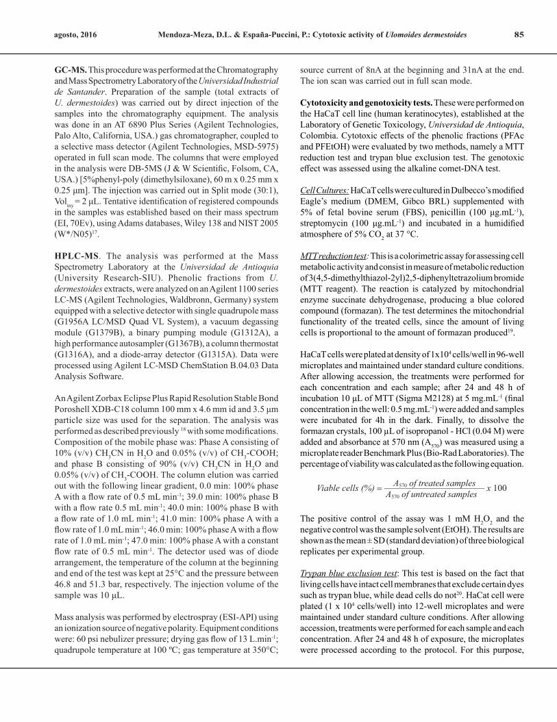
By HPLC-MS, the following compounds were tentatively identified: 3,4-dihydroxybenzoic or protocatechuic acid, dinitrosalicylic acid and ethylresorcinol (Table II). Protocatechuic acid (abbreviation PCA) was common in both fractions (Figure 1); this compound was found in the cuticle of arthropods, such as cockroaches (Blatella orientalis and Periplaneta americana)22-24 and beetles (Pachynoda epphipiata and Tenebrio molitor)25. PCA seems to play the important role in obscuring the cuticle and capturing free radicals. This compound is widely distributed in plant kingdom22, being extracted by their pharmacological properties as antioxidant agent26-27.
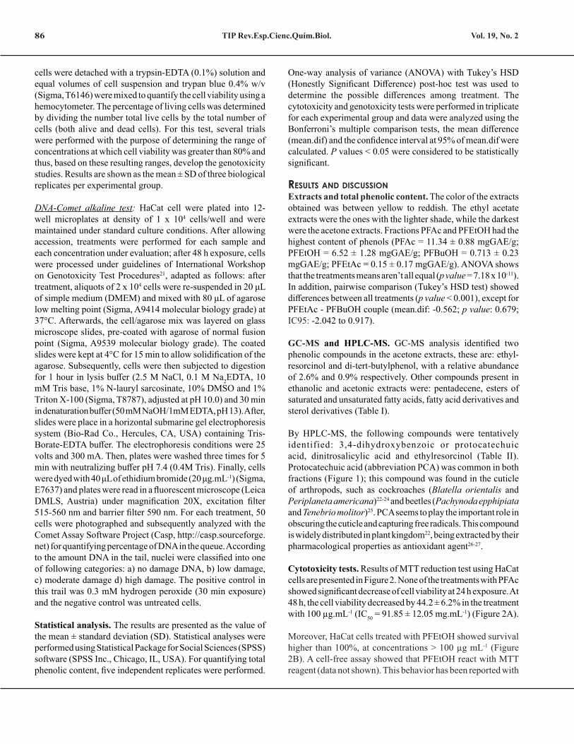
Cytotoxicity tests. Results of MTT reduction test using HaCat cells are presented in Figure 2. None of the treatments with PFAc showed significant decrease of cell viability at 24 h exposure. At 48 h, the cell viability decreased by 44.2 ± 6.2% in the treatment with 100 µg.mL-1 (IC50 = 91.85 ± 12.05 mg.mL-1) (Figure 2A).
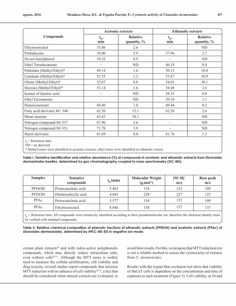
Moreover, HaCat cells treated with PFEtOH showed survival higher than 100%, at concentrations > 100 µg mL-1 (Figure 2B). A cell-free assay showed that PFEtOH react with MTT reagent (data not shown). This behavior has been reported with
Mendoza-Meza, D.L. & España-Puccini, P.: Cytotoxic activity of Ulomoides dermestoides 87agosto, 2016
CompoundsAcetonic extracts Ethanolic extracts
tR,min
Relativequantity, %
tR,min
Relativequantity, %
Ethyiresorcinol 35.86 2.6 - NDPentadecene 38.00 5.9 37.96 2.7Di-tert-butylphenol 38.52 0.9 - NDEthyl Tetradecanoate - ND 46.19 0.4Palmitate (Methyl/Ethyl)* 49.14 1.6 50.13 18.0Linoleate (Methyl/Ethyl)* 52.55 1.2 53.87 30.9Oleate (Methyl/Ethyl)* 52.67 0.8 54.01 38.1Stereate (Methyl/Ethyl)* 53.14 3.6 54.48 3.6Isomer of linoleic acid - ND 58.35 0.8Ethyl Eicosanoate - ND 59.10 1.1Heneicosenoate 60.40 1.0 60.44 0.5Fatty acid derivates M+ 348 62.20 15.1 62.20 2.6Mono stearine 65.83 58.1 - NDNitrogen compound M+337 67.90 3.6 - NDNitrogen compound M+351 71.78 3.9 - NDSterol derivates 81.69 4.0 81.78 1.3
tR = Retention time.ND = no detected.* Methyl esters were identified in acetonic extracts; ethyl esters were identified en ethanolic extract.
Samples Tentative compounds tR (min) Molecular Weight
(g.mol-1)[M−H]−
m/zBase peak
m/zPFEtOH Protocatechuic acid 5.463 154 153 109PFEtOH Dinitrosalicylic acid 4.843 228 227 123
PFAc Protocatechuic acid 5.577 154 153 109PFAc Ethylresorcinol 8.846 138 137 137
tR = Retention time. All compounds were tentatively identified according to their pseudomolecular ion, therefore the chemical identity must be verified with standard compounds.
Table I. Tentative identification and relative abundance (%) of compounds in acetonic and ethanolic extracts from Ulomoides dermestoides beetles, determined by gas chromatography coupled to mass spectrometry (GC-MS).
Table II. Relative chemical composition of phenolic fractions of ethanolic extracts (PFEtOH) and acetonic extracts (PFAc) of Ulomoides dermestoides, determined by HPLC-MS-ESI in negative ion mode.
certain plant extracts28 and with redox-active polyphenolic compounds, which may directly reduce tetrazolium salts, even without cells29-31. Although the MTT assay is widely used to measure the cellular proliferation, cell viability and drug toxicity, several studies report compounds that increase MTT reduction with no enhance of cell viability30,32, a fact that should be considered when natural extracts are evaluated, to
avoid false results. For this, we propose that MTT reduction test is not a reliable method to assess the cytotoxicity of extracts from U. dermestoides.
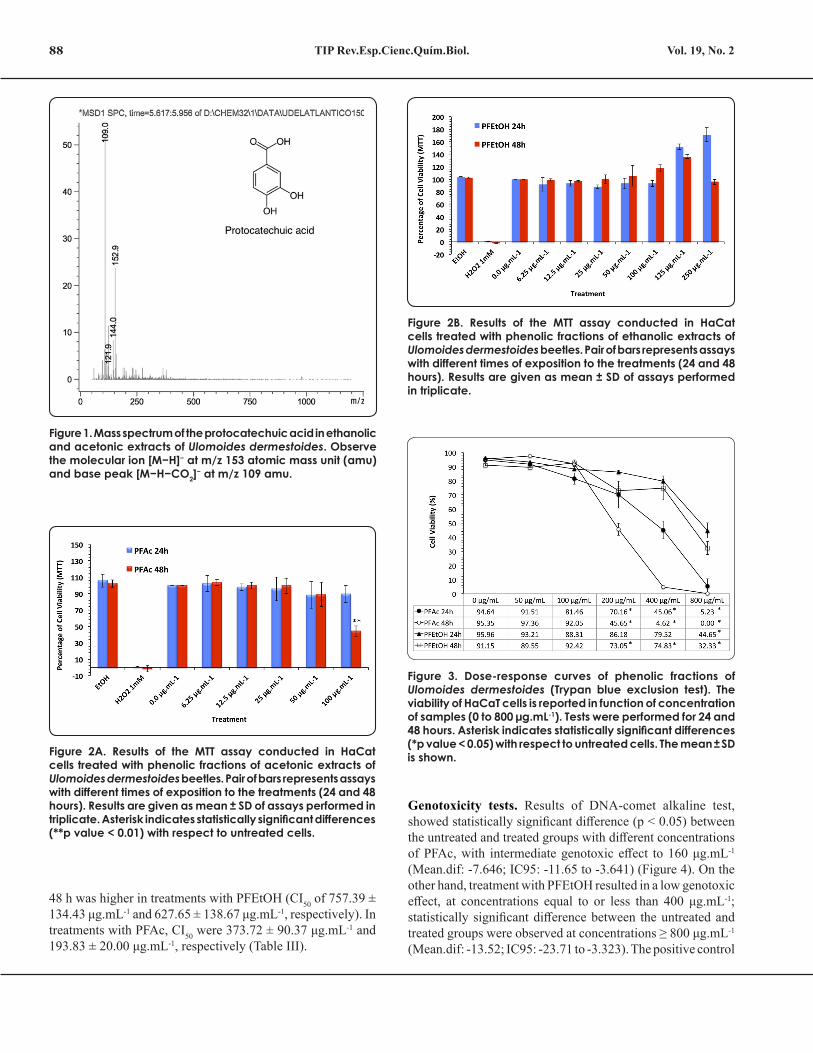
Results with the trypan blue exclusion test show that viability of HaCaT cells is dependent on the concentration and time of exposure to each treatment (Figure 3). Cell viability, at 24 and
TIP Rev.Esp.Cienc.Quím.Biol.88 Vol. 19, No. 2
48 h was higher in treatments with PFEtOH (CI50 of 757.39 ± 134.43 μg.mL-1 and 627.65 ± 138.67 μg.mL-1, respectively). In treatments with PFAc, CI50 were 373.72 ± 90.37 μg.mL-1 and 193.83 ± 20.00 μg.mL-1, respectively (Table III).
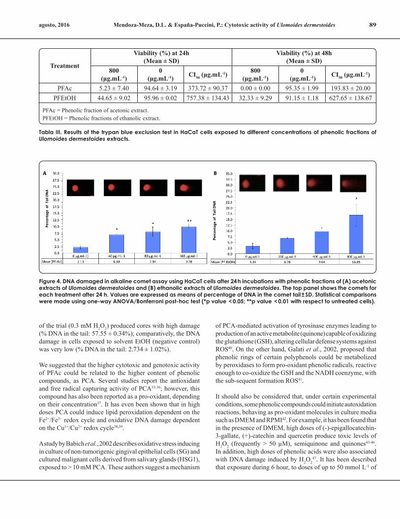
Genotoxicity tests. Results of DNA-comet alkaline test, showed statistically significant difference (p < 0.05) between the untreated and treated groups with different concentrations of PFAc, with intermediate genotoxic effect to 160 μg.mL-1 (Mean.dif: -7.646; IC95: -11.65 to -3.641) (Figure 4). On the other hand, treatment with PFEtOH resulted in a low genotoxic effect, at concentrations equal to or less than 400 μg.mL-1; statistically significant difference between the untreated and treated groups were observed at concentrations ≥ 800 μg.mL-1 (Mean.dif: -13.52; IC95: -23.71 to -3.323). The positive control
Figure 1. Mass spectrum of the protocatechuic acid in ethanolic and acetonic extracts of Ulomoides dermestoides. Observe the molecular ion [M−H]− at m/z 153 atomic mass unit (amu) and base peak [M−H−CO2]− at m/z 109 amu.
Figure 2A. Results of the MTT assay conducted in HaCat cells treated with phenolic fractions of acetonic extracts of Ulomoides dermestoides beetles. Pair of bars represents assays with different times of exposition to the treatments (24 and 48 hours). Results are given as mean ± SD of assays performed in triplicate. Asterisk indicates statistically significant differences (**p value < 0.01) with respect to untreated cells.
Figure 2B. Results of the MTT assay conducted in HaCat cells treated with phenolic fractions of ethanolic extracts of Ulomoides dermestoides beetles. Pair of bars represents assays with different times of exposition to the treatments (24 and 48 hours). Results are given as mean ± SD of assays performed in triplicate.
Figure 3. Dose-response curves of phenolic fractions of Ulomoides dermestoides (Trypan blue exclusion test). The viability of HaCaT cells is reported in function of concentration of samples (0 to 800 µg.mL-1). Tests were performed for 24 and 48 hours. Asterisk indicates statistically significant differences (*p value < 0.05) with respect to untreated cells. The mean ± SD is shown.
Mendoza-Meza, D.L. & España-Puccini, P.: Cytotoxic activity of Ulomoides dermestoides 89agosto, 2016
of the trial (0.3 mM H2O2) produced cores with high damage (% DNA in the tail: 57.55 ± 0.34%); comparatively, the DNA damage in cells exposed to solvent EtOH (negative control) was very low (% DNA in the tail: 2.734 ± 1.02%).
We suggested that the higher cytotoxic and genotoxic activity of PFAc could be related to the higher content of phenolic compounds, as PCA. Several studies report the antioxidant and free radical capturing activity of PCA33-36; however, this compound has also been reported as a pro-oxidant, depending on their concentration37. It has even been shown that in high doses PCA could induce lipid peroxidation dependent on the Fe2+/Fe3+ redox cycle and oxidative DNA damage dependent on the Cu1+/Cu2+ redox cycle38,39.
A study by Babich et al., 2002 describes oxidative stress inducing in culture of non-tumorigenic gingival epithelial cells (SG) and cultured malignant cells derived from salivary glands (HSG1), exposed to > 10 mM PCA. These authors suggest a mechanism
of PCA-mediated activation of tyrosinase enzymes leading to production of an active metabolite (quinone) capable of oxidizing the glutathione (GSH), altering cellular defense systems against ROS40. On the other hand, Galati et al., 2002, proposed that phenolic rings of certain polyphenols could be metabolized by peroxidases to form pro-oxidant phenolic radicals, reactive enough to co-oxidize the GSH and the NADH coenzyme, with the sub-sequent formation ROS41.
It should also be considered that, under certain experimental conditions, some phenolic compounds could initiate autoxidation reactions, behaving as pro-oxidant molecules in culture media such as DMEM and RPMI42. For example, it has been found that in the presence of DMEM, high doses of (-)-epigallocatechin-3-gallate, (+)-catechin and quercetin produce toxic levels of H2O2 (frequently > 50 µM), semiquinone and quinones43-46. In addition, high doses of phenolic acids were also associated with DNA damage induced by H2O2
47. It has been described that exposure during 6 hour, to doses of up to 50 mmol L-1 of
Figure 4. DNA damaged in alkaline comet assay using HaCaT cells after 24 h incubations with phenolic fractions of (A) acetonic extracts of Ulomoides dermestoides and (B) ethanolic extracts of Ulomoides dermestoides. The top panel shows the comets for each treatment after 24 h. Values are expressed as means of percentage of DNA in the comet tail ± SD. Statistical comparisons were made using one-way ANOVA/Bonferroni post-hoc test (*p value < 0.05; **p value < 0.01 with respect to untreated cells).
Treatment
Viability (%) at 24h (Mean ± SD)
Viability (%) at 48h (Mean ± SD)
800(μg.mL-1)
0 (μg.mL-1) CI50 (μg.mL-1) 800
(μg.mL-1)0
(μg.mL-1) CI50 (μg.mL-1)
PFAc 5.23 ± 7.40 94.64 ± 3.19 373.72 ± 90.37 0.00 ± 0.00 95.35 ± 1.99 193.83 ± 20.00PFEtOH 44.65 ± 9.02 95.96 ± 0.02 757.38 ± 134.43 32.33 ± 9.29 91.15 ± 1.18 627.65 ± 138.67
PFAc = Phenolic fraction of acetonic extract.PFEtOH = Phenolic fractions of ethanolic extract.
Tabla III. Results of the trypan blue exclusion test in HaCaT cells exposed to different concentrations of phenolic fractions of Ulomoides dermestoides extracts.
TIP Rev.Esp.Cienc.Quím.Biol.90 Vol. 19, No. 2
purified flavonoids, could cause chromosomal translocation and clastogenicity in human cell lines48,49.
Given that our experiments were performed in cells cultured in DMEM, it is possible that the decrease in cell viability is related to H2O2 production from auto-oxidation of phenolic compounds present in extracts of U. dermestoides analyzed.
ConClusionsEthanolic and acetonic extracts from whole body of Ulomoides dermestoides are a source of redox-reactive phenolic compounds that could be of biomedical interest. In both extracts was tentatively identified the protocatechuic acid, but further analysis with the standard compound would be required to confirm this assignment. Additionally, experimental design developed in this study suggests that the beetle’s ethanol extracts shown low toxicity in HaCaT cell line, at the tested concentrations. We suggest that the greater cytotoxicity/genotoxicity of acetonic extracts could be related to a higher concentration of redox-reactive compounds present in the sample and the time of exposure to these compounds.
referenCes1. Costa Neto, E.M., Ramos-Elorduy, J.& Pino, J.M. Los insectos
medicinales de Brasil: primeros resultados. Boletín Sociedad Entomologica Aragonesa. 38: 395−414 (2006).
2. Chu, G.S., Palmieri, J.R. & Sullivan, J,T. Beetle-eating: a Malaysia folk medical practice and its public health implications. Tropical and Geographical Medicine. 29: 422-427 (1977).
3. Costa-Neto, E.M. The use of insects in folk medicine in the state of Bahia Northeastern Brazil, with notes on insects reported elsewhere in Brazilian folk medicine. Human Ecolology. 30: 245-263 (2002).
4. Flores, G.E., Padin, S.B. & Stetson, R.E. First records of the Oriental species Ulomoides dermestoides (Coleoptera: Tenebrionidae) in Argentina. Revista de la Sociedad Entomologica Argentina. 61: 48-50 (2002).
5. Santos, R.C., Lunardelli, A., Caberlon, E., Bastos, C.M., Nunes, F.B., Pires, M.G., et al. Anti-inflammatory and immunomodulatory effects of Ulomoides dermestoides on induced pleurisy in rats and lymphoproliferation in vitro. Inflammation. 33: 173-179 (2010).
6. Crespo, R., Villaverde, M.L., Girotti, J.R., Guerci, A., Juárez, M.P. & de Bravo, M.G. Cytotoxic and genotoxic effects of defence secretion of Ulomoides dermestoides on A549 cells. Journal of Ethnopharmacology. 136: 204-209 (2011).
7. Tobón, F.A., Gutiérrez, G.P. & Mejia, M.L. Evaluación del perfil neurofarmacológico del aceite de Ulomoides dermestoides (Coleoptera: Tenebrionidae). Revista Colombiana de Entomología. 37: 251-255 (2011).
8. Mendoza, D., Salgado, M. & Durant, L. Capacidad antioxidante de extractos metanólicos de cuerpo entero del escarabajo Ulomoides dermestoides (Chevrolat, 1893). Revista Cubana de Investigaciones Biomédicas. 32: 402-410 (2013).
9. Long, D., Defu, C., Beibei, Z., Xiaocan, L. & Jia, Y. Optimization of extraction conditions for superoxide dismutase from Martianus dermestoides. Journal of Northeast Forestry University. 37: 69-70 (2009).
10. Yu, W.G., Zhang, B.B., Shen, Y.J., Li, Y., Tian, Y.B. & Jiang, M.H. Purification and Characterization of Superoxide Dismutase from Martianus dermestoides Chevrola. Advanced Materials Research. 773: 336-341(2013).
11. Al-Tameemi, W., Dunnill, C., Hussain, O., Komen, M.M., van den Hurk, C.J., Collett, A. & Georgopoulos, N.T. Use of in vitro human keratinocyte models to study the effect of cooling on chemotherapy drug-induced cytotoxicity. Toxicology in Vitro. 28: 1366-1376 (2014).
12. Hewitt, N.J., Edwards, R.J., Fritsche, E., Goebel, C., Aeby, P., Scheel, J., et al. Use of human in vitro skin models for accurate and ethical risk assessment: metabolic considerations. Toxicology Science. 133:209-217 (2013).
13. Odraska, P., Mazurova, E., Dolezalova, L. & Blaha, L. In vitro evaluation of the permeation of cytotoxic drugs through reconstructed human epidermis and oral epithelium. Klin Onkol. 24:195-202 (2011).
14. Boukamp, P., Popp, S., Altmeyer, S., Hulsen, A., Fasching, C., Cremer, T. & Fusenig, N.E. Sustained nontumorigenic phenotype correlates with a largely stable chromosome content during long-term culture of the human keratinocyte line HaCaT. Genes and Chromosomes Cancer. 19:201-214 (1997).
15. Rosler, K.H. & Goodwin, R.S. A general use of Amberlite XAD-2 resin for the purification of flavonoids from aqueous fractions. Journal of Natural Products. 47: 188 (1984).
16. Ozgen, M., Wyzgoski, F. & Tulio, A. Antioxidant capacity and phenolic antioxidants of Midwestern black raspberries grown for direct markets are influenced by production site. Hortscience: A Publication of the American Society for Hortcultural Science. 43: 2039-2047 (2008).
17. Adams, R.P. Identification of essential oil components by Gas Chromatography/Mass Spectroscopy. US (United States): Allured Publ Corp Carol Stream, IL, USA (1995).
18. Simonovska, B., Vovk, I., Andrensek, S., Valentová, K. & Ulrichová, J. Investigation of phenolic acids in yacon (Smallanthus sonchifolius) leaves and tubers. Journal of Chromatography A. 1016: 89-98 (2003).
19. Mosmann, T. Rapid colorimetric assay for cellular growth and survival: Application to proliferation and cytotoxicity assay. Journal of Immunological Methods. 65: 55-63 (1983).
20. Storber, W. Trypan blue exclusion test of cell viability. Current Protocols in Immunology. Appendix 3:Appendix 3B (2001).
21. Tice, R.R., Agurell, E., Anderson, D., Burlinson, B., Hartmann, A., Kobayashi, H., et al. Single cell gel/comet assay: guidelines for in vitro and in vivo genetic toxicology testing. Environmental and Molecular Mutagenesis. 35: 206-221 (2000).
22. Pryor, M.G.M., Rusell, P.B. & Todd, A.R. Phenolic substance concerned in hardening the insect cuticle. Nature. 159: 399-400 (1947).
23. Whitehead, D.L. Haemocytes play a commensal role in the synthesis
Mendoza-Meza, D.L. & España-Puccini, P.: Cytotoxic activity of Ulomoides dermestoides 91agosto, 2016
of the dihydrobenzoate required as a precursor for sclerotization (ootheca) in the cockroach Periplaneta americana (L). Bulletin of Entomological Research. 101: 251-259 (2011).
24. Czapla, T.H., Hopkins, T.L., Kramer, K.J. & Morgan, T.S. Diphenols in hemolymph and cuticle during development and cuticle tannig of Periplaneta americana (L.) and other cockroach species. Archives of Insect Biochemistry and Physiology. 7:13-28 (1988).
25. Andersen, S.O. Cuticular sclerotization in the beetles Pachynoda epphipiata and Tenebrio molitor. Journal of Insect Physiology. 21: 1225-1232 (1975).
26. Sahil, K. & Souravh, B. A review on protocatechuic acid and its pharmacological potential. ISRN Pharmacology. 952943: 1-9 (2014).
27. Tanaka, T., Tanaka, T. & Tanaka, M. Potential cancer chemopreventive activity of protocatechuic acid. Journal of Experimental and Clinical Medicine. 3: 27-33 (2011).
28. Shoemaker, M., Cohen, I. & Campbell, M. Reduction of MTT by aqueous herbal extracts in the absence of cells. Journal of Ethnopharmacology. 93: 381-388 (2004).
29. Peng, L., Wang, B. & Ren, P. Reduction of MTT by flavonoids in the absence of cells. Colloids and Surfaces B: Biointerfaces. 45: 108-111 (2005).
30. Han, M., Li, J., Tan, Q., Sun, Y. & Wang, Y. Limitations of the use of MTT assay for screening in drug discovery. Journal of Chinese Pharmaceutical Sciences. 19: 195-200 (2010).
31. Denizot, F. & Lang, R. Rapid colorimetric assay for cell growth and survival, Modifications to the tetrazolium dye procedure giving improved sensitivity and reliability. Journal of Immunological Methods. 89: 271–277 (1986).
32. Bruggisser, R., von Daeniken, K., Jundt, G., Schaffner, W. & Tullberg-Reinert, H. Interference of plant extracts, phytoestrogens and antioxidants with the MTT tetrazolium assay. Planta Medica. 68:445-448 (2002).
33. Masella, R., Cantafora, A., Modesti, D., Cardilli, A., Gennaro, L., Bocca, A., et al. Antioxidant activity of 3,4-DHPEA-EA and protocatechuic acid: a comparative assessment with other olive oil biophenols. Redox Report. 4: 113-121 (1999).
34. Yoshino, M. & Murakami, K. Interaction of iron with polyphenolic compounds: application to antioxidant characterization. Analytical Biochemistry. 257: 40-44 (1998).
35. Lodovici, M., Guglielmi, F., Meoni, M. & Dolara, P. Effect of natural phenolic acids on DNA oxidation in vitro. Food and Chemical Toxicology. 39: 1205-1210 (2001).
36. Ueda, J.I., Saito, N., Shimazu, Y. & Ozawa, T. A comparison of scavenging abilities of antioxidants against hydroxyl radicals. Archives of Biochemistry and Biophysics. 333:377-384 (1996).
37. Fukumoto, L.R. & Mazza, G. Assessing antioxidant and prooxidant
activities of phenolic compounds. Journal of Agricultural and Food Chemistry. 48: 3597-3604 (2000).
38. Nakamura, Y., Torikai, K. & Ohigashi, H. A catechol antioxidant, protocatechuic acid, potentiates inflammatory leukocyte-derived oxidative stress in mouse skin via a tyrosinase bioactivation pathway. Free Radical Biology and Medicine. 30: 967-978 (2001).
39. Nakamura, Y., Torikai, K. & Ohigashi, H. Toxic dose of a simple phenolic antioxidant, protocatechuic acid, attentuates the glutathione level in ICR mouse liver and kidney. Journal of Agricultural and Food Chemistry. 49: 5674-5678 (2001).
40. Babich, H., Sedletcaia, A. & Kenigsberg, B. In vitro cytotoxicity of protocatechuic acid to cultured human cell from oral tissue: Involvement in oxidative stress. Pharmacology and Toxicology. 91:245-253 (2002).
41. Galati, G., Sabzevari, O., Wilson, J.X. & O’Brien, P.J. Prooxidant activity and cellular effects of the phenoxyl radicals of dietary flavonoids and other polyphenolics. Toxicology. 177: 91-104 (2002).
42. Halliwell, B. Are polyphenols antioxidants or pro-oxidants? What do we learn from cell culture and in vivo studies? Archives of Biochemistry and Biophysics. 476:107-112 (2008).
43. Sang, S., Yang, I., Buckley, B., Hoc, C. & Yang, C. Autoxidative quinone formation in vitro and metabolite formation in vivo from tea polyphenol (−)-epigallocatechin-3-gallate: studied by real-time mass spectrometry combined with tandem mass ion mapping. Free Radical Biology and Medicine. 43: 362-371 (2007).
44. Halliwell, B. Oxidative stress in cell culture: an under-appreciated problem? FEBS Letters. 540: 3-6 (2003).
45. Halliwell, B., Clement, M.V., Ramalingam, J. & Long, L.H. Hydrogen peroxide. Ubiquitous in cell culture and in vivo? IUBMB Life. 50: 251-257 (2000).
46. Long, L.H., Clement, M.V. & Halliwell, B. Artefacts in cell culture: Rapid generation of hydrogen peroxide on addition of (-)-epigallocatechin, (-)-epigallocatechin gallate, (+)-catechin and quercetin to commonly-used cell culture media. Biochemical and Biophysical Research Communications. 273: 50-53 (2000).
47. Ferguson, L.R. Role of plant polyphenols in genomic stability. Mutation Research. 475: 89-111 (2001).
48. Strick, R., Strissel, P.L., Borgers, S., Smith, S.L. & Rowley, J.D. Dietary bioflavonoids induce cleavage in the MLL gene and may contribute to infant leukemia. Proceedings of the National Academy of Sciences of the United States of America. 97: 4790-4795 (2000).
49. Lambert, J.D., Sang, S. & Yang, C.S. Possible controversy over dietary polyphenols: Benefits vs risks. Chemical Research in Toxicology. 20:583-585 (2007).